To investigate the mechanical properties and bone integration capability of bionic gradient porosity Gyroid-structured porous implants. We fabricated porous implants using selective laser melting and large particle sandblasting acid etching techniques, and conducted systematic evaluations through mechanical testing and animal experiments. Mechanical tests demonstrated that the elastic modulus of uniform porosity and gradient porosity Gyroid-structured implants were similar after SLA processing, while the yield strength and compressive strength of gradient porosity Gyroid-structured implants were significantly lower than those of uniform porosity structures. However, all mechanical properties of gradient porosity Gyroid-structured implants matched those of human cancellous bone. Histological examination revealed that at 4 weeks post-implantation, the apical region of gradient Gyroid-structured implants exhibited more new tissue formation compared to uniform porosity Gyroid-structured implants, with no significant difference observed in the neck region. At 8 weeks, bone tissue ingrowth was significantly greater in the neck and apical regions of gradient Gyroid-structured implants compared to uniform porosity Gyroid-structured implants. Additionally, no abnormal inflammatory responses were observed on either type of implant surface. Gradient porosity Gyroid-structured implants demonstrated excellent mechanical properties and enhanced bone integration capabilities, providing a reference for the design of novel implants.
To investigate the ability of novel Gyroid-shaped titanium alloy (TC4) porous bioscaffolds to induce angiogenesis and osteogenesis in bone defect areas. This study employed selective laser melting (SLM) technology to fabricate Gyroid shaped and Cube-shaped TC4 porous bioscaffolds, using the commonly used cube shape as a control. The unit cell size was 4 mm, with a wall thickness or rod diameter of 300 μm and a porosity of approximately 80%. These scaffolds were implanted into rabbit mandibular defect sites (10 mm × 7 mm × 5 mm) to evaluate the angiogenic and osteogenic potential of the Gyroid-shaped scaffold. Material characterization revealed that sandblasted and acid-etched (SLA) TC4 scaffolds met design specifications, exhibiting uniformly distributed micrometer-scale pores and enhanced surface hydrophilicity. Histological staining revealed that compared to the Cube-shaped scaffold, the Gyroid-shaped scaffold induced greater angiogenesis and new bone formation within the bone defect area. Both scaffolds demonstrated good biocompatibility. Western Blot and RT-qPCR results indicated that the Gyroid-shaped scaffold possessed superior angiogenesis potential (compared to the Cube-shaped scaffold). During the early implantation phase (1-2 weeks), Gyroid-shaped scaffolds exhibited higher expression of platelet-endothelial cell surface adhesion molecule 1 (CD31) and endothelial mucin (EMCN). Concurrently, vessel distribution within the scaffold showed spatial variation. Additionally, gene expression of hypoxia-inducible factor 1α (HIF-1α) and vascular endothelial growth factor A (VEGFA) was elevated in the early bone defect area. Imaging analysis confirmed successful implantation of both scaffolds, with the Gyroid-shaped scaffold exhibiting a higher proportion of new bone formation. Consequently, the novel Gyroid-shaped TC4 porous bioscaffold demonstrates excellent potential for angiogenesis and osteogenesis, providing a reference for Gyroid-shaped scaffold-based bone defect repair.
BACKGROUND:Robot-assisted implant surgery has been shown to achieve high levels of accuracy. However, there is currently a paucity of clinical studies evaluating the accuracy of marker-based intraoral scanner (IOS) registration (IR) methods. PURPOSE:The purpose of this study was to compare the accuracy of the marker-based cone beam computed tomography (CBCT) registration (CR) method and the IR method in the dental implant in the robotic computer-aided implant system (R-CAIS). MATERIALS AND METHODS:This retrospective study included 20 participants, with 10 undergoing implant surgery using the CR method within a robotic system, and the remaining 10 receiving implants using the IR method. Preoperative CBCT images used for implant planning were aligned with the postoperative CBCT images to assess and quantify positional deviations in implant placement. The primary outcome measures were FRE, entry deviation, apical deviation, and angular deviation. A Student's t-test was performed to compare differences between the two groups, with a p-value of < 0.05 considered statistically significant. RESULTS:The mean ± standard deviation values for FRE were 0.027 ± 0.007 mm for the CR group and 0.031 ± 0.006 mm for the IR group (p = 0.149). The mean ± standard deviation values for entry deviation were 0.58 ± 0.11 mm for the CR group and 0.53 ± 0.15 mm for the IR group (p = 0.072). The mean ± standard deviation values for apical deviation were 0.52 ± 0.12 mm for the CR group and 0.50 ± 0.14 mm for the IR group (p = 0.730). The mean ± standard deviation values for apical deviation were 1.10 ± 0.34 mm for the CR group and 1.17 ± 0.23 mm for the IR group (p = 0.730). CONCLUSIONS:In R-CAIS, the IR method demonstrated accuracy comparable to that of the CR method, with both methods yielding clinically satisfactory outcomes.
Statement of problem. A unified standard for measuring robot implantation errors has not yet been established. A coordinate measuring machine (CMM) measures the coordinates of an object with high accuracy. However, evaluations of the accuracy of a robotic computer-assisted implant system (R-CAIS) using CMM are lacking. Purpose. The purpose of this in vitro study was to evaluate the accuracy of an optics-based R-CAIS using a CMM and to assess the accuracy of cone beam computed tomography (CBCT), a laboratory scanner (LS), and an intraoral scanner (IOS) in measuring the accuracy of the R-CAIS. Material and methods. Two 60x50x40-mm cubic models were prepared for the experiment. One master model and several replica models were used for the first part. Employing a robotic system software, virtual planning was performed on the digital imaging and communications in medicine (DICOM) image of the master model, and spatial mapping was performed by using an optical tracking marker (OT-marker) to ensure that virtual planning of the master model could be executed when replica casts were drilled and placed the implants. The actual placements of the implants in the replica casts were measured by using CMM. The errors between the actual and virtual-planned positions were calculated. In the second part, virtual planning was performed on the experimental model, and an optics-based R-CAIS was used to drill holes and place the implants. The actual positions of the implants were measured by using CMM, CBCT, LS, and IOS. The errors between the actual and virtual-planned positions were calculated, and the error results among groups were compared by 1-way analysis of variance or a nonparametric test. The Dunnett test was used for post hoc comparison (alpha=.05). Results. In the first part, the entry, apical, and angle deviations were 0.33 +/- 0.10 mm, 0.41 +/- 0.11 mm, and 0.33 +/- 0.13 degrees, respectively. In the second part, as compared with CMM, no statistically significant differences were observed in the LS group (P>.05), whereas significant differences were observed in entry-depth, entry, apical-depth, apical, and angle deviations in the IOS group, as well as in entry-depth and apical-depth deviations in the CBCT group (all P<.05). Conclusions. The optical-based R-CAIS exhibited high accuracy. The application of CBCT for clinical implantation may be close to that of the true deviation.
Statement of problem Although the accuracy of implant placement in robotic computer-assisted implant systems (R-CAISs) can be influenced by registration techniques, surgeon experience, and human-machine interactions, clinical factors such as alveolar bone condition at the implant site have been less studied. The extent to which these factors affect placement accuracy remains uncertain. Purpose The purpose of this in vitro study was to investigate the effect of guide drills with different macrogeometries and bone inclinations on the accuracy of R-CAIS. Material and methods A simulated bone block was fixed, and 3 brands of guide drills—Straumann (gdS) (Ø1.6×41 mm, 026.0056), Nobel (gdN) (Ø2×33 mm, 36118), and Dentium (gdD) (Ø2.2×35 mm, XLD2235)—were mounted on an implant handpiece controlled by a robotic arm (Remebot; Beijing Baihui Weikang Technology Co, Ltd). Drilling was performed at a constant force of 9 N and a speed of 50 mm/second with a vertical displacement of 10 mm and horizontal displacement of 5 mm. Each type of guide drill was tested 20 times. Movement durations were recorded to assess drilling efficiency. Then, 4 experimental models with different simulated bone inclinations were prepared. Implant positions were virtually planned and divided into 3 groups based on drill geometry, each with 4 implant sites. Robot-assisted implant placement was conducted, followed by coordinate measurement using a coordinate measuring machine (CMM, O-Inspect543; Carl Zeiss Industrielle Messtechnik GmbH). Deviations between actual and planned positions were calculated, and the error results among groups were compared using a 2-way ANOVA. Post hoc comparisons between bone inclinations were conducted using the least significant difference (LSD) test (α=.05). Results Cutting efficiency was highest for gdD, followed by gdN and gdS. Two-factor analysis of variance (ANOVA) results indicated that bone inclination had a more significant impact on all 7 deviation indices than the guide drill (P<.001). Significant differences were observed across bone inclinations for all indices (P<.001), whereas the guide drill significantly influenced only angular deviation. A significant interaction between bone inclination and guide drill was found solely for angular deviation (P=.001), with no significant interactions for the remaining indices (P>.05). Conclusions The alveolar bone inclination at the implant site affected the accuracy of R-CAIS implantation. Moreover, in the process of robot-assisted implantation, the selection of guiding drills may affect the accuracy of R-CAIS implantation.
This study aimed to prepare high-quality sandblasted large-grit acid-etched surfaces of pure titanium and to evaluate their physicochemical and biological properties. We investigated the acid-etching parameters of pure titanium under two sandblasting conditions, focusing on acid concentration, temperature, and time, utilizing an orthogonal experimental design. The optimal conditions for each sandblasted titanium surface were determined through range analysis, enabling the production of high-quality sandblasted large-grit acid-etched surfaces. Surface properties, including morphology and elemental composition, were assessed, along with biocompatibility tests for cell adhesion, proliferation, and osteogenic differentiation. For sandblasted group a, the optimal acid-etching parameters were a mixture of 4.6 mol/L sulfuric acid and 0.814 mol/L hydrochloric acid at 60 °C for 60 min, which yielded a moderately rough surface (Group A: Sa = 1.30 ± 0.07μm). The optimal acid etching parameters for sandblasting group b were etching at 80 °C for 30 min in the same mixed solution to form a rough surface (Group B: Sa = 2.60 ± 0.10μm). Both surface treatments resulted in hydrophilicity and superior cell proliferation and osteogenic differentiation compared with those of the controls. Different parameters of large-particle sandblasting and acid etching treatments can create pure titanium surfaces with varying degrees of roughness. However, both of which demonstrate favorable physicochemical and biological properties.
OBJECTIVES:This study evaluated the accuracy of intraoral scanning (IOS) and cone beam computed tomography (CBCT) data registration (ICR) in robotic computer-assisted implant surgery (r-CAIS) using six different intraoral scanners, considering the impact of surgeon experience. METHODS:A total of 112 standardized mandibular partially edentulous models were assigned to six experimental groups and one control group based on the intraoral scanner used. In the control group, preoperative CBCT data were registered with CBCT data from a model containing an optical tracking marker (OT-Marker). In the experimental groups, IOS data from models with OT-Markers were registered with preoperative CBCT data. Each experimental group was further divided based on the surgeon's experience: one subgroup had a surgeon with over 5 years of IOS experience, while the other had a novice surgeon. Following registration, two implants were placed in each model using r-CAIS. Postoperative CBCT images were analyzed to measure and compare three-dimensional (3D) and two-dimensional (2D) deviations of implant positioning. RESULTS:No significant differences were observed in angular, entry level, or apical deviations among the ICR methods using the six intraoral scanners (P > 0.05). However, different scanners found substantial variations in entry, apical, entry depth, and apical depth deviations (P < 0.05). Furthermore, no considerable differences were found in implant deviation indices between surgeons with different levels of scanning experience (P > 0.05). CONCLUSION:The ICR method demonstrates high accuracy across six intraoral scanners, regardless of the surgeon's IOS experience. However, accuracy varies among scanners. CLINICAL SIGNIFICANCE:This in vitro study provides valuable insights for surgeons in selecting an appropriate intraoral scanner for the ICR method. Moreover, it confirms that the method's accuracy is independent of the surgeon's experience, supporting its broader clinical adoption.
Statement of problem. The static computer-aided implant system (S-CAIS), dynamic computer-aided implant system (D-CAIS), and robotic computer-aided implant system (R-CAIS) have been used to improve the accuracy of implant placement. However, the accuracy of freehand (FH),S-CAIS, D-CAIS, and R-CAIS implant placement has not been compared and verified under identical conditions. Purpose. The purpose of this in vitro study was to compare the accuracy of dental implant placement using S-CAIS, D-CAIS, R-CAIS, and FH techniques under identical conditions. Material and methods. A total of 60 standardized polyurethane resin models with missing mandibular teeth were prepared and divided into 4 groups: FH, S-CAIS, D-CAIS, and R-CAIS, each consisting of 15 models. Preoperative implant planning was performed using cone beam computed tomography (CBCT), and 2 implants were placed in each model using the FH, S-CAIS, D-CAIS, and R-CAIS techniques, respectively. Postoperatively, CBCT scans were made for analysis of the entry, apical, and angle deviations. The error results among groups were compared using 1-way analysis of variance or a nonparametric test. The Dunnett test was used for post hoc comarison (alpha=.05). Results. The mean +/- standard deviation values for entry deviation were 1.09 +/- 0.33 mm for the FH group, 0.72 +/- 0.33 mm for S-CAIS, 0.69 +/- 0.29 mm for D-CAIS, and 0.48 +/- 0.18 mm for R-CAIS (P<.05). The mean (quartiles) apical deviations were 1.01 (0.94 -1.22) for the FH group, and the mean +/- standard deviation values were 0.87 +/- 0.07 mm for the S-CAIS group, 0.64 +/- 0.05 mm for D-CAIS, and 0.47 +/- 0.03 mm for R-CAIS (P<.05). The mean +/- standard deviation values for angle deviation for the FH group were 2.74 +/- 0.84 degrees, 1.99 +/- 0.76 degrees for S-CAIS, 0.85 +/- 0.46 degrees for D-CAIS, and 0.53 +/- 0.20 degrees for R-CAIS (P<.05). Conclusions. R-CAIS is a reliable implant placement method, demonstrating better implant accuracy compared with the S-CAIS, D-CAIS, and FH techniques.
Titanium alloys are widely used in dental implants due to their superior biocompatibility and mechanical strength. However, these alloys are prone to corrosion and wear in the oral environment, thereby shortening their clinical lifespan. This study investigates the enhancement of titanium alloy surface properties using magnetic abrasive finishing (MAF) and examines the influence of magnetic needle diameters (0.2–1.5 mm) on surface modification. Titanium alloy samples were processed by MAF and systematically evaluated for surface morphology, grain size, surface hardness, residual stress, electrochemical corrosion behavior, and tribological performance. Results demonstrated that MAF improves surface morphology, significantly refines grain size, and enhances surface hardness and compressive residual stress, thereby optimizing surface properties. The 1.0 mm magnetic needle group demonstrated the best performance, achieving a Vickers hardness of 376.71 ± 12.48 HV and a compressive residual stress of −579.1 ± 8.49 MPa. In addition, this group showed a higher self-corrosion potential (−0.5661 V), a lower corrosion current density (0.0114 μA·cm−2), and the lowest wear rate ((4.49 ± 0.42) × 10−4 mm3/N·m) in artificial saliva, demonstrating superior corrosion and wear resistance. Overall, MAF technology markedly enhances the surface integrity of titanium alloys in artificial saliva through the synergistic effects of grain refinement and stress modulation. These findings provide valuable experimental evidence supporting future efforts to optimize the surface properties of titanium alloy dental implants.
OBJECTIVES:A randomized controlled study was conducted to compare the accuracy of three different robotic-computer aided implant surgical protocols (fully guided surgery, partially guided surgery performed by a novice surgeon, and partially guided surgery conducted by an experienced surgeon) under the same conditions. METHODS:A total of n = 81 implants were placed in porcine ribs according to three different protocols (n = 27 implants per group). All cases were digitally planned, and the final implant positions were captured through cone-beam computed tomography and compared to the planned positions. The primary outcome variable was angular deviation. The secondary outcome variables were global, horizonal, and vertical deviations at the coronal and apical levels. RESULTS:In terms of implant insertion, the novice surgeon exhibited significantly larger angular deviation compared to both the experienced surgeon and the robot, with no significant difference observed between the latter two. Regarding vertical deviation, the robot showed worse performance than both surgeons, though no significant difference was found between the novice and experienced surgeons. CONCLUSIONS:For novice surgeons, using the robot to guide the whole process of implantation can obtain smaller angular deviation, while the vertical deviation may increase; for experienced surgeons, using the robot to guide only the osteotomy process and adopting a freehand implant insertion manner can obtain smallest angular and vertical deviation. CLINICAL SIGNIFICANCE:This study highlights the potential of improving angular precision for novice surgeons through a fully guided surgical protocol, while a partially guided approach may optimize both angular and vertical precision for experienced surgeons, offering tailored strategies based on surgical expertise.
Chondrocytes undergo endoplasmic reticulum stress (ERS)-induced apoptosis under abnormal stimulation. However, the underlying molecular mechanism remains unclear. We investigated the regulatory effect of the PI3K/AKT signaling pathway on ERS and its effect on chondrocyte apoptosis. In addition, we established a unilateral anterior crossbite (UAC) model in rats to induce temporomandibular joint osteoarthritis (TMJOA). Chondrocytes were isolated from the temporomandibular joints and treated with lipopolysaccharide (LPS) in vitro. Protein expression of ERS and apoptosis markers (GRP78 and CASP12) was analyzed by immunohistochemistry and western blotting. The expression of GRP78, CASP12, p-PI3K, and p-AKT significantly increased in the UAC group. LY294002, a PI3K/AKT signaling pathway inhibitor, reduced the protein expression of GRP78, ATF4, CHOP, and CASP12, whereas 740 Y-P, an activation agent, elevated the expression of proteins GRP78, ATF4, CHOP, and CASP12. In the present study, UAC and LPS stimulation induced apoptosis of chondrocytes in the ERS pathway. Inhibition of the PI3K/AKT signaling pathway reduced ERS-induced chondrocyte apoptosis.
OBJECTIVE:To compare the accuracies among three oral implant surgical techniques: freehand (FH), static computer-assisted implant surgery (sCAIS), and robotic computer-assisted implant surgery (rCAIS). METHODS:The polyurethane and bovine femur implant models were fabricated, and 126 and 96 implant sites were designed on them. The implant sites were divided into three groups: FH, sCAIS, and rCAIS, according to the implantation method. The deviation between the actual implant position and the planned position was analyzed and compared by cone beam computed tomography. RESULTS:In the polyurethane model test, the entry deviation, entry-level deviation, apical deviation, apical level deviation, and angle deviation in sCAIS and rCAIS groups were significantly reduced compared with those in the FH group (P<0.05). No significant differences were observed in all kinds of deviations between the sCAIS and rCAIS groups (P>0.05). In the bovine femur model test, the entry deviation, entry-level deviation, apical deviation, apical level deviation, and angle deviation in both sCAIS and rCAIS groups were significantly reduced compared with those in the FH group (P<0.05). No significant differences were observed in all kinds of deviations between the sCAIS and rCAIS groups (P>0.05). CONCLUSION:This in vitro study shows that the rCAIS technique is superior to the freehand, but has the same accuracy as the sCAIS.
Several methods exist for repairing mandibular segmental bone defects, typically employing the implant method to accomplish the repair. Following a comparative analysis, the Ti6Al4V structural scaffold implant was chosen for bone defect repair. Triply periodic minimal surface (TPMS) is characterized by a high surface area to volume ratio, an average curvature of zero, and other notable advantages, providing a new line of thinking for bone tissue scaffolds. In this work, the in vitro osteogenesis of a cell unit measuring 4 mm was investigated. First, the finite element analysis (FEA) method and the mechanical experiment method were employed to screen the elasticity modulus of the cancellous bone of the mandible. Subsequently, the selective laser melting (SLM) technique was adopted to prepare three different structures precisely - Gyroid, octahedron, and cube - each with wall thicknesses of 0.3 mm, 0.4 mm, and 0.5 mm. In the in vitro osteogenic experiments, it was observed through confocal laser scanning microscopy (CLSM) that each scaffold displayed favorable cell spreading at 1 day and 3 days. Moreover, osteoblast cell adhesion and proliferation assays revealed improved cell adhesion and proliferation with prolonged co-cultivation time, signifying the excellent biocompatibility of the structural titanium alloy scaffolds. Furthermore, findings from cell differentiation and bioactivity assays indicated that the Gyroid structure exhibited superior osteogenesis compared to the cube and octahedron structures. However, no statistically significant difference was noted between varying wall thicknesses within the same structure.
STATEMENT OF PROBLEM:The static computer-aided implant system (S-CAIS), dynamic computer-aided implant system (D-CAIS), and robotic computer-aided implant system (R-CAIS) have been used to improve the accuracy of implant placement. However, the accuracy of freehand (FH),S-CAIS, D-CAIS, and R-CAIS implant placement has not been compared and verified under identical conditions. PURPOSE:The purpose of this in vitro study was to compare the accuracy of dental implant placement using S-CAIS, D-CAIS, R-CAIS, and FH techniques under identical conditions. MATERIAL AND METHODS:A total of 60 standardized polyurethane resin models with missing mandibular teeth were prepared and divided into 4 groups: FH, S-CAIS, D-CAIS, and R-CAIS, each consisting of 15 models. Preoperative implant planning was performed using cone beam computed tomography (CBCT), and 2 implants were placed in each model using the FH, S-CAIS, D-CAIS, and R-CAIS techniques, respectively. Postoperatively, CBCT scans were made for analysis of the entry, apical, and angle deviations. The error results among groups were compared using 1-way analysis of variance or a nonparametric test. The Dunnett test was used for post hoc comparison (α=.05). RESULTS:The mean ±standard deviation values for entry deviation were 1.09 ±0.33 mm for the FH group, 0.72 ±0.33 mm for S-CAIS, 0.69 ±0.29 mm for D-CAIS, and 0.48 ±0.18 mm for R-CAIS (P<.05). The mean (quartiles) apical deviations were 1.01 (0.94 -1.22) for the FH group, and the mean ±standard deviation values were 0.87 ±0.07 mm for the S-CAIS group, 0.64 ±0.05 mm for D-CAIS, and 0.47 ±0.03 mm for R-CAIS (P<.05). The mean ±standard deviation values for angle deviation for the FH group were 2.74 ±0.84 degrees, 1.99 ±0.76 degrees for S-CAIS, 0.85 ±0.46 degrees for D-CAIS, and 0.53 ±0.20 degrees for R-CAIS (P<.05). CONCLUSIONS:R-CAIS is a reliable implant placement method, demonstrating better implant accuracy compared with the S-CAIS, D-CAIS, and FH techniques.
ObjectivesThis observational study aimed to evaluate the accuracy of robotic computer-assisted implant surgery (r-CAIS) for full-arch immediate restoration and to analyse possible factors contributing to deviations.MethodsThree edentulous patients (five arches) underwent r-CAIS. Osteotomies were performed using an autonomous robot under the surgeon's supervision, and implant placement was performed in a freehand or robotic manner. Prefabricated provisional prostheses were delivered immediately after surgery. Postoperative cone beam computed tomography scans were performed to assess the deviations between the planned and placed implants. Statistics were compared with deviations of s-CAIS outlined in a meta-analysis.ResultsA sum of 28 implants were used. The mean global coronal and apical deviations measured 0.91 ±0.43 mm and 1.01 ±0.45 mm, respectively, and the mean angular deviation measured 1.21 ±1.24 º. The r-CAIS showed significantly better precision than the s-CAIS in full-arch cases (P <0.001). The implants inserted using the robotic arm exhibited fewer deviations than those placed in the freehand manner. Eighty percent of prefabricated provisional prostheses were successfully delivered.ConclusionsWithin the limitations of the present study, our data suggest that autonomous r-CAIS is a feasible approach for simultaneous immediate restoration in edentulous patients, showing better accuracy than s-CAIS. Further large-scale studies are necessary to verify the advantages and disadvantages of this novel technique and to explore possible factors that influence its accuracy.Clinical SignificanceAutonomous r-CAIS can provide clinically acceptable implant placement accuracy in edentulous patients, significantly surpassing s-CAIS. This level of accuracy may represent a viable therapeutic approach for simultaneous immediate full-arch restoration.
口腔种植机器人是近年来口腔种植领域新的临床技术,鉴于其高精度及高稳定性,现已应用于口腔医学多个专业领域,如口腔种植、口腔颌面外科等.本共识在临床应用及文献检索的基础上,就口腔种植机器人的相关术语定义、临床应用要求、操作规范以及风险防范等方面进行探讨,并依据国内部分专家的临床操作经验提出共识观点.
目的 研究口腔种植机器人在美学区辅助种植即刻修复的临床应用效果.方法 回顾我院进行美学区口腔种植机器人导航种植手术且术后行即刻修复的病例,分析种植体术前设计与术后实际位置之间的偏差.结果 本研究为10 名患者在口腔种植机器人辅助下植入12 颗种植体,术后即刻戴入种植体支持的临时修复体.植入点总偏差为(0.488±0.268)mm(范围0.110~0.810 mm),根尖点总偏差为(0.573±0.278)mm(范围0.180~1.040 mm),角度偏差为1.166°±0.439°(范围0.290°~1.690°).植入点与根尖点的总偏差与横向偏差高度相关,与深度偏差弱相关.结论 种植机器人导航精度高,可在术中精准实现术前设计的种植体位置,适宜在美学区辅助进行种植窝洞预备和种植体植入,有利于快速完成即刻修复等临床程序.
已经有大量文献支持穿牙槽嵴上颌窦底提升和侧壁开窗上颌窦底提升可以获得较高的种植体总体留存率.本文将上颌窦底剩余骨高度、骨增量材料等因素对于上颌窦底提升种植体留存率的影响、上颌窦底提升两大术式的临床指征与优缺点、上颌窦底提升的种植体负荷方案等做一总结性概述和临床建议,希望为广大口腔种植医生的相关临床应用提供参考.
Stomatognathic system rehabilitation (SSR) is an important component of dental implant therapy, involving multiple disciplines and factors. This article focuses on the importance of clinical issues, such as mandibular position, vertical distance, occlusion and temporomandibular joint in SSR, in order to provide reference for dentists in clinical diagnosis and treatment.
 Chen Deping (陈德平)合作论文数15Zhou Libo合作论文数11Liu Qian (刘倩)合作论文数11Wei Geng (耿威)合作论文数Beijing Stomatological Hospital, Capital Medical University/Graduate School of Capital Medical University11Xue-min PI (皮雪敏)合作论文数10Pan Hong (潘红)合作论文数9RunTai Lin (林润台)合作论文数9Gang Xu (徐刚)合作论文数6Ren Bin (任斌)合作论文数5Lingyan Peng合作论文数5Xiu-ying JIANG (姜秀瑛)合作论文数5
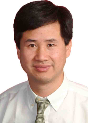 Linhu Ge (葛林虎)合作论文数School & Hospital of Stomatology, Guangzhou Medical University5Gaoyi Wu合作论文数5Weiwei Teng (滕微微)合作论文数4
 Lei Chen (陈磊)合作论文数School of Stomatology, Shandong University4
 Liping Wang (王丽萍)合作论文数School & Hospital of Stomatology, Guangzhou Medical University4
 Yanmin Zhou (周延民)合作论文数Implant Center,Stomatology Hospital,Jilin University4Yong-Jin Chen (陈永进)合作论文数3Xinyu Wang合作论文数3Wang Xia (汪霞)合作论文数3Ye Bi合作论文数3Xueqi Guo合作论文数3Ying Fang合作论文数3GuoXiong Zhu (朱国雄)合作论文数3
 Baosheng Tan (谭包生)合作论文数Beijing Stomatological Hospital Capital Medical University3Shu Qianyi (舒倩怡)合作论文数2Lei Qin合作论文数2lichun wu (武丽春)合作论文数2qing huang (黄庆)合作论文数2Yu Dong (董豫)合作论文数2Xue-jing Zhang (张雪净)合作论文数2YUAN Qiu-hua (苑秋华)合作论文数2Ling-xiao WANG (王凌霄)合作论文数2zhenyu song (宋振宇)合作论文数2jianfeng zhou (周建锋)合作论文数2Qian Liu合作论文数2
 Mubarak Ahmed Mashrah合作论文数Key Lab Oral Med, Guangzhou Med Univ2yanling ren (任燕玲)合作论文数2jia rao (饶佳)合作论文数2ZhanXin Wang (王战鑫)合作论文数2Feng Duan (段峰)合作论文数2Juan Du (杜鹃)合作论文数2
 Yi Man (满毅)合作论文数Hospital of Stomatology, Sichuan University2Liu Changying合作论文数2
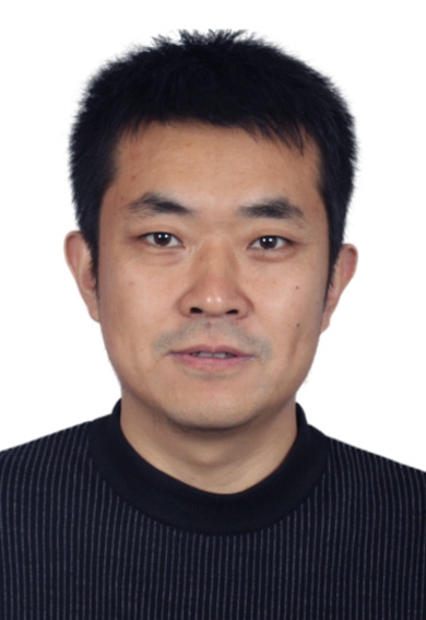 Shujun Li (李述军)合作论文数中国科学院金属研究所2
 Jun Li (李钧)合作论文数Beijing Stomatological Hospital, Capital Medical University;Graduate School of Capital Medical University2Zhen Li (李珍)合作论文数2
 Yan-Liang Wang合作论文数School of Life Sciences, Henan University2Jun Zha (查骏)合作论文数1




