目的 比较年龄相关性白内障患者Symfony连续视程人工晶状体(IOL)与ZMB00多焦点IOL植入术后的视力及视觉质量.方法 纳入行白内障超声乳化吸除联合IOL植入术后的患者共51例(60眼),Symfony组30例(36眼)植入Symfony连续视程IOL(Tecnis Symfony ZXR00),ZMB00组21例(24眼)植入ZMB00多焦点IOL(Tecnis ZMB00).观察两组术后6个月远、中、近视力,术后6个月离焦曲线,通过iTrace视觉功能分析仪检查获得患者术后6个月的高阶像差、传递调制函数(MTF值),采用视功能指数量表(VF-14)评估患者术后生活质量.结果 术后6个月,两组均可提供清晰的远视力;中距离视力(80 cm)Symfony组优于ZMB00组(P<0.001);近距离视力(33 cm)ZMB00组优于Symfony组(P<0.001).两组离焦曲线显示:Symfony组在+1.0~-2.0 D区间趋于平稳,可实现这一区间的连续视程,而在-2.5 D开始呈现下降趋势;ZMB00 IOL离焦曲线呈现双峰状,峰值分别在0 D和-3.0 D.iTrace检查结果显示Symfony组高阶像差及三叶草差低于ZMB00组(均P<0.001).Symfony组的MTF与ZMB00组均较高,差异无统计学意义(P>0.05);Symfony组术后VF-14量表评分高于ZMB00组,差异有统计学意义(P=0.001).结论 两种人工晶状体植入均可有效矫正老视,术后满意度及脱镜率较高,Symfony连续视程IOL可为老年患者提供清晰高质量的全程视力,而ZMB00多焦点IOL的近视力更优.
目的 研究介入技术对恶性气道狭窄患者的治疗效果.方法 综合分析2014年6月~2018年1月期间接受介入治疗的恶性气道狭窄患者27例的临床资料,分析其病因、接受介入治疗的方式、介入治疗的效果及安全性.结果 恶性气道狭窄病因包括肺鳞癌、小细胞肺癌、肺腺癌、食管癌、腺样囊性癌及淋巴瘤等,接受介入治疗的方式有支架、冷冻、氩气刀、高频电刀.恶性气道狭窄患者经介入治疗后,气促评分较治疗前改善明显.在临床疗效评价中,显效7例,部分有效15例,轻度有效5例,总有效率81.48%.介入治疗安全性较高,无严重并发症发生.结论 肺鳞癌是引起恶性气道狭窄的主要病因,支气管镜下接受介入治疗,能有效缓解患者症状,安全性高,并能提高患者生活质量.
资料 患者男性,53岁,因右眼渐进性视力下降1年就诊于我院.1年前无明显诱因出现右眼渐进性视物不清,无畏光眼痛等不适.既往史:1年前于外院行左眼超声乳化白内障摘除联合人工晶状体(intraocular lens,IOL)植入术,植入美国Alcon公司的非球面多焦点散光矫正型IOL(ART IOL).视力:右眼0.05,-2.00 DS →0.1;左眼1.0.眼压:右眼18 mmHg(1 mmHg=0.133 kPa),左眼17 mmHg.右眼晶状体混浊(N2C3P2,LOCSⅡ分类),眼底视不清.左眼瞳孔圆,IOL在位居中,眼底未见异常.光学相干层析成像(optical coherence tomography,OCT):双眼黄斑区形态大致正常.
随着白内障超声乳化手术的广泛开展,白内障手术从复明手术进入到屈光手术时代,与人工晶状体设计不断改进、功能不断完善、技术不断更新密不可分,近些年多焦点人工晶状体在临床工作中越来越多的应用,使白内障患者术后拥有了更好的远中近距离视力和视觉质量,进而满足了患者的生活、工作需要.本文针对在临床运用的几种主要的多焦点人工晶状体,从设计原理、优势、不足之处、术后视力、对比敏感度以及离焦曲线进行综述,了解各种多焦点人工晶状体的特性,针对不同患者进行人工晶状体个性化选择,使患者获得更好的视觉质量.
目的 探讨多焦点人工晶体植入矫正白内障术后老视的视觉质量.方法 19例(26眼)接受白内障超声乳化联合多焦点人工晶体(AMO,美国,ZMB00)植入患者(多焦组),观察术后3个月裸眼远视力、最佳矫正远视力和裸眼近视力、最佳矫正近视力、最佳矫正远视力下的近视力,同时测量调制传递函数(MTF)值,问卷调查术后脱镜率及不良视觉现象发生率.同期接受白内障超声乳化联合单焦点人工晶体(AMO,美国,ZCB00)植入术治疗患者26例(26眼)为单焦对照组.结果 多焦组在裸眼远视力、最佳矫正远视力和最佳矫正近视力与单焦对照组相比没有显著差异,但是多焦组裸眼近视力及最佳矫正远视力下的近视力显著优于单焦对照组(P=0.000).多焦组MTF平均值略低于单焦对照组,但差异没有统计学意义.术后问卷调查术后视近脱镜率(P=0.000)及视觉干扰发生率(P=0.048)多焦组显著高于单焦对照组.结论 白内障手术联合多焦点人工晶体植入很好地改善了老视状态,解决了白内障术后对眼镜的依赖,同时使患者获得了较好的视觉质量.
年龄相关性白内障也被称为老年性白内障,在我国发病率极高,随着年龄的增加,发病率越来越高。白内障超声乳化手术可以使老年性白内障患者重新恢复视力,随着超声乳化手术设备及技术的不断发展,手术时间不断缩短,手术安全性大大提高,但是术中术后还是不可避免的会发生一些不可预知的严重并发症,如后囊膜破裂、晶状体核坠落入玻璃体腔、爆发性脉络膜出血等等。近年来,白内障超声乳化术中房水迷流综合征越来越受到手术医生的关注。本文回顾分析了5例术中发生房水迷流综合征患者的术中突发表现,总结诊断及鉴别诊断要点及危险因素,现报道如下。
白内障是目前最常见的致盲眼病,随着手术技术的提高、设备的改进和人工晶状体(Intraocular lens,IOL)种类的不断创新,白内障患者术后对视力的期望也越来越高[1]。术后获得较好的视觉质量和多种因素息息相关,如术者的手术技巧、患者眼部状态、IOL的性能[2]。其中充分了解IOL的特性,为患者选择合适的、个性化的IOL,减少IOL植入术后的并发症,将为患者带来福音。
晶状体不全脱位可分为先天性、外伤性和其他眼部疾病所致,脱位后引起的高眼压,如果不进行合理的治疗,可能会导致视神经萎缩,是造成此类患者失明的主要原因。目前晶状体不全脱位继发青光眼的患者在临床上越来越多见,在诊断上存在一定的难度,需要与其他类型的青光眼相辨别。这类患者由于眼部病理改变不典型,增加了诊治的难度,
At present, the occurrence and development of cataract in patients operated by vitrectomy is more common. The patient, who would be in cataract surgery, is prone to suffering from higher risks and complications in eyes if he had vitrectomy before than those without vitrectomy. Consequently, it is a significant challenge for ophthalmologists to determine the choice of surgery and the operation during surgery. Compared with cataract surgery after vitrectomy without perfusion, phacoemulsification with 25-gauge perfusion is an effective method, which can maintain the intraocular pressure, stabilize the anterior chamber, and reduce the intraoperative and postoperative complications. In conclusion, phacoemulsification with 25-gauge perfusion is a preferred method for cataract patients after vitrectomy.
研究表明青光眼手术会加速白内障的发生、发展.青光眼术后的患者眼部结构具有一定的特殊性,如滤过泡的存在、角膜内皮细胞计数低、浅前房、虹膜后粘连、瞳孔变形、晶状体核硬度大及悬韧带损伤等,这增加了白内障手术的难度和风险,对于眼科医师来讲是很大的挑战.临床医师应详细评估青光眼术后白内障患者术前眼部情况,正确选择手术时间及手术方案,减少术中不利于滤过泡的操作,术后积极抗炎对症治疗并适当延长糖皮质激素类药物的使用,使患者视力恢复的同时,尽量减少眼压升高的风险,提高患者的生活质量.该文对青光眼手术加速白内障的发生、发展及抗青光眼术后白内障手术难点进行了综述,以期为临床医师提供参考.
Ocular surface disease, particularly dry eye disease (DED), is one of the most common conditions influencing quality of life, and morbidity is increasing with changes in the environment, way of life and age composition, especially in females and old people. Since age-related cataract is the most common reason for patients to undergo phacoemulsification, the overlapping demographics aggravate the incidence and severity of DED even further after cataract surgery. Furthermore, cataract surgery has been shown to lead to worsening DED, which may lead to a dissatisfaction with vision quality and an increase in complications. This review discusses the related factors of DED in the perioperative period of cataract surgery and the latest advances in treatment.
Objective To observe the effect of the use of three-dimensional (3 D)printing technology in the standard-ized training of ear,nose and throat (ENT)resident physicians,as well as exploring its feasibility and effectiveness. Methods The ear,nose,throat,etc models were 3 Dprinted and was used in combination with the standardized train-ing which ENT residents usually receive to teach training theory.Both operation guidance and practice were taught using the aforementioned techniques.Results The 3 D printed model helped the physicians to form a spatial stereoscopic awareness,better understand and master complex anatomical structures in a short time,improve their surgical skills and shortentheirtrainingperiod.Conclusion 3Dprintingtechnologyisagoodprospectinimprovingthestandardized training of ENT resident physicians.
目的 探讨合并特发性肺纤维化(IPF)的糖尿病患者的临床特点、治疗及预后.方法 回顾分析45例合并IPF的糖尿病患者的临床资料,分析患者临床表现、肺功能、胸部影像学、实验室检查、病理检查等特点,总结治疗及预后情况.结果 患者均有渐进性呼吸困难、咳嗽,部分(75.6%)存在低氧血症;75.6%(34/45)、64.4%(29/45)、60.0%(27/45)患者的肺功能分别存在限制型通气障碍、弥散功能障碍、小气道阻塞.患者肺CT均表现为双肺多发网格状及条索状影.22例行经支气管镜肺活检,病理检查均可见较多中性粒细胞浸润.40例患者接受了口服降糖药物或皮下注射胰岛素治疗控制血糖,36例患者静脉应用糖皮质激素;40例患者(88.9%)症状改善,5例患者(11.1%)因呼吸衰竭而死亡.结论 对于糖尿病患者,应定期检查肺功能及胸片,以及早发现IPF.对于合并IPF的糖尿病患者,在控制血糖的基础上,推荐静脉使用或雾化吸入小剂量糖皮质激素,以改善预后.
AIM: To investigate the clinical effect of cataract combined lens dislocation greater than 2 quadrants` patients after having phase I intracapsular cataract extraction and anterior vitrectomy,postoperative corrected vision greater than 0.3,and then taking phase II small incision suture-fixation of intraocular lens suspension.METHODS: Totally 34 cases with cataract combined lens dislocation greater than 2 quadrants had been treated in our hospital from July 2014 to December 2016.We investigated their 1wk,1 and 3mo postoperative complications and the uncorrected visual acuity,best corrected visual acuity,intraocular pressure,corneal astigmatism who took phase II small incision suture-fixation of intraocular lens suspension with corrected vision greater than 0.3 after having phase I intracapsular cataract extraction and anterior vitrectomy.RESULTS: Along with the recovery time extension,patients` uncorrected visual acuity and best corrected visual acuity increased obviously than preoperative in each phase.On the 3mo postoperatively,the best corrected visual acuity of 1 eyes was between 0.3 and 0.1,8 eyes was between 0.5 and 0.3,16 eyes was between 0.7 and 0.5,and 9 eyes was better than 0.7.The best corrected visual acuity was achieved or near the best corrected visual acuity before surgery.Intraocular pressure rechecked became in the normal range at 1wk,1 and 3mo postoperative.Surgery did not significantly increase the corneal astigmatism.CONCLUSION: It is a safe and effective way in improving eyesight effectively,with stable intraocular pressure and fewer complications towards cataract combined lens dislocation greater than 2 quadrants` patients having phase I intracapsular cataract extraction and anterior vitrectomy,whose postoperative corrected vision greater than 0.3,and then phase II small incision suture-fixation of intraocular lens suspension.
At present,surgical treatment of cataract is becoming challenging due to a high prevalence of complication with lens dislocation.Surgical interventions mainly include phacemulsification,capsular tension ring implantation combined with intraocular lens implantation,Artisan iris-claw intraocular lens implantation, suspensory intraocular lens implantation in ciliary groove and multiple modified techniques,etc.However,the range of suspensory ligament rupture can affect the surgical complexity and selection of surgical approach.The range of lens dislocation,suspensory ligament defect or rupture can be classified into less than 1 quadrant,1 to 2 quadrants and >2 quadrants.In this paper,how to choose surgical approaches for different ranges of lens dislocation and the advantages and disadvantages of each surgery were summarized.
In recent years,with the extensive application of trachea intubation in the emergency medicine and surgery under general anesthesia,the incidence of tracheal stenosis has increased.And its treatment methods emerge in more endlessly.However,some patients may undergo tracheal stenosis again after relevant treatments.It is still bothering clinicians how to select an effective treatment method for patients with recurrent tracheal stenosis.This paper reports a case of recurrent tracheal stenosis caused by trachea intubation who is cured by balloon dilation combined with T-tube implantation,which can provide a reference for the clinical treatment of recurrent tracheal stenosis.
目的 检测支气管扩张症合并哮喘患者外周血清白介素-4(IL-4)、γ干扰素(IFN-γ)的表达水平,并分析其临床意义.方法 按照支气管扩张及哮喘的诊断标准将41例支气管扩张和哮喘患者分为A、B、C组,其中14例支气管扩张患者为A组,12例哮喘患者为B组,15例支气管扩张合并哮喘患者为C组,7例健康体检者为D组.应用酶联免疫吸附试验(ELISA)方法测定上述各组研究对象血清中IL-4、IFN-γ的表达水平.结果 (1)外周血清IL-4的表达水平:支扩合并哮喘组IL-4的表达水平317.02±53.53ng/L高于支扩组279.02±44.24ng/L,低于哮喘组367.60±45.10ng/L,差异有统计学意义(P<0.05).(2)外周血清IFN-γ的表达水平:支扩合并哮喘组患者血清中IFN-γ的表达水平241.68±50.33 ng/L低于支扩组295.65±77.10ng/L,高于哮喘组153.76±26.69 ng/L,差异有统计学意义(?P<0.05,△P<0.01).结论 IL-4,IFN-γ在支气管扩张症合并哮喘的发病中可能起一定作用,检测IL-4,IFN-γ对支气管扩张症合并哮喘的辅助诊断有重要参考价值.
Objective:To explore the clinical features of the patients with Kartagener syndrome (KS),and to review the associated literatures about its diagnosis and treatment,and to improve the understanding of the clinician for KS.Methods:The clinical data of one KS patient was collected and the therapeutic effect was analyzed. Results:The patient was clearly diagnoed as KS after relevant examinations. The symptoms such as cough, expectoration,and shortness of breath of the patient were improved after symptomatic treatment, such as anti-inflammation,relieving cough and reducing sputum,and spasmolysis.Conclusion:KS is a congenital autosomal recessive disease.The clinical treatment can only relieve the symptoms,but the structural change is irreversible.The genetics of KS needs further study to reduce its occurrence.
Objective To explore the causes of primary airway stenosis and to choose appropriate treatment modes.Methods The causes of 40 patients with primary airway stenosis in the Second Hospital of Jilin University were collected,and the effects of different treatment modes were observed.Results Among the 40 patients with primary airway stenosis,15 patients (37.5%) had a lump in the trachea,11 patients(27.5 %) had tuberculosis,nine patients (22.5 %) underwent tracheal intubation and incision,three patients (7.5 %) had tracheal mucosal inflammation,one patient (2.5 %) had tracheal amyloidosis,and one patient (2.5 %) underwent had radiotherapy after operation for esophageal cancer.In terms of treatment modes,except for 19 patients who were given conservative treatment,nine patients were given tracheotomy,six patients were given balloon dilatation,five patients were given T-tube insertion,seven patients were given cryotherapy,and six patients were given argon knife treatment.The symptoms like dyspnea of patients who had accepted tracheotomy,balloon dilatation,T-tube insertion,cryotherapy and argon knife therapy all relieved obviously.Conclusions Tracheal intubation and tracheotomy both make the morbidity of airway stenosis increase.The symptoms can be rapidly relieved through tracheotomy.The combination of intervention techniques and T tube implantation has already become an effective method besides surgical procedures.
目的:评价透明晶状体摘除联合后房型折叠人工晶状体植入术矫正中老年超高度近视的有效性及安全性。方法对32例60只超高度近视眼在表面麻醉下行透明晶状体皮质吸出联合后房型折叠人工晶状体植入术。患者年龄40~65〔平均(52.33±11.19)〕岁。双眼28例,单眼4例,随访3~18个月。眼轴长平均(30.03±2.04)mm。比较术前术后视力、最佳矫正视力、散光度数,随访术后并发症情况。结果患眼术后3个月视力≥0.5者32眼(53.3%);最佳矫正视力≥0.5者46眼(76.7%)。术中无1例发生后囊破裂。术后无1例发生网脱,术后有8例14眼(23.3%)发生后囊混浊,行钕:钇-铝-石榴石激光( Nd:YAG)后囊切开术。随访期内未发现黄斑水肿、出血、青光眼、人工晶状体偏心等并发症。结论透明晶状体摘除治疗中老年超高度近视术后效果满意、安全,但熟练掌握超声乳化技术及术前完善的预防性治疗工作至关重要,是开展此项技术的关键,并且术后需长期随访,注意远期并发症的发生。
 XingLong Wang (王兴龙)合作论文数7
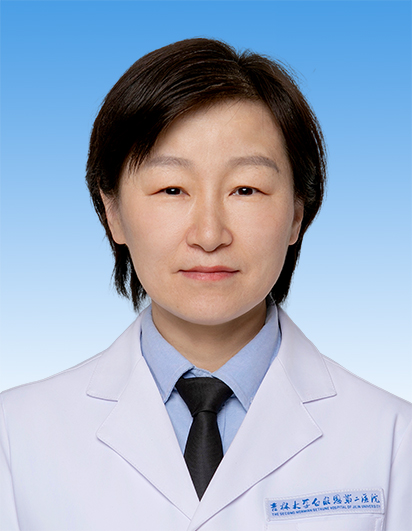 He Zou (邹贺)合作论文数The Second Norman Bethune Hospital, Jilin University6Ying Dou (窦莹)合作论文数6SuJie Lin (林苏杰)合作论文数5
 Junling Yang (杨俊玲)合作论文数The Second Norman Bethune Hospital of Jilin University5LiJuan Bian (边立娟)合作论文数4
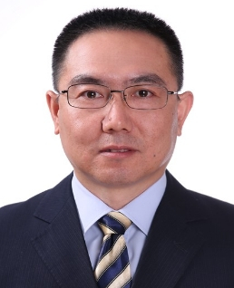 Linzhu Ren (任林柱)合作论文数College of Animal Science, Jilin University4LiJin Cui (崔丽瑾)合作论文数3Kai Liu (刘锴)合作论文数3Xu Ding (丁旭)合作论文数3Qing Zhang (张晴)合作论文数3Meijiao Song合作论文数2jian Yang (杨健)合作论文数2Wenwen Dou (窦文文)合作论文数2huiran bai (白慧然)合作论文数2xuemei liu (刘雪梅)合作论文数2YuChen Dong (董宇晨)合作论文数2LianLian Zhang (张连莲)合作论文数2Wei Zhong (仲炜)合作论文数2ZhenXiang Yu (于振香)合作论文数2Yue Song (宋跃)合作论文数2LiYan Pan (潘丽艳)合作论文数2HaiNing Zhang (张海宁)合作论文数2XiaoYan Ding (丁晓燕)合作论文数2
 Qi Wang (王琦)合作论文数The Second Norman Bethune Hospital of Jilin University2
 Bingdi Yan (闫冰迪)合作论文数The Second Hospital of Jilin University2
 Rui Tian (田蕊)合作论文数The Second Norman Bethune Hospital, Jilin University2
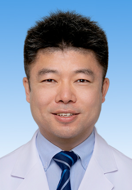 Liangliang Zhao (赵亮亮)合作论文数The Second Norman Bethune Hospital of Jilin University2
 Meisheng Zhao (赵梅生)合作论文数The Second Norman Bethune Hospital of Jilin University2Ailing Zhang (张爱玲)合作论文数2Xiaomin Hu合作论文数1Yingyuan Guo合作论文数1tong sun (孙同)合作论文数1ZHANG Rui (张睿)合作论文数1mingming xie (谢明明)合作论文数1shuxian liu (刘淑贤)合作论文数1Hui LI (李卉)合作论文数1yanxia zhang (张艳霞)合作论文数1LIU Lu (刘璐)合作论文数1Guangmou Yan合作论文数1shuping gong (宫淑萍)合作论文数1Yan-hui HE (何艳慧)合作论文数1Yin-duo LI (李银朵)合作论文数1xiuji zhang (张秀洁)合作论文数1CAO Jian (曹剑)合作论文数1Hai-ying SHI (石海英)合作论文数1Jiang Chen (姜晨)合作论文数1LIU Zhi-tao (刘志涛)合作论文数1wei zhong (仲伟)合作论文数1




