Introduction and aims: Increasing evidence suggests an association between peri-implantitis and osteoporosis, both of which are prevalent diseases that significantly affect patient quality of life. This study aimed to investigate the shared genes and molecular mechanisms underlying the co-pathogenesis of peri-implantitis and osteoporosis. Methods: Transcriptomic data from blood samples of patients with peri-implantitis and osteoporosis. were downloaded from the Gene Expression Omnibus database and shared differentially expressed genes (DEGs) were identified. Analysis of Gene Ontology and Kyoto Encyclopedia of Genes and Genomes pathways was next conducted for shared DEGs. Then, an mRNA–miRNA network of the hub genes was established based on the miRTarbase database. Potential drugs related to the hub genes were predicted using the Genecards database. Finally, the CIBERSORT core algorithm was used to calculate immune cell infiltration levels. Results: Utilizing several gene expression datasets (GSE33774, GSE106090, GSE35956, and GSE35958), we identified a total of 10,839 DEGs. Notably, we identified 3 hub genes that exhibited differential expression in 4 datasets. Functional enrichment analysis revealed that intersected DEGs are involved in regulating oxidative stress, lipid metabolism, and osteoclast differentiation, while immune cell infiltration analyses highlighted correlations between gene expression and various immune cell types. Furthermore, our miRNA–mRNA interaction network analysis suggested complex regulatory mechanisms related to the hub genes. Finally, potential drug targets associated with these hub genes are identified. Conclusion: This study provides valuable insights into the molecular mechanisms underlying peri-implantitis and osteoporosis, highlighting the potential for further investigation into targeted therapies that may improve clinical outcomes for affected patients. Clinical Relevance: The clinical translational value ranges from biomarkers to treatment targets. Shared genes may act as diagnostic markers, informing both the risk of osteoporosis and the bone integration ability of implants. Drugs targeting common genes may offer the potential for 'one drug, two effects'.
INTRODUCTION:Metastasis is an important cause of high mortality and lethality of oral cancer. Fusobacterium nucleatum (Fn) can promote tumour metastasis. Outer membrane vesicles (OMVs) are secreted by Fn. However, the effects of Fn-derived extracellular vesicles on oral cancer metastasis and the underlying mechanisms are unclear. OBJECTIVES:We aimed to determine whether and how Fn OMVs mediate oral cancer metastasis. METHODS:OMVs were isolated from brain heart infusion (BHI) broth supernatant of Fn by ultracentrifugation. Tumour-bearing mice were treated with Fn OMVs to evaluate the effect of OMVs on cancer metastasis. Transwell assays were performed to determine how Fn OMVs affect cancer cell migration and invasion. The differentially expressed genes in Fn OMV-treated/untreated cancer cells were identified by RNA-seq. Transmission electron microscopy, laser confocal microscopy, and lentiviral transduction were used to detect changes in autophagic flux in cancer cells stimulated with Fn OMVs. Western blotting assay was performed to determine changes in EMT-related marker protein levels in cancer cells. Fn OMVs' effects on migration after blocking autophagic flux by autophagy inhibitors were determined by in vitro and in vivo experiments. RESULTS:Fn OMVs were structurally similar to vesicles. In the in vivo experiment, Fn OMVs promoted lung metastasis in tumour-bearing mice, while chloroquine (CHQ, an autophagy inhibitor) treatment reduced the number of pulmonary metastases resulting from the intratumoral Fn OMV injection. Fn OMVs promoted the migration and invasion of cancer cells in vivo, leading to altered expression levels of EMT-related proteins (E-cadherin downregulation; Vimentin/N-cadherin upregulation). RNA-seq showed that Fn OMVs activate intracellular autophagy pathways. Blocking autophagic flux with CHQ reduced in vitro and in vivo migration of cancer cells induced by Fn OMVs as well as reversed changes in EMT-related protein expression. CONCLUSION:Fn OMVs not only induced cancer metastasis but also activated autophagic flux. Blocking autophagic flux weakened Fn OMV-stimulated cancer metastasis.
This study aims to investigate whether the combination of resin infiltration and nanohydroxyapatite (nHA) can provide a synergistic treatment for post-orthodontic white spot lesions (WSL), thus establishing an experimental basis for its treatment. For this study, a total of 60 bicuspid teeth that required extraction due to orthodontics were collected. Orthodontic models were established for each tooth sample, and demineralization staining was conducted using Coca-Cola. The dental samples were randomly divided into 4 groups: control group, nHA group, resin infiltration group, and resin infiltration+nHA group. The demineralized samples were remineralized with 10% nHA and Icon resin infiltration, respectively. The samples were subjected to color difference observation, microhardness test, energy-dispersive X-ray spectrum analysis, and surface roughness detection. The results showed that the tooth surface of the control group was rough and dull after demineralization. The area around the bracket exhibited a brown color, greatly contrasting with the color of the surrounding tooth enamel. After the remineralization treatment with nHA and resin infiltration, the color difference decreased significantly and the microhardness increased. A significant difference was observed between the nHA group and the resin infiltration group (P < 0.05). However, the combination of resin infiltration and nHA exhibited superior staining resistance and improved WSL microhardness value. Additionally, nHA was found to enhance the mass ratio and molar ratio of calcium to phosphorus in WSL samples, while also reducing the surface roughness Ra, demonstrating a greater effectiveness compared to resin infiltration. The use of nHA following resin infiltration led to a significant improvement in mineral deposition and a substantial reduction in surface roughness. In conclusion, both resin infiltration and nHA play a crucial role in promoting enamel remineralization, offering their respective advantages in reducing color difference, increasing microhardness and calcium and phosphorus content, and reducing surface roughness in WSL samples. Remarkably, the combined application of resin infiltration and nHA enables resin infiltration to improve the remineralization and anti-coloring ability in WSL, while preserving its aesthetic effects. This combination demonstrates a positive application outcome.
Background: Head and neck squamous cell carcinomas (HNSCs) are a diverse collection of tumors that originate in the oral cavity, pharynx, and larynx and pose a severe threat to human health, contributing to a fast-rising burden of cancer morbidity and mortality. The search for prognostic biomarkers of HNSC has been a hot topic. Spindle and kinetochore-associated (SKA) complex, including three members SKA1/2/3, which stabilize the spindle microtubules at the kinetosite during mitosis metaphase, has been demonstrated to be associated with poor prognosis of different cancers. The function of SKA1/2/3 in HNSC remains to be investigated. We used a vast variety of public datasets and web-based technologies to investigate SKA complex expression and its link to patient prognosis, and discovered multiple pathways by which the SKA complex is regulated in HNSC. Methods: The Cancer Genome Atlas (TCGA) database was used to determine SKA1/2/3 expression level in HNSC. SKA1/2/3-related proteins level and immune cells infiltration level were identified. Metascape was used for Gene Ontology (GO) and Kyoto Encyclopedia of Genes and Genomes (KEGG) pathway analysis. GSE31056 from Gene Expression Omnibus (GEO) database was used as an external dataset for data validation. A nomogram containing SKA1/2/3 for the prognosis of HNSC patients was established. Results: SKA1/2/3 were highly expressed in HNSC. High expression of SKA2 was significantly related to the poor overall survival (OS) and poor disease-free survival (DFS) of HNSC patients. SKA1/2/3 and the related proteins were enriched in cell division, chromosome segregation, and mitotic cell cycle. SKA1/2/3 expression was obviously positively correlated to several immune cells' infiltration. The expression values of SKA1/2/3 were higher in tumors than in healthy tissues in GSE31056. Conclusions: SKA1/2/3 were shown to be related to the prognosis and immune cell infiltration of HNSC, which could be used as biological markers and therapeutic targets for HNSC.
目的:观察激光照射联合药物脱敏剂应用于牙体硬组织非龋性疾病的临床疗效.方法:将2019年5月-2020年5月于厦门大学附属第一医院就诊的66例患者(158颗患牙)纳入研究,将患者随机分为观察组(33例,83颗患牙)和对照组(33例,75颗患牙).对照组采用药物脱敏剂进行治疗,观察组激光照射联合药物脱敏剂治疗,对比两组视觉模拟评分法(VAS)评分、生活质量(DHEQ-15)及疗效,观察激光照射联合药物脱敏剂对缓解牙体硬组织非龋性疾病导致的牙本质敏感的作用.结果:治疗前两组VAS评分和DHEQ-15评分比较,差异均无统计学意义(P>0.05).治疗后即刻、1、4及8周,观察组经冷气和经机械刺激的VAS评分均低于对照组(P<0.05),且观察组总有效率高于对照组(P<0.05),治疗后8周观察组DHEQ-15评分均低于对照组(P<0.05).结论:激光照射联合药物脱敏剂应用于牙体硬组织非龋性疾病可有效缓解其导致的牙本质敏感,快速降低患者不适感,且治疗简便,值得临床广泛应用.
Objectives: Oral cancer is the ninth most common cancer worldwide and a leading cause of cancer-related death. Oral squamous cell carcinoma (OSCC) accounts for 90% of all oral can-cers. Autophagy is a conserved essential catabolic process related to OSCC. The aim of this study was to elucidate diagnostic and prognostic autophagy-related biomarkers in OSCC.Methods: The OSCC gene expression data set was obtained from the Gene Expression Omni-bus (GEO) database. Differentially expressed genes (DEGs) between the OSCC samples and adjacent healthy tissues were identified by R software. The Human Autophagy Database was screened, which revealed 222 autophagy-related genes. The autophagy-related DEGs were identified. Gene Ontology (GO) and Kyoto Encyclopedia of Genes and Genomes (KEGG) pathway analyses were applied. Protein-protein interaction network analysis was per-formed in the STRING database. cytoHubba in the Cytoscape software was applied to deter-mine the top 10 hub genes. The data set of patients with OSCC from The Cancer Genome Atlas (TCGA) was used to evaluate the prognostic value of the 10 hub genes. The associa-tion between prognosis-related hub genes and immune infiltrates was explored.Results: Twenty-seven autophagy-related DEGs were identified. The top 10 hub genes were CCL2, CDKN2A, CTSB, CTSD, CXCR4, ITGA6, MAP1LC3A, MAPK3, PARP1, and RAB11A. ITGA6 was identified as the most efficient biomarker. Receiver operating characteristic curve analysis indicated that ITGA6 had the highest diagnostic accuracy for OSCC (area under the curve = 0.925). ITGA6 expression was significantly related to immune infiltrates.Conclusions: The autophagy-related gene ITGA6 might be an efficient diagnostic and prog-nostic biomarker in OSCC.(c) 2022 The Authors. Published by Elsevier Inc. on behalf of FDI World Dental Federation. This is an open access article under the CC BY-NC-ND license (http://creativecommons.org/licenses/by-nc-nd/4.0/)
OBJECTIVES:Periodontitis is a chronic inflammatory illness that may lead to tooth loosening and even loss, and its pathogenesis is not fully understood. Ferroptosis is an iron-dependent, regulated cell death. The present study aims to find the key ferroptosis-related genes (FRGs) in periodontitis and develop an mRNA-miRNA-lncRNA network to deeply explore the pathogenesis of periodontitis. METHODS:Data from the Gene Expression Omnibus (GEO) database and FerrDb database were downloaded to discover the differentially expressed mRNA, miRNA, and FRGs. Functional enrichment analysis was conducted for the differentially expressed FRGs (DE-FRGs), including gene ontology, Kyoto Encyclopedia of Genes and Genomes (KEGG) pathway, and protein-protein interaction (PPI) network analysis. Targetscan and miRtarbase were used to estimate the miRNAs that DE-FRGs may interact with, whilst StarBase v3.0 was used for lncRNA-miRNA interaction. RESULTS:Seven DE-FRGs were identified through differential expression analysis. Interleukin 1 beta (IL1B) interacted with XBP1 and MMP13 in the PPI network. After taking the intersection between DE-miRNAs and predicted miRNAs, a ceRNA network containing IL1B, has-miR-185, has-miR-204, has-miR-211, has-miR-4306, and 28 lncRNAs was established. CONCLUSIONS:Seven FRGs in periodontitis were identified, which might promote deeper understanding of ferroptosis in periodontitis.
目的:观察窝沟封闭联合含氟凝胶预防儿童龋齿的效果.方法:从时间为2017年01月至2019年12月之间本院接收的预防龋齿的儿童中随机抽取200例进行调查.将200例预防龋齿的儿童按照随机信封分组法分为两个不同组.参考组儿童单纯使用窝沟封闭进行龋齿预防,实践组儿童使用窝沟封闭联合含氟凝胶进行龋齿预防.对比两组儿童进行预防后的患龋情况、窝沟封闭保存率和家属满意度.结果:在对儿童进行龋齿预防后,实践组儿童的防龋效果优于参考组(P<0.05),对比有意义;实践组儿童的窝沟封闭保存率高于参考组(P<0.05),对比有意义;实践组儿童的家属满意度高于参考组(P<0.05),对比有意义.结论:在对儿童进行龋齿预防时,使用窝沟封闭联合含氟凝胶能够有效降低儿童龋齿发生情况,保障儿童健康成长,提高窝沟封闭保存率,提升家属满意度,值得在临床上研究推广.
目的 探讨疱疹病毒感染与慢性牙周炎活动性的关系及其对牙周基础治疗临床效果的影响.方法 选取2019年1月—2021年1月厦门大学附属第一医院收治的80例接受牙周基础治疗的慢性牙周炎患者为研究对象,其中活性期组34例和静止期组46例,检测患者单纯疱疹病毒1型(HSV-1)、EB病毒(EBV)、人巨细胞病毒(HCMV)阳性率和病毒载量;分析血清肿瘤坏死因子-α(TNF-α)、降钙素原(PCT)、C-反应蛋白(CRP)、白细胞介素-6(IL-6)水平;分析患者治疗后1个月的总有效率和菌斑指数(PLI)、龈沟出血指数(SBI)、牙周袋深度(PD)、附着丧失(AL)情况.结果 活动期组患者HSV-1、EBV和HCMV阳性率均高于静止期组(P<0.05),活动期组患者HSV-1、EBV、HCMV病毒载量和血清PCT、IL-6、CRP和TNF-α水平均高于静止期组(P<0.05);治疗后,活动期患者中,感染组治疗总有效率与未感染组比较差异无统计学意义,静止期患者中,感染组治疗总有效率低于未感染组;感染组和未感染组PLI、SBI、AL及PD均低于治疗前(P<0.05),且感染组PLI、SBI、AL及PD均高于未感染组(P<0.05).结论 疱疹病毒HSV-1、EBV、HCMV感染可能共同参与了慢性牙周炎的发生、发展,并且对牙周基础治疗效果产生影响.
Periodontitis is a chronic inflammatory oral disease that affects nearly 50% of all adults. Fusobacterium nucleatum (F. nucleatum) is known to be involved in the formation and development of periodontitis. Outer membrane vesicles (OMVs) harboring toxic bacterial components are continuously released during F. nucleatum growth and regulate the extent of the inflammatory response by controlling the functions of immune and non-immune cells in tissues. Macrophages are important immune cells in periodontal tissue that resist pathogen invasion and play an important role in the pathophysiological process of periodontitis. However, the role of the interaction between F. nucleatum OMVs and macrophages in the occurrence and development of periodontitis has not been studied. The purpose of this study was to clarify the effect of F. nucleatum OMVs on the polarization of macrophages and the roles of this specific polarization and F. nucleatum OMVs in the pathophysiology of periodontitis. The periodontitis model was established by inducing ligation in C57BL/6 mice as previously described. Micro-CT, RT-qPCR, hematoxylin-eosin (H&E) and tartrate acid phosphatase (TRAP) staining assays were performed to analyze the periodontal tissue, alveolar bone loss, number of osteoclasts and expression of inflammatory factors in gingival tissue. The changes in the state and cytokine secretion of bone marrow-derived macrophages (BMDMs) stimulated by F. nucleatum OMVs were observed in vivo by confocal microscopy, flow cytometry, Western blot and ELISA. Mouse gingival fibroblasts (MGFs) were isolated and then cocultured with macrophages. The effects of F. nucleatum OMVs on the proliferation and apoptosis of MGFs were analyzed by flow cytometry and lactate dehydrogenase (LDH) assays. The periodontitis symptoms of mice in the F. nucleatum OMVs + ligation group were more serious than those of mice in the simple ligation group, with more osteoclasts and more inflammatory factors (IL-1β, IL-6, and TNF-α) being observed in their gingival tissues. M0 macrophages transformed into M1 macrophages after the stimulation of BMDMs with F. nucleatum OMVs, and the M1 macrophages then released more inflammatory cytokines. Analysis of the coculture model showed that the MGF apoptosis and LDH release in the inflammatory environment were increased by F. nucleatum OMV treatment. In conclusion, F. nucleatum OMVs were shown to aggravate periodontitis, alveolar bone loss and the number of osteoclasts in an animal model of periodontitis. F. nucleatum OMVs promoted the polarization of macrophages toward the proinflammatory M1 phenotype, and the inflammatory environment further aggravated the toxicity of F. nucleatum OMVs on MGFs. These results suggest that M1 macrophages and F. nucleatum OMVs play roles in the occurrence and development of periodontitis.
Pulpitis is one of the common diseases indicated by the department of stomatology that is located in the tooth and contains abundant nerve vessels. In order to evaluate the pain degree and functional recovery of patients after treatment by visual analogue pain scale (VAS) and temporomandibular joint function score, a retrospective analysis was performed on 128 patients diagnosed with pulpitis who received root canal treatment in the department of stomatology from January 2020 to March 2021. The results show that 3%NaClO combined with 0.9% sodium chloride injection can effectively relieve the pain degree of patients after treatment, and the antibacterial effect is significantly better than 3%H2O2 combined with 0.9% normal saline. Meanwhile, it can effectively improve the temporomandibular joint function and reduce the recurrence rate, which has good clinical application value.
Background Occlusal trauma can aggravate periodontitis, but the mechanism remains unclear. Yes-associated protein (YAP), a mechanical stressor protein, may play an important role in this process. Methods Western blot and quantitative real-time polymerase chain reaction (qRT-PCR) were applied to detect the expression of YAP and inflammatory factors in patients with periodontitis accompanied with or without occlusal trauma. Through local administration of Porphyromonas gingivalis and composite resin bonding on maxillary molars in mice, we established periodontitis and occlusal trauma models. Treatment with or without XAV939, to inhibit YAP activation, was performed in these models. Micro-computed tomography, immunofluorescence (IF), and qRT-PCR were used to explore the YAP pathway in periodontitis with occlusal trauma. Cyclic stress and lipopolysaccharide (LPS) stimuli were applied to the L929 mouse fibroblast cell line with or without XAV939. Western blot, IF, and qRT-PCR were used to verify the in vivo results. Results Activated dephosphorylated YAP and increased expression of inflammatory factors were observed in patients with periodontitis accompanied with occlusal trauma. In the mouse model of periodontitis with occlusal trauma, YAP transferred into the nucleus, resulting in Jun N-terminal kinases (JNK) related pro-inflammatory pathway up-regulation. L929 cell cyclic stress and LPS stimulation results confirmed the in vivo results. Application of XAV939 inhibited YAP protein dephosphorylation and reduced JNK pro-inflammatory pathway factor expression in vivo and in vitro. Conclusions Occlusal trauma can activate YAP nuclear transfer, resulting in the up-regulation of the JNK pro-inflammatory pathway. This can be inhibited by the XAV939 YAP inhibitor.
Objective:To study the clinical effect of dynamic reconstruction of lower lip defect after squamous cell cancer resection by using unilateral Gillies fan flap and perioral muscle restoration.Methods:From Feburary 2017 to January 2020, 8 patients (6 female and 2 male, aged 55-81, 62.9 in average)diagnosed lower lip squamous cell carcinoma were accepted treatment in the Department of Otolaryngology Head & Neck Surgery, the First Affiliated Hospital of Xiamen University. All the moderate lower lip defect after cancer resection were repaired with unilateral Gillies fan flap with perioral muscle restoration under general anesthesia. The length of the vermilion of the upper lip in the Gillies fan flap was designed to be 1/3 of the length of the vermilion of the lower lip defect. The composite axial flap was prepared with the blood supply from contralateral upper lip artery-ipsilateral upper lip artery-mouth corner part of facial artery-ipsilateral lower lip artery.The end of all the perioral muscles were also prepared. After reconstruction of the continuity of the orbicularis oris, the ends of these perioral radiation muscles were restored in the position where they should be in the reconstructed lip.These perioral muscles training were undergone for 6 months after operation. After the reconstructed lip tend to be stable, measurement and calculation were carried out to evaluate the appearance, opening and closing functions of the mouth.Results:In our series of 8 cases, the length of the vermilion of the upper/lower lip was 52.4 mm(46.0-60.0 mm) in average; the circumference of the vermilion was 104.8 mm(92-120 mm)in average; the length of the defect of lower lip was 27.6 mm (21.0-35.0 mm)in average, 52.5% (42.0-67.3%) of the vermilion of lower lip. Six cases were using the right-side flap, and the other 2 cases were left. The length of the vermilion of the upper lip in the Gillies fan flap was 9.5 mm(7.0-12.0 mm)in average.All cases healed well with inconspicuous scar. No infection, hematoma or other surgical complications.No local or distant recurrence of cancer was observed during the follow-up period. Numbness of the lower lip happened posto-peration, and gradually disappeared 6 months later.Evaluation was conducted according to the measurement result preoperatively and 6 months postoperatively. (1) Appearance: the reconstructed lips were naturally symmetrical. Both side of the mouth corner were in the same horizontal level. The upper lip was lengthened with average increase of 6.8 mm and average increase rate of 15.9%. The lower lip was lengthened with average increase of 15.4 mm and average increase rate of 45.3%. In the rest situation, the circumference of the lips decreased by 5.5 mm in average compared with that preoperatively, which was 94.9% of the lip preoperatively, basically rehabilitate to the shape preoperatively.(2) Mouth-opening function: 1 week postoperation, the opening shape of mouth in all the cases was skewed with an about 1.5 fingers mouth-opening. Six months later, the opening shape of mouth was basically symmetrical round with a 3 fingers mouth-opening; in maximum mouth opening situation, the circumference of the lips decreased by 46.5 mm in average compared with that preoperatively, which was 73.1% of the lip in maximum mouth opening situation preoperatively, basically meeting physiological needs. (3) Mouth-closing function: All patient shave complete mouth-closing function and speech function.No leakage when making closed lip sound and whistling. All patients were satisfied with the oral appearance and the function of opening and closing mouth.Conclusions:Unilateral Gillies fan flap with perioral muscle restoration is a reliable method to repair the moderate full-thickness defect of the lower lip after cancer resection. After dynamic reconstruction of the perioral muscle group, the insufficient tissue of lips can be extended to obtain sufficient mouth opening, natural corner of the mouth and symmetrical lip appearance.
目的 对微种植钉在正畸治疗双颌前突伴露龈笑中的应用效果进行调查.方法 以我院48例双颌前突伴露龈笑患者为调查样本,将其随机分为对照组、观察组,各24例.对照组给予传统支抗治疗,观察组给予微种植钉正畸治疗,对两组患者正畸各项指标进行比较,统计患者正畸时间、并发症发生率.结果 治疗前两组患者UI-E1、L1-E1、GSL水平无统计差异(P>0.05);治疗后观察组患者UI-E1、L1-E1、GSL水平下降程度高于对照组,统计有差异(P<0.05).治疗前两组患者UI-SN、LI-MP、UI-LI、UI/PP、LI/MP水平无统计差异(P>0.05);治疗后观察组患者UI-SN、LI-MP、UI/PP水平下降程度超过对照组,UI-LI、LI/MP上升水平超过对照组,统计有差异(P<0.05).对照组并发症发生率为16.7%;观察组无并发症病例,统计有差异(P<0.05);观察组患者矫正时间为(17.4±3.2)个月,对照组为(24.6±1.9)个月,统计有差异(P<0.05).结论 微种植钉支抗正畸能够缩短正畸时间,正畸效果更理想.
目的:比较使用不同方法修复根管治疗后的下颌第一磨牙疗效的差异.方法:选取2017年1月~10月根管治疗后需行冠部修复的下颌第一磨牙201颗,缺损范围分为邻牙合面洞型MO/OD组(107颗)和近中-牙合面-远中MOD组(94颗).两组患牙根据冠部修复的方案继续分为充填治疗组、高嵌体治疗组、全冠治疗组以及桩核冠治疗组.比较修复后24个月4种不同冠部修复方式及不同缺损类型患牙的治疗效果.结果:修复完成24个月后,4种冠部修复方法治疗总体效果无显著性差异(P>0.05);充填治疗成功率(79.4%)最低.MO/OD组整体成功率为90.7%高于MOD组(88.3%),但两种缺损总体治疗效果差异无统计学意义(P>0.05);采用同种方法修复时,MO/OD组和MOD组缺损修复治疗效果差异亦均无统计学意义(P>0.05).结论:下颌第一磨牙根管治疗后缺损为MO/OD及MOD的患牙,无论采取何种方式进行冠部修复,短期内均可以取得较好的疗效.
目的 对上颌前牙区采用不同即刻种植术的效果及对牙槽骨宽度、 软组织美学的影响进行分析,并探讨安全性.方法 方便2016年1月—2018年8月在该院口腔科接治的38例上颌前牙区即刻种植患者纳入观察研究中,根据唇侧不同骨壁厚度分成试验组和对照组,均为19例;其中对照组采取翻瓣+唇侧GBR即刻种植术,试验组采取不翻瓣+唇侧间隙植骨即刻种植术,对比两组种植区域修复后即刻、术后1年不同时间探诊深度、软组织美学效果;术后即刻、术后1年牙槽骨宽度变化.结果 试验组和对照组所有植体均没有脱落,骨愈合良好,完成修复.试验组术后即刻的牙槽骨宽度、探诊深度分别为(3.23±0.20)mm、(2.62±0.13)mm,对照组分别为(4.58±0.38)mm、(2.51±0.27)mm,差异有统计学意义(t=9.203、8.121,P<0.01),术前、术后1年差异无统计学意义(P>0.05).试验组术后即刻、术后3个月、术后1年的PES评分分别为(9.16±1.10)分、(10.18±1.33)分、(11.30±1.92)分,分别高于对照组的(7.22±1.17)分、(8.54±1.40)分、(9.25±1.84)分,差异有统计学意义(t=7.384、8.092、8.433,P<0.05).结论 在上颌前牙区根据不同骨壁厚度选择不翻瓣和翻瓣即刻种植术的成功率基本一致,1年后骨壁厚度基本一致,但翻瓣组较不翻瓣组美学效果差.
目的:分析多次根管治疗与一次性根管治疗外伤性牙体牙髓病的效果.方法:选择2019年1月-2020年5月在笔者所在医院进行治疗的外伤性牙体牙髓病患者140例开展本次研究,随机分成对照组与试验组,每组70例,对照组给予多次根管治疗,试验组采用一次性根管治疗,对比两组治疗效果.结果:试验组治疗总有效率为95.7%,高于对照组的85.7%,差异有统计学意义(P<0.05);试验组并发症发生率、疼痛评分均低于对照组,治疗时间短于对照组,差异均有统计学意义(P<0.05).结论:采取一次性根管治疗外伤性牙体牙髓病,比多次根管治疗具有更佳的疗效,安全性更高,有效减少患者不良并发症的发生,值得推荐.
目的:探究即刻种植与延期种植对上颌前牙缺损患者植体周围软组织的影响.方法:2017年6月-2018年6月对笔者所在医院收治的80例上颌前牙缺损患者展开研究,采用简单随机法将80例上颌前牙缺损患者随机分为对照组和观察组,每组40例.对照组患者采取延期种植,观察组患者采取即刻种植,对比两组患者种植体周围软组织情况.结果:两组患者修复前红色美学评分(PES)比较,差异无统计学意义(P>0.05);修复后1年,观察组患者PES评分明显高于对照组,差异均有统计学意义(P<0.05).观察组患者修复后1个月改良龈沟出血指数(mBI)低于对照组,差异有统计学意义(P<0.05);观察组患者修复后1个月的探诊深度(PD)与对照组比较,差异无统计学意义(P>0.05).随访1年,观察组种植满意度显著高于对照组,差异有统计学意义(P<0.05).结论:对上颌前牙缺损患者采取即刻种植能够提高患者红色美学指数,提升患者治疗满意度,改善其龈沟出血情况,对患者治疗具有积极意义,值得推广.
目的 分析三维数字化技术辅助颏成形术矫治颏部畸形对颏部形态的影响,总结手术计划.方法 2012年1月~2018年2月,医院颅领面外科共采用三维数字化技术辅助颏成形术,矫治颏部畸形46例常规正畸治疗无效患者,回顾性分析,分析并发症发生情况、随访复发情况、患者满意率,14例对象进行了体像障碍心理健康评估,采用美容心理状态自评量表(Cosmetic Mental States-Scale,CMSS)评价是否出现体像障碍,对比设计、术后即刻、术后随访检测各测量值水平.结果 未见愈合延迟、感染、神经与血管损伤、关节疼痛、下颌关节紊乱等并发症,24例出现面部感觉麻木表现,持续1个月~5个月不等,未见复发、患者手术满意率达到100%,出现1名体像障碍.所有的对象手术都达到预期的目标,美观度、功能恢复达到兼顾,获得患者的好评.三维数字化设计、术后即刻、术后随访的Go-Me/SN、Pog-NB、SNB、Si-EP、N-ANS、ANS-Me、ULL/LLL、Gm-Ns-Pogs、Sn-Gns-C水平差异无统计学意义(P>0.05),术后即刻N-Me高于三维数字化设计、术后随访低于术后即刻,差异有统计学意义(P< 0.05).结论 三维数字化技术辅助颏成形术矫治颏部畸形疗效肯定,设计完成率高,在鼻根点、颏下点距离控制上设计完成率有待提升,但是可以通过后期的正畸等治疗弥补.
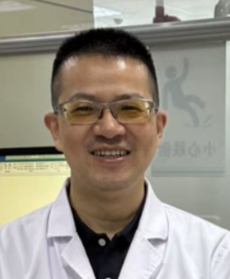 Jianguo Ke (柯建国)合作论文数The First Affiliated Hospital of Xiamen University11
 Xiao-yun CHEN (陈晓芸)合作论文数5
 Lili Xue (薛丽丽)合作论文数The First Affiliated Hospital, Xiamen University;Department of Stomatology, School of Medicine, Xiamen University5Churen Zhang合作论文数4
 Feixiang Wang (王飞翔)合作论文数The First Affiliated Hospital of Xiamen University4
 Xianyang Luo (骆献阳)合作论文数The First Affiliated Hospital of Xiamen University3
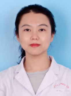 Ou Du (杜欧)合作论文数The First Affiliated Hospital, Xiamen University;Xiamen Medical College3
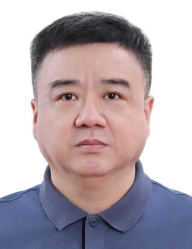 Dongchen Ou (欧东晨)合作论文数The First Affiliated Hospital, Xiamen University;Department of Stomatology, School of Medicine, Xiamen University3
 Wenjin Li (李文进)合作论文数The First Affiliated Hospital, Xiamen University3
 Bing Yan (闫冰)合作论文数The First Affiliated Hospital of Xiamen University;School of Medicine, Xiamen University3Gang Chen合作论文数2Jinhua Tu (涂金花)合作论文数2SuHua Zhang (张素花)合作论文数2QingQuan Fang (方庆全)合作论文数2
 Ruowei Su (苏若为)合作论文数The First Affiliated Hospital of Xiamen University2
 Qiuyan Yang (杨秋燕)合作论文数The First Affiliated Hospital of Xiamen University2
 Hongwei Zhou (周宏伟)合作论文数Zhujiang Hospital, Southern Medical University;The Great Bay Area Center for Brain Research, Southern Medical University2Yina LIN (林艺娜)合作论文数1
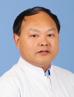 Yubo Xu (徐余波)合作论文数School of Medicine, Tongji University;Department of Stomatology, East Hospital Affiliated to Tongji University1Xiao-hua ZENG (曾晓华)合作论文数1
 Jiajia Wang (王佳佳)合作论文数Center for Drug Safety Evaluation and Research, School of Pharmacy, Zhejiang University1
 Xichun Wang合作论文数Beckman Research Institute, Diabetes and Metabolism Research Institute of the City of Hope1Liangyu Tan合作论文数1Cai-yun LUO (罗彩云)合作论文数1Jie Liu合作论文数1chunbo zhang (张春波)合作论文数1Shan Jiang合作论文数1Jia Li合作论文数1
 Jia Qiao合作论文数1Sufeng CHEN (陈素凤)合作论文数1Pei-qiong CHEN (陈培琼)合作论文数1MingKun Zhan (詹明坤)合作论文数1Can Xiao (肖灿)合作论文数1Rongrong Li合作论文数1Yuan Yue合作论文数1
 Qin Yang (杨琴)合作论文数College of Biomass Science and Engineering, Sichuan University1
 Min Wang (王旻)合作论文数中国药科大学生命科学与技术学院1Linyu Xu (许潾于)合作论文数福建医科大学附属口腔医院正畸科1
 Limei Guan (关丽梅)合作论文数The First Affiliated Hospital of Xiamen University1
 Cheng Chen (陈呈)合作论文数The First Affiliated Hospital of Xiamen University;Department of Stomatology, School of Medicine, Xiamen University1Yi Li合作论文数1
 Hong Chen (陈宏)合作论文数Department of Environmental Science and Engineering, Fudan University1
 Liang Hao (郝亮)合作论文数West China Hospital of Stomatology, Sichuan University1
 Feng Niu (牛峰)合作论文数Plastic Surgery Hospital, Chinese Academy of Medical Science1
 Buling Wu (吴补领)合作论文数Nanfang Hospital, Southern Medical University1
 Ping Gong (宫苹)合作论文数West China School, Sichuan University1
 Jinle Li (李津乐)合作论文数West China Hospital of Stomatology, Sichuan University1
 Pei Sun (孙佩)合作论文数The First Affiliated Hospital of Xiamen University1Chunna Gao (高春娜)合作论文数1




