This study aimed to preliminarily evaluate the value of 18F-FAPI-04 PET/CT in assessing target lesions viability and treatment response in patients with unresectable hepatocellular carcinoma (uHCC) following Yttrium-90(90Y) microsphere selective internal radiation therapy (SIRT). Patients with uHCC who underwent 90Y-SIRT and subsequently received post-SIRT 18F-FDG and/or 18F-FAPI-04 PET/CT evaluations from February 2024 to June 2025 were retrospectively included. Visual assessments of post-SIRT target lesions were performed based on CECT/MRI, 18F-FDG PET/CT, and 18F-FAPI-04 PET/CT, and lesion detection efficacy was compared among the three methods. The SUVmax, peak standardized uptake value corrected for lean body mass (SULpeak), metabolic/FAPI-avid tumor volumetric parameters of target and whole-body lesions before and after SIRT were measured, and the percentage changes (Δ
IntroductionRenal fibrosis is a common pathological feature of chronic kidney disease and a major driver of progression to end-stage renal disease, but its molecular mechanisms remain incompletely understood.MethodsWe integrated multi-omics datasets from GEO and published studies, including mRNA, protein, miRNA, and circRNA data from unilateral ureteral obstruction (UUO) models, TGF-β-induced in vitro fibrosis models, and human umbilical cord mesenchymal stem cell-derived exosomes (HucMSC-Exo). Differential expression analysis, functional enrichment, immune infiltration analysis, fuzzy c-means clustering, weighted gene co-expression network analysis, and ceRNA network construction were performed, with selected findings further validated experimentally.ResultsWe identified stable fibrosis-associated genes and proteins, with metabolic dysregulation emerging as a prominent feature of renal fibrosis. Time-series analysis revealed dynamic transcriptional changes during UUO progression. Comparative analysis showed that in vitro fibrosis models reproduced only part of the in vivo molecular landscape. Immune analyses consistently highlighted macrophages, especially M2-like macrophages, and also suggested a potential role for B cells. In addition, we identified immune-related hub genes and constructed fibrosis-associated ceRNA networks linked to macrophage regulation. Several miRNAs enriched in HucMSC-Exo, particularly miR-30a-5p, were predicted to counteract fibrosis, and exosome treatment alleviated renal injury, macrophage infiltration, and fibrotic marker expression.ConclusionThese findings provide a comprehensive view of the molecular and immune landscape of renal fibrosis, clarify key differences between in vivo and in vitro fibrosis models, and suggest potential therapeutic targets for antifibrotic intervention.
Abstract The fibroinflammatory liver microenvironment (FILM), characterized by collagen-rich stroma and immunosuppressive inflammation, is prevalent in hepatocellular carcinoma (HCC) and correlates with poor response to programmed cell death protein 1 (PD-1) blockade. Here, we show that FILM suppresses gasdermin E (GSDME)-dependent pyroptosis and promotes immune suppression and anti-PD-1 resistance. Mechanistically, FILM-associated cancer-associated fibroblasts recruit and polarize macrophages toward a nitric oxide synthase 2 (NOS2)⁺ inflammatory phenotype. NOS2+ macrophage-derived nitric oxide induces SP1 S-nitrosylation, impairs SP1 binding to the peroxisome proliferator-activated receptor alpha (PPARA) promoter and transcriptionally represses PPARA in HCC cells. PPARα downregulation reduces pyruvate dehydrogenase kinase 4 (PDK4) expression, mitochondrial reactive oxygen species production, caspase-3 activation and GSDME cleavage. Conversely, ligand activation of tumor intrinsic PPARα restores the PDK4–ROS–caspase-3–GSDME axis, enhances dendritic cell and CD8⁺ T cell activation, and sensitizes HCC to anti–PD-1 therapy. The clinically approved PPARα agonist fenofibrate enhances anti-PD-1 efficacy in HCC models in male mice and is associated with improved clinical benefit in a retrospective cohort of patients with HCC. We propose a FILM–NOS2–SP1–PPARα–PDK4 axis that controls pyroptotic immunogenicity and immunotherapy response, supporting PPARα activation as a strategy to overcome FILM-associated immune resistance in HCC.
We present a label-efficient pipeline for CT auto-segmentation in resource-constrained settings. The framework couples a semi-supervised segmentation backbone with a foundation model-guided regularizer to strengthen the learning from scarce annotations. To better exploit volumetric context, we introduce a multi-view collaborative learning procedure that performs view-specific inference to form a unified supervision signal that suppresses view-dependent noise and improves mask fidelity. We evaluate on a public CT benchmark with varying numbers of labeled scans. In the highly label-limited regime, the approach yields strong accuracy with average Dice 83.79% for the liver and 60.08% for the tumor using 20 labeled cases, outperforms existing segmentation methods. By reducing contouring from hours to seconds, improving small-structure recovery and boundary fidelity, and requiring no interactive prompts, the method offers a plug-and-play path to deployment and a reliable basis for downstream radiomics and longitudinal monitoring.
Background:90Y microsphere selective internal radiation therapy (90Y-SIRT) has been widely used for the treatment of unresectable primary or metastatic liver tumors. 99mTc-macroaggregated albumin single-photon emission computed tomography-computed tomography (99mTc-MAA SPECT/CT) and 90Y positron emission tomography-computed tomography (90Y PET/CT) are performed before and after 90Y-SIRT, respectively, for pretreatment dose distribution assessment and posttreatment verification of microsphere distribution. The purpose of this study was to evaluate the predictive value of 99mTc-MAA SPECT/CT for dose distribution assessment and analyze its potential impact on the calculation of absorbed dose to normal liver tissue. To achieve this, the differences in tumor imaging, liver tumor-to-normal ratio (TNR), and normal liver tissue absorbed dose (Dliver) between 99mTc-MAA SPECT/CT and 90Y PET/CT were analyzed. Methods:Thirty-eight patients with primary or metastatic liver cancer who underwent 90Y-SIRT were retrospectively enrolled. Their 99mTc-MAA SPECT/CT and 90Y PET/CT images were analyzed, and the TNR measured by 99mTc-MAA SPECT/CT (TNRSPECT) and 90Y PET/CT (TNRPET) was compared. The absolute differences in TRN (ΔTNR) and percent differences in TNR (%TNR) between the two values were also calculated. Correlation analysis was conducted with patient's age, body mass index, total liver volume, and target tumor volume. The absolute differences in Dliver (ΔDliver) and percent differences in Dliver (%Dliver) as calculated by TNRSPECT and TNRPET were respectively compared. Results:TNRSPECT and TNRPET were not significantly different across imaging modes (P=0.118) and were highly correlated (r=0.854; P<0.01). When stratification by tumor type (primary: n=27; metastatic: n=37) and location (left lobe: n=20; right lobe: n=44) was applied, significant TNR differences emerged only in the left-lobe group (TNRSPECT 5.9 vs. TNRPET 4.7; P=0.015). Bland-Altman analysis demonstrated good agreement in both total and subgroup TNR measurements. The mean ΔTNR was 0.4±1.7, with %TNR ≤92.1%. Dliver calculations showed no significant intermodal difference (P=0.198). Analyses of ΔDliver and %Dliver revealed maximum overestimations of 37.3 Gy (ΔDliver >0) and underestimations of 44.5 Gy (ΔDliver <0), with peak %Dliver reaching 92.0%. Conclusions:Comparison with 90Y PET/CT imaging indicated that 99mTc-MAA SPECT/CT could accurately reflect TNR and Dliver in 90Y-SIRT. However, TNRSPECT may have the tendency to overestimate the true TNR, and appropriate dose adjustments should be considered during treatment planning. The differences in TNR observed in the left-lobe group and the potential differences in the primary group should be further analyzed in larger-sample studies.
PurposeTo compare the efficacy and safety of TACE combined with TKIs and PD-1 inhibitors between HCC patients with and without prior TIPSMethodsThis retrospective propensity score matching (PSM) study included advanced HCC patients treated with prior TIPS followed by TKIs, PD-1 inhibitors, and TACE between January 2021 and January 2023. Patients were matched with a control group of HCC patients who had not undergone TIPS (non-TIPS). Outcome measures included objective response rate (ORR) using modified RECIST (mRECIST v1.1), disease control rate (DCR), progression-free survival (PFS), overall survival (OS), and safety assessed by CTCAE v5.0.ResultsA total of 172 patients were included before PSM. After PSM, 42 patients with prior TIPS were matched with 71 non-TIPS patients. ORR was 31.0% in the TIPS group and 57.7% in the non-TIPS group (p = 0.007), Both PFS and OS were longer in the non-TIPS group, with a median PFS of 7.9 months for TIPS patients versus 12.3 months for non-TIPS patients (hazard ratio [HR] = 2.253, p < 0.001), and a median OS of 13.5 months versus 21.1 months, respectively (HR = 2.282, p = 0.002). Treatment-related adverse events showed no significant differences between the two groups.ConclusionTACE combined with TKIs and PD-1 inhibitors showed lower efficacy in HCC patients with prior TIPS, but it remains a viable option, providing a favorable safety profile and effective disease control.
This study aimed to evaluate the diagnostic value of 18F-FAPI-04 PET/CT in unresectable hepatocellular carcinoma(uHCC) and with a comparative analysis against 18F-FDG PET/CT. Patients with uHCC who underwent paired 18F-FDG and 18F-FAPI-04 PET/CT for pre-treatment staging or post-treatment restaging were retrospectively included. The two tracers were compared in terms of their ability to detect intrahepatic and extrahepatic lesions, including sensitivity, maximum standardized uptake value (SUVmax), and tumour-to-background ratio (TBR). Eighty-five patients (mean age: 53.9 ± 10.0 years) were included. In patient-based analysis, 18F-FAPI-04 showed higher intrahepatic lesion detection sensitivity (98.8
GINS1 is correlated with a poor prognosis in numerous cancers including liver hepatocellular carcinoma (LIHC). Here, efforts have been made to explore the function and underlying mechanism in LIHC through bioinformatics analysis. The mRNA and protein expression data of GINS1 were downloaded from The Cancer Genome Atlas (TCGA) database, the Clinical Proteomic Tumor Analysis Consortium (CPTAC), the University of Alabama at Birmingham CANcer Data Analysis Portal (UALCAN), and the Human Protein Atlas (HPA) database. Moreover, the protein expression of GINS1 was further substantiated by immunohistochemistry staining from 116 clinical samples. Subsequently, the diagnostic and prognostic role of GINS1 in LIHC patients were determined using receiver operating characteristic (ROC) analysis and the Kaplan-Meier plotter (KM-plotter) database. GeneMANIA and STRING databases were respectively used to construct gene and protein-protein interaction (PPI) networks of GINS1. Enrichment analyses were conducted to investigate the functions of GINS1. To assess the genetic alterations, methylation, and prognostic value, cBioPortal, and MethSurv databases were utilized. Additionally, Tumor Immune Estimation Resource (TIMER), Tumor-Immune System Interaction Database (TISIDB), and Gene Expression Profiling Interactive Analysis (GEPIA) were used to explore the correlation with tumor immune. Differential expression analyses validated the upregulation of GINS1 in LIHC. Furthermore, the prognostic and diagnostic values of GINS1 were substantiated by the ROC curve, Kaplan-Meier plotters, and forest plots. Further enrichment, methylation, and tumor immune microenvironment analyses showed an intimate connection with GINS1. In conclusion, GINS1 which is correlated with methylation and immune escape may predict the prognosis of LIHC.
Compared with epirubicin, idarubicin increased survival rates in participants with Barcelona Clinic Liver Cancer stage B hepatocellular carcinoma undergoing drug-eluting bead transarterial chemoembolization without an increase adverse event incidence.
Remnant gastric cancer (RGC) is a serious long-term complication following gastrectomy, for which radical resection remains the standard treatment. With continued advancements in laparoscopic techniques, laparoscopic radical gastrectomy (LRG) has emerged as an important therapeutic option for early-stage and select advanced cases of RGC. This procedure offers reduced surgical trauma, faster postoperative recovery, and a lower incidence of complications. However, several challenges remain in its clinical application. On the one hand, postoperative anatomical changes and intra-abdominal adhesions can limit the surgical field, complicating lymphadenectomy and digestive tract reconstruction. On the other hand, no consensus has been reached regarding the optimal surgical approach, the extent of lymphadenectomy, or the anastomotic technique, resulting in variation in procedural standardization. Nonetheless, with ongoing refinement of laparoscopic techniques, this approach has been increasingly adopted across multiple centers. Its successful implementation largely depends on the surgeons' technical proficiency and clinical experience. This study aims to systematically introduce the experiences of LRG for RGC and provide supplementary information for the existing research data on these techniques.
Myofibroblasts constitute a significant component of the tumor microenvironment (TME) and play a pivotal role in the progression of hepatocellular carcinoma (HCC). Integrin α5 (ITGA5) is a crucial regulator in myofibroblasts of malignant tumors. Therefore, the potential of ITGA5 as a novel target for the therapeutic strategy of HCC should be investigated. Digital scanning and analysis of the HCC tissue microarray were performed to locate the distribution of ITGA5 and conduct the prognosis analysis. CRISPR Cas9-mediated ITGA5 knockout was performed to establish the ITGA5-KO myofibroblast cell line. Extracellular vesicles (EVs) derived from LX2 were extracted for the treatment of HCC cells. Subsequently, the sphere-forming ability and the stemness markers expression of the treated HCC cells were examined. An orthotopic HCC mouse model with fibrotic injury was constructed to test the outcomes of ITGA5-targeting therapy and its efficacy in the programmed death-ligand 1 (PD-L1) treatment. Co-immunoprecipitation/mass spectrometry and transcriptome data were integrated to delve into the mechanism. The tissue microarray results revealed that ITGA5 was highly enriched in the stromal myofibroblasts of HCC tissues and contributed to enhanced tumor progression and poor prognosis. Notably, ITGA5 transmission via extracellular vesicles (EVs) from myofibroblasts to HCC cells induced the acquisition of cancer stem cell-like properties. Mechanistically, ITGA5 directly bind to YES1, facilitating the activation of YES1 and its downstream pathways, thereby enhancing the stemness of HCC cells. Furthermore, the blockade of ITGA5 impeded tumor progression driven by ITGA5+ myofibroblasts and enhanced the efficacy of treatment with PD-L1 in a mouse model of HCC. Our findings elucidated a novel mechanism by which the EV-mediated transfer of ITGA5 from myofibroblasts to tumor cells augmented HCC stemness. ITGA5-targeting therapy helped prevent the progression of HCC and improved the efficacy of PD-L1 treatment.
Background:Globally, liver cancer as one of the most frequent fatal malignancies, hits hard and fast. And the lack of effective treatments for liver hepatocellular carcinoma (LIHC), activates the researchers to promote promising precision medicine. Interestingly, emerging evidence proves that cellular senescence is involved in the progression of cancers and is recognized for its hallmark-promoting capabilities. Hence, efforts have been made to construct and validate the senescence risk score signature (SRSS) model as a novel prognostic biomarker for LIHC. Methods:The existing databases were mined for the following bioinformatics analyses. GSE22405, GSE57957, and senescence-related genes (SRGs) from public databases were utilized as a training set and the validation set was constituted by LIHC and pancreatic adenocarcinoma (PAAD) from The Cancer Genome Atlas (TCGA). After overlapping differentially expressed genes (DEGs) with SRGs, differentially expressed SRGs were identified with the progression of liver cancer through univariate and multivariate Cox regression and enrichment analyses. The model that utilized three SRGs was constructed using the least absolute shrinkage and selection operator (LASSO) regression algorithm. Next, to evaluate the predictive performance of the SRSS model, the overall survival (OS) and survival rates were assessed through Kaplan-Meier (KM) and the receiver operating characteristic (ROC) curves. The predictive value for LIHC prognosis was further evaluated by capitalizing on risk score, nomograms, decision curve analysis (DCA) curves, and clinical information including tumor stages, gender, age, and race. Results:DEGs were revealed as enriching in multiple tumor-related biological processes (BPs) and pathways. IGFBP3, SOCS2, and RACGAP1 were identified as the three considerable SRGs for the model. The high-risk group had a worse prognosis [both hazard ratio (HR) >1, P<0.001] and ROC curves showed a reliable predictive model with area under the curve (AUC) predictive values ranging from 0.673-0.816 for different-year survival rates respectively. The univariate and multivariate Cox regression analyses exhibited that risk score was the only credible prognostic predictor (HR >1, P<0.001) among clinical features such as tumor stage, age, etc., in LIHC. The nomograms, and DCA curves, combined with multiple clinical information, proved that the predictive ability of SRSS was strongest, followed by nomogram and traditional tumor node metastasis (TNM) stage was the weakest. Conclusions:In summary, comprehensive analyses supported that the SRSS model can better predict survival and risk in LIHC patients. Promisingly, it may point out a brand-new direction for LIHC therapy.
Hypersplenism and esophageal variceal hemorrhage caused by portal hypertension are common and serious complications of decompensated cirrhosis. In recent years, with the widespread application of various therapeutic methods such as drugs, endoscopy, splenic artery embolization, transjugular intrahepatic portal shunt, and liver transplantation, the role of surgery in the treatment of portal hypertension has gradually diminished, and the indications for surgical treatment have become more strictly defined. However, according to the clinical practice in China, surgical treatment of portal hypertension still holds an important role that other treatments cannot fully replace. In fact, surgical treatment of portal hypertension is widely performed in hospitals at all levels in China, saving numerous lives. Splenectomy combined with pericardial devascularization (SPD) is the most common surgical method for treating hypersplenism and esophageal variceal hemorrhage caused by portal hypertension. Long-term clinical practice has proven that SPD is a safe and effective treatment for hypersplenism and esophageal variceal rupture and hemorrhage due to portal hypertension. With the rapid development of laparoscopic techniques, the minimally invasive advantages of laparoscopic splenectomy combined with pericardial devascularization (LSPD) have become increasingly evident. However, the successful performance of LSPD mainly depends on the skill and proficiency of the surgeon. In this context, this article presents detailed techniques for LSPD.
BACKGROUND:A 43-year-old female patient was found to have an abnormal liver function, abnormally elevated alpha-fetoprotein and space-occupying lesions in the liver on routine screening. The patient came to our hospital for further diagnosis and treatment. CASE PRESENTATION:Investigations: Laboratory investigations, digital subtraction angiography (DSA) of the hepatic artery, abdominal ultrasound examination, and magnetic resonance imaging (MRI) scan were conducted using pathological staining and immunohistochemistry. DIAGNOSIS:Clinical diagnosis: cT3NxM0. Barcelona clinic liver cancer (BCLC) staging: BCLC stage C. China liver cancer (CNLC) staging: CNLC IIIa. DISCUSSION:The patient was hospitalized for the first time for transcatheter arterial chemoembolization (TACE) and FOLFOX-based hepatic arterial infusion chemotherapy (HAIC). Then, the second and third hospital admissions were given HAIC based on FOLFOX. Camrelizumab and oncolytic virus were also injected into the liver cancer through the microcatheter in the first three treatments. On the fourth admission, the patient's indicators were improved, and the tumor shrank. Furthermore, as the patient suffered adverse reactions the first few times, we suspended the treatment of FOLFOX and the oncolytic virus. Before surgical treatment, lenvatinib was used throughout the treatment. On the fifth admission, the patient underwent liver cancer resection. CONCLUSION:It proves the value of multiple combination therapy, which can provide guidance for patients with advanced hepatocellular carcinoma that cannot be surgically removed.
This study showcases a comprehensive treatment protocol for high-risk hepatocellular carcinoma (HCC) patients, focusing on the combined use of Y-90 transarterial radioembolization (TARE) and Programmed Cell Death-1 (PD-1) inhibitors as neoadjuvant therapy. Highlighted through a case report, it offers a step-by-step reference for similar therapeutic interventions. A retrospective analysis was conducted on a patient who underwent hepatectomy following Y-90 TARE and PD-1 inhibitor treatment. Key demographic and clinical details were recorded at admission to guide therapy selection. Y-90 TARE suitability and dosage calculation were based on Technetium-99m (Tc-99m) macroaggregated albumin (MAA) perfusion mapping tests. Lesion coverage by Y-90 microspheres was confirmed through single photon emission computed tomography/computed tomography (SPECT/CT) fusion imaging, and adverse reactions and follow-up outcomes were meticulously documented. The patient, with a 7.2 cm HCC in the right hepatic lobe (T1bN0M0, BCLC A, CNLC Ib) and an initial alpha-fetoprotein (AFP) level of 66,840 ng/mL, opted for Y-90 TARE due to high recurrence risk and initial surgery refusal. The therapy's parameters, including the lung shunting fraction (LSF) and non-tumor ratio (TNR), were within therapeutic limits. A total of 1.36 GBq Y-90 was administered. At 1 month post-therapy, the tumor shrank to 6 cm with partial necrosis, and AFP levels dropped to 21,155 ng/mL, remaining stable for 3 months. After 3 months, PD-1 inhibitor treatment led to further tumor reduction to 4 cm and AFP decrease to 1.84 ng/mL. The patient then underwent hepatectomy; histopathology confirmed complete tumor necrosis. At 12 months post-surgery, no tumor recurrence or metastasis was observed in follow-up sessions. This protocol demonstrates the effective combination of Y-90 TARE and PD-1 inhibitor as a bridging strategy to surgery for HCC patients at high recurrence risk, providing a practical guide for implementing this approach.
GINS1 regulates DNA replication in the initiation and elongation phases and plays an important role in the progression of various malignant tumors. However, the role of GINS1 in hepatocellular carcinoma (HCC) remains largely unclear. In this study, we investigated the role and underlying mechanisms of GINS1 in contributing to HCC metastasis. We found that GINS1 was significantly upregulated in HCC tissues and cell lines, especially in HCC tissues with vascular invasion and HCC cell lines with highly metastatic properties. Additionally, high expression of GINS1 was positively correlated with the progressive clinical features of HCC patients, including tumor number (multiple), tumor size (>5 cm), advanced tumor stage, vascular invasion and early recurrence, suggesting that GINS1 upregulation was greatly involved in HCC metastasis. Moreover, Kaplan-Meier survival analysis revealed that high GINS1 expression predicted a poor prognosis. Both in vitro and in vivo, silencing of GINS1 inhibited proliferation, migration, invasion and metastasis, while overexpression of GINS1 induced opposite effects. Mechanistically, we found that ZEB1 was a crucial regulator of GINS1-induced epithelial-mesenchymal transition (EMT), and GINS1 promoted EMT and tumor metastasis through β-catenin signaling. Overall, the present study demonstrated that GINS1 promoted ZEB1-mediated EMT and tumor metastasis via β-catenin signaling in HCC.
For recurrent choledocholithiasis, abdominal adhesions in previous surgeries lead to changes in anatomical structures, and a secondary injury occurs easily when performing another operation for laparoscopic common bile duct exploration (LCBDE), which was once considered a relative contraindication. In view of the limitations of the current surgical technique, this study summarized the surgical approaches and crucial anatomical landmarks for reoperation for LCBDE. Four general surgical approaches were proposed to expose the common bile duct, including the ligamentum teres hepatis approach, the anterior hepatic duodenal ligament approach, the right hepatic duodenal ligament approach, and the hybrid approach. Additionally, this study highlighted seven crucial anatomical landmarks: the parietal peritoneum, the gastrointestinal serosa, the ligamentum teres hepatis, the inferior margin of the liver, the gastric antrum, the duodenum, and the hepatic flexure of the colon, which were helpful to safely separate abdominal adhesions and expose the common bile duct. Moreover, to shorten the time of choledocholithotomy, a sequential method was innovatively applied for the removal of the stones in common bile duct. Mastering the above surgical approaches, including identifying crucial anatomical landmarks and adopting the sequential method will improve the safety of reoperation for LCBDE, shorten the operation time, promote the fast recovery of patients, reduce postoperative complications, and contribute to the popularization and application of this technique.
Hypertrophic lysosomes are critical for tumor progression and drug resistance; however, effective and specific lysosome-tar-geting compounds for cancer therapy are lacking. Here we con-ducted a lysosomotropic pharmacophore-based in silico screen in a natural product library (2,212 compounds), and identified polyphyllin D (PD) as a novel lysosome-targeted compound. PD treatment was found to cause lysosomal damage, as evi-denced by the blockade of autophagic flux, loss of lysophagy, and the release of lysosomal contents, thus exhibiting anti-cancer effects on hepatocellular carcinoma (HCC) cell both in vitro and in vivo. Closer mechanistic examination revealed that PD suppressed the activity of acid sphingomyelinase (SMPD1), a lysosomal phosphodieserase that catalyzes the hy-drolysis of sphingomyelin to produce ceramide and phospho-choline, by directly occupying its surface groove, with Trp148 in SMPD1 acting as a major binding residue; this suppression of SMPD1 activity irreversibly triggers lysosomal injury and initiates lysosome-dependent cell death. Furthermore, PD-enhanced lysosomal membrane permeabilization to release sorafenib, augmenting the anticancer effect of sorafenib both in vivo and in vitro. Overall, our study suggests that PD can potentially be further developed as a novel autophagy inhibitor, and a combination of PD with classical chemotherapeutic anti-cancer drugs could represent a novel therapeutic strategy for HCC intervention.
Objectives To identify the most significantly differentially expressed circular RNAs (circRNAs) in colorectal cancer (CRC) tissues in terms of their expression levels and circularity, and to analyze the relationship between their expression levels and the clinical characteristics of patients. Methods circRNA RNA-seq technology was used to screen differentially expressed circRNAs in CRC. Sanger sequencing was used to identify circRNA back-splice junction sites. The relative expression levels of hsa_circ_0003761 (circMSH3) in CRC tissues and cell lines were detected by quantitative real-time fluorescence PCR technology. An RNA-protein pull-down assay was used to detect protein binding to circRNAs. Dual-luciferase reporter gene vectors were constructed to verify that circRNAs bind to microRNAs. Results Four hundred twenty circRNAs were found to be upregulated, and 616 circRNAs were downregulated. circMSH3 was derived from the MutS homolog 3 (MSH3) gene and was formed by a loop of exons 9, 10, 11, and 12. In 110 pairs of CRC and adjacent tissues, circMSH3 expression was 4.487-fold higher in CRC tissues. circMSH3 was also highly expressed in the HT-29 and LOVO CRC cell lines. The expression level of circMSH3 was associated with distant metastasis in CRC patients (P = 0.043); the area under the curve (AUC) of circMSH3 for CRC diagnosis was 0.75, with a sensitivity and specificity of 70.9% and 66.4%, respectively. circMSH3 could bind to a variety of proteins, mainly those involved in RNA transcription, splicing, cell cycle, and cell junctions. Furthermore, circMSH3 could bind to miR-1276, miR-942-5p, and miR-409-3p. Conclusion circMSH3 is a potential biomarker for the diagnosis of CRC and affects the distant metastasis of CRC. Multiple RNA-binding protein binds to circMSH3, and circMSH3 binds to miR-1276, miR-942-5p, and miR-409-3p, thereby affecting the expression of circMSH3.
目的:探讨超声引导下经皮肝穿刺胆管取石术(PTCL)治疗肝移植术后胆道远期并发症的疗效及优势.方法:选择2例肝移植术后胆道远期并发症患者,运用超声引导下PTCL进行胆管取石、胆管狭窄扩张及胆道支架取出.分析2例患者的皮肤瘙痒改善程度、肝功能指标、感染指标及胆管狭窄变化.结果:2例患者治疗后皮肤瘙痒症状显著减轻,肝功能及感染指标显著降低,放置的引流管既能通畅引流又能支撑胆管狭窄.结论:超声引导下PTCL能有效解决肝移植术后胆道远期并发症,该方法安全可行.
 Xuejun Lao (劳学军)合作论文数The Fifth Affiliated Hospital of Jinan University19
 JianWei Jiang (蒋建伟)合作论文数14
 Zhilong Liu (刘志龙)合作论文数College of Plant Protection, China Agricultural University14Yuchuan Jiang合作论文数13
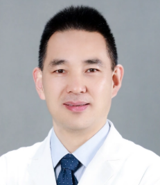 Jin Gong (龚瑾)合作论文数The First Affiliated Hospital of Jinan University12Junjie Liang合作论文数10
 Youzhu Hu (胡友主)合作论文数Jinan University;The Affiliated Shunde Hospital of Jinan University10
 Qiang Li (李强)合作论文数Tianjin Medical University Cancer Hospital10Kangshou Liu合作论文数9bocheng luo (罗伯诚)合作论文数8YuHong Luo (罗羽宏)合作论文数8
 Zhongping Liang (梁忠平)合作论文数The Affiliated Qingyuan People's Hospital of Guangzhou Medical University8Fan Zhang (张帆)合作论文数8
 Jian Hong (洪健)合作论文数School of Medicine, Jinan University8Zeping Han (韩泽平)合作论文数8
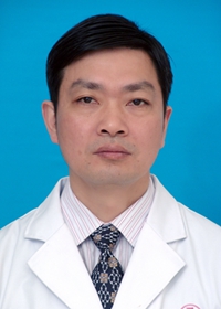 Shaohui Tang (汤绍辉)合作论文数The First Affiliated Hospital of Jinan University7
 Jinhua He (何金花)合作论文数The Affiliated Panyu Central Hospital of Guangzhou Medical University7Jinying Li合作论文数6Min Hu合作论文数6Yu-Guang Li合作论文数6
 Leyang Xiang (相乐阳)合作论文数The First Affiliated Hospital of Jinan University6
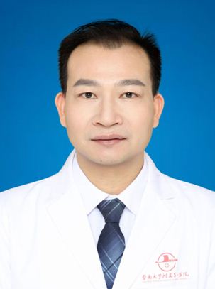 Jiexing Li (李接兴)合作论文数The First Affiliated Hospital of Jinan University6Nan Yao合作论文数6Zhiwei Chen合作论文数5Jia-Bin Zhou合作论文数5
 Hongke Zhou (周鸿科)合作论文数The First Affiliated Hospital of Jinan University5
 Jianwei Jiang (蒋建伟)合作论文数School of Medicine, Jinan University5Lv Yu Bing合作论文数5Rongce Zhao (赵荣策)合作论文数4Yuan Cheng合作论文数4
 Liang-He Lu合作论文数4Lie Zheng合作论文数4Jiming Liu合作论文数4
 Chen Qu合作论文数4MaoXian Zou (邹茂贤)合作论文数4ShengLan Wu (吴胜兰)合作论文数4Hong Zhang (张红)合作论文数4
 Jie Mei (梅洁)合作论文数College of Economics and Management, Nanjing Forestry University4
 Chong Zhong (钟崇)合作论文数广州中医药大学第一临床医学院4
 Yunlong Pan (潘运龙)合作论文数MOE Key Laboratory of Tumor Molecular Biology, Jinan University;The First Affiliated Hospital of Jinan University4
 Shaohua Li合作论文数4Yulong Liu (刘玉龙)合作论文数4
 Rongping Guo (郭荣平)合作论文数Sun Yat-Sen University Cancer Center4Tianqi Lai合作论文数3Jingwen Zou合作论文数3Qijun Cai合作论文数3Qiaoxuan Wang合作论文数3




