目的 探讨损伤控制骨科(damage control orthopaedics,DCO)理论在骨科严重多发伤中的可行性和有效性.方法 回顾性分析采用DCO理论指导一体化救治模式后骨科严重多发伤的救治疗效.自2009-01~2011-12在院前、院内一体化救治模式下,骨科严重多发伤采用DCO理论进行救治,比较连续三年的疗效变化,包括来院至接受确定性手术的时间、ICU住院时间、总住院时间、并发症和死亡率等.结果 DCO理论指导一体化救治模式具有可行性,临床效果明显,缩短了ICU的住院时间,显著缩短了严重多发伤患者入院至接受确定手术的时间,明显降低了多发伤患者的并发症和死亡率.结论 运用DCO理论指导骨科多发创伤的一体化治疗,可以降低患者的死亡率,提高救治成功率.
BACKGROUND:Bone tissue engineering scaffold beta-tricalcium phosphate/calcium polyphosphate fiber/poly-L-lactic acid(β-TCP/CPPF/PLLA) composite has good biocompatibility.OBJECTIVE:To explore the effect of β-TCP/CPPF/PLLA scaffold and bone marrow mesenchymal stem cells(BMSCs) in repairing rabbit long segment bone defects.METHODS:Forty New Zealand white rabbits were divided into BMSCs/β-TCP/CPPF/PLLA group(group A),pure β-TCP/CPPF/PLLA group(group B) and contrast group(group C) randomly.Animal models of long segment radius bone defect were established.The BMSCs/β-TCP/CPPF/PLLA were implanted into the right side of the radius defect(group A),while the pure β-TCP/CPPF/PLLA were implanted into the left side(group B) as control.We did nothing with the contrast group.The rabbits were sacrificed respectively after 4,8,12,16 weeks and the X-ray film was performed at the same time to evaluate the repair effect in different groups.RESULTS AND CONCLUSION:X-ray film showed there was a small quantity of clouding bony callus in defect region after 2 weeks in group A.Marked osteogenesis screenage could be seen after 4 weeks.The defect region was filled with neonate osseous tissue completely and the bone medullary cavity was completely recanalized during 12-16 weeks.The defect region was filled partly in group B and group C,while group B was significant than group C(P < 0.01).X-ray score revealed that group A was much higher than group B at any time(P < 0.01).The BMSCs/β-TCP/CPPF/PLLA composite were capable of repairing long segment bone defects.
Objective To understand the effect of hyaluronic acid (HA) on the proliferation and apoptosis of chondrocytes cultured in vitro with Kashin-Beck disease(KBD) to provide the experimental evidences for treating KBD diseases with HA. Methods The articular cartilage samples collected from KBD patients were selected according to Diagnosis for Kaschin-Beck Disease(GB 16003-1995). And the normal cartilage samples were collected from victims of incidence (control). Chandrocytes were separated and cultured in vitro. Then varying dosages of HA were administered to chondrocytes and individed into 0,100,500 mg/L group, according to HA doages. The effect of HA on the proliferation and apoptosis of chondrocytes cultured/n vitro both KBD and the controls were investigated by methyl thiazolyl tetrazolium(MTT), Annexin V/PI staining on 2~(nd), 4~(th), 6~(th) day. Results In the control group, 500 mg/L group(0.140 ± 0.049) promoted chondrocyte proliferation significantly than 0 mg/L group (0.116 ± 0.021 ) at the 4~(th) day(P < 0.05), similar phenomenon was observed in KBD group in the 6~(th) day between 500 and 0 mg/L group(0.179 ± 0.081,0.128 ± 0.017, P< 0.05). In the KBD group, compared with 0 mg/L (12.860 ± 2.159), both 100 and 500 mg/L( 10.458 ± 1.143,7.877 ± 1.346) inhibited chondrocyte apoptosis rate (P < 0.05). In control, apoptosis rate of 500 mg/L group(4.045 ± 1.204) descreased compared with 0 mg/L group (7.128 ± 1.244, P < 0.05). Conclusion HA can promote the proliferation and inhibit the apoptosis of KBD chondrocytes cultured in vitro, and 500 mg/L HA play more effective role than that of 100 mg/L in promoting proliferation and inhibiting poptosis.
目的:观察骨髓间充质干细胞(MSCs)在TGF-β2诱导下向软骨细胞表型转化的能力,探讨其作为软骨组织工程种子细胞的可能性.方法:抽取兔髂骨骨髓液3~4ml,进行原代和传代培养,传代后实验组以高糖DMEM无血清特定培养液诱导(含TGF-β210ng/ml、地塞米松10-7M、维生素C 50μmol/L),对照组以高糖DMEM无血清培养液培养,相差显微镜下观察细胞形态变化,免疫组织化学染色检测软骨特异性Ⅱ型胶原表达.结果:诱导后细胞体外扩增能力显著降低,细胞形态由成纤维样梭形向多角形、多边形或类圆形转变,诱导21天后细胞形态变化最为显著,Ⅱ型胶原免疫组化染色深而均匀.结论:TGF-β2可有效诱导MSCs向软骨细胞表型转化,分泌软骨细胞特异性基质,有可能成为软骨组织工程较理想的种子细胞来源.
BACKGROUND:Intraarticular hyaluronic acid (HA)has been used as one common method in the treatment of osteoarthritis. But its application in treating Kashin-Beck disease (KBD) is generalizing. The effect of HA on chondrocyte metabolism in KBD patients remains unclear.OBJECTIVE:To investigate the effects of HA on the synthesis of collagen Ⅱ and aggrecan in KBD chondrocytes cultured in vitro to understand its clinical treatment for KBD.DESIGN,TIME AND SETTING:Comparative observation. The experiment was performed in the Laboratory Center of Biomedicine and Endemic Disease Laboratory of College of Medicine,Xi'an Jiaotong University from September 2006 to January 2009.MATERIALS:Samples of KBD were from cartilage and corpus liberum excised from 6 KBD patients undergoing knee joint operation,and normal samples of fresh knee cartilages were provided by 6 people undergoing amputation or death by accident METHODS:Chondrocytes of KBD and normal control were separated and cultured in vitro. Varying dosages of HA (100,500 gm/L) were administered to normal and KBD chondrocytes cultured in vitro for 6 days. Negative control was not treated with HA.MAIN OUTCOME MEASURES:Effects of HA on collagen Ⅱ and aggrecan mRNA expression in KBD patients and normal people by RT-PCR.RESULTS:Compared with normal control group,the mRNA level of collagen Ⅱ and aggrecan were lower in KBD (P<0.05).After administration of HA (100,500 mg/L) the mRNA level of collagen Ⅱ and aggrecan were significantly increased,and the effects of 500 mg/L was more significant.CONCLUSION:HA can increase the collagen Ⅱ and aggrecan mRNA level of KBD chondrocytes cultured in vitro. In particular,500 mg/L HA significantly promoted chondrocyte metabolism compared with 100 mg/L HA.
AIM:To investigate the influence of preoperative limited fluid resuscitation on traumatic shock patients.METHODS:Ninety-six traumatic shock patients complicated with uncontrolled hemorrhage were randomly divided into 3 groups according to the level of systolic blood pressure(SBP) sustained by different fluid resuscitation:group A(around 70 mmHg),group(around 80 mmHg) and group C(over 90 mmHg).General conditions and fluid resuscitation measurement before operation,and the incidence of organ dysfunction(OD) and mortality during hospital stay were compared.RESULTS:(1)There was no significant difference in age,gender,injury severity score(ISS),initiated resuscitation time,initiated operation time and base deficit among the three groups.Before operation,there was significant difference in the volume of fluid resuscitation and the volume of infused erythrocyte suspension between the 3 groups.The volume of fluid resuscitation of group A,group B and group C was respectively(2487±96),(2696±87) and(3200±95) mL(P<0.05),with no significant difference between group A and group B(P>0.05).The volume of infused erythrocyte suspension of Group A,group B and group C was respectively(300±210),(416±163) and(800±130) mL(P<0.05),with no significant difference between group A and group B(P>0.05).(2)The hemoglobin level in group C(90±45) g/L was lower than that in group A(115±32) g/L,and group B(110±34) g/L,(P<0.05),with no significant difference between group A and group B(P>0.05).(3)The incidence of OD in group C was higher than that in group A and group B(45.5%,15.6%,22.5%,P<0.05),with no significant difference between group A and group B(P>0.05).The mortality in group C was higher than that in group A and group B,(39.3%,9.37%,16.1%,P<0.05),with no significant difference between group A and group B(P>0.05).CONCLUSION:The results of our study show that the preoperative limited fluid resuscitation can reduce the volume of bleeding,the incidence of OD and mortality rate in traumatic shock patients with uncontrolled hemorrhage.
Objective To investigate the bone mineral density(BMD) in part of the population in Xi'an district and provide the basis for prevention,diagnosis and treatment of osteoporosis.Methods The BMDs of 1478 inhabitants of different age group in Xi'an district were measured using dual energy X-ray absorptiometry(DEXA).Results The peak BMDs(PBM) lie in the age group of 20-24 for women,and in the age group of 30-34 for men.but two small BMD peaks occurred in the age group of 30~34、50 in females and of 40、60~64 in men.The bone loss rate and prevalence of osteoporosis increased after 50 years old in women and 55 in men.Conclusions The prevalence of osteoporosis increases with age,especially after 50 years old in women and 55 years old in men.
目的体外培养大骨节病(Kashin-Beckdisease,KBD)患者和正常对照的关节软骨细胞并对比观察细胞形态、I、Ⅱ、Ⅲ和X型胶原表型及基质金属蛋白酶13(MMP-13)的表达变化,以明确KBD软骨细胞的生长特性。方法依据我国“大骨节病临床诊断标准”和病理学变化特征,选择KBD患者为KBD组(n=8),并设立正常对照组(n=8)。在无菌手术条件下采集关节软骨,胶原酶等消化、分离后进行软骨细胞培养,光镜及透射电子显微镜观察形态,免疫组织化学及流式细胞术观察细胞I、Ⅱ、Ⅲ、X型胶原和MMP-13的体外表达变化,对比研究KBD软骨细胞表型表达的特点。结果KBD软骨细胞内空泡及核变形现象较正常增多,并有绒毛及胶原纤维不规则排列等超微结构变化。两组间I、Ⅱ、Ⅲ、X型胶原表型表达差异显著,其中KBD组II型胶原表型表达显著低于对照组(t=-5.54,P〈O.001),而MMP-13表达显著增高(t=3.70,P〈O.01)。结论KBD软骨细胞I、Ⅱ、Ⅱ、X型胶原表型和MMP-13的表达不同于正常对照组,以软骨细胞的退行性和增生性变化为著。
目的体外培养大骨节病(Kashin-Beck disease,KBD)患者和正常对照的关节软骨细胞并对比观察细胞形态、Ⅰ、Ⅱ、Ⅲ和Ⅹ型胶原表型及基质金属蛋白酶13(MMP-13)的表达变化,以明确KBD软骨细胞的生长特性。方法依据我国"大骨节病临床诊断标准"和病理学变化特征,选择KBD患者为KBD组(n=8),并设立正常对照组(n=8)。在无菌手术条件下采集关节软骨,胶原酶等消化、分离后进行软骨细胞培养,光镜及透射电子显微镜观察形态,免疫组织化学及流式细胞术观察细胞Ⅰ、Ⅱ、Ⅲ、Ⅹ型胶原和MMP-13的体外表达变化,对比研究KBD软骨细胞表型表达的特点。结果KBD软骨细胞内空泡及核变形现象较正常增多,并有绒毛及胶原纤维不规则排列等超微结构变化。两组间Ⅰ、Ⅱ、Ⅲ、Ⅹ型胶原表型表达差异显著,其中KBD组Ⅱ型胶原表型表达显著低于对照组(t=-5.54,P<0.001),而MMP-13表达显著增高(t=3.70,P<0.01)。结论KBD软骨细胞Ⅰ、Ⅱ、Ⅱ、Ⅹ型胶原表型和MMP-13的表达不同于正常对照组,以软骨细胞的退行性和增生性变化为著。
目的:探讨抗风湿颗粒对佐剂性关节炎大鼠血清肿瘤坏死因子(Tumor necrosis factor-a,TNF-a)的影响.方法:40只Sprague Dawley(SD)大鼠,随机分为五组,A空白对照组;B模型组;旭痹颗粒组;D抗风湿颗粒小剂量组;E抗风湿颗粒大剂量组.用完全弗氏佐剂(CompIete Freund's adjuvant,CFA)造模成功后,分别灌服相应药物,28天后在腹腔麻醉下处死动物采集血样采用放免检测血清TNF-a含量.结果:[1]三种药物治疗组的大鼠血清TNF-a含量均低于模型组(2.2062±0.2349 ng/ml),高于空白组(1.1700±0.3145 ng/ml);[2]抗风湿颗粒大剂量组大鼠血清TNF-a含量(1.4500±0.3386 ng/ml)最低,其次为(九王)痹颗粒组(1.7525±0.3890 ng/ml)和抗风湿颗粒小剂量(1.8025±0.2032 ng/ml).结论:抗风湿颗粒具有明显降低佐剂性关节炎大鼠血清TNF-a含量的作用,可抑制TNF-a的分泌.
Objective To research on the molecular pathway of substance P-induced osteoblast differentiation and discuss the mechanism of osteoporosis after nerve injury. Methods Mesenchymal stem cells were isolated and cultured;the cell growth curve was drawn.Substance P receptor(NK1) antagonist or Substance P was used to interfere with osteoblast differentiation respectively;total RNA was extracted and Ostrix gene expression was evaluated by RT-PCR. Results Mesenchymal stem cells logarithmic proliferation time was 4~6 d,the osteoblast was confirmed by positive alkaline phosphatase(ALP) dyeing.Substance P receptor(NK1) antagonist interfere with osteoblast differentiation though downregulate the important transcription fator Ostrix gene expression.which depress mesenchymal stem cells differentiate to osteoblast and leading to osteoporosis ultimately. Conclusion Reduction of substance P may downregulate Ostrix gene expression;depress mesenchymal stem cells differentiate to osteoblast.It is one important pathway of disuse osteoporosis after trauma of nerve.
Objective To investigate the feasibility of chondrogenic phenotype differentiation of rabbit bone marrow stem cells(MSCs) under the induction of transforming growth factor-β2 as seeding cells in cartilage tissue engineering.Methods About 3~4 ml of bone marrow were aspirated from bilateral femoral,and then primary and subcultures of MSCs were done.In the experimental group,the subcultures of MSCs were cultured in serum-free chondroinductive medium containing TGF-β2,dexamethasone and ascorbic acid.In the control group,the cells were cultured in serum-free medium.The growth of the cultured MSCs was observed by phase-microscope.Immunohistochemical staining was used to detect the expression of collagen type Ⅱ.Results The cells of the experimental group changed from a spindle-like fibroblastic appearance to a polygonal shape,and the capability of proliferation was decreased markedly.Immunohistochemical staining of collagen Ⅱ were positive for the pass age,especially on the 21st days.Conclusion Under the induction of TGF-β2,MSCs could be differentiated into chondrogenic phenotype and secrete specificity matrix of cartilage in vitro.MSCs can likely be served as optimal autogenous cell source for cartilage tissue engineering.
<正>目前中国拥有两种医学,中医学和西医学。中医学在2000多年的历史中为中国人民作出了极大的贡献,但是随着现代科学的发展,中医学发展出现停滞不前的局面;几百年来现代医学
Objective: To explore the effect of percutaneous vertebroplasty for the treatment of osteoporotic vertebral body compression fractures. Methods:17 patients with compression fractures at 21 different level underwent percutaneous verte bro-plasty PVP with the guidance of C-arm image intensifier, bone needle was inserted into the fracture vertebral bodies via a unilateral transpedicular approach. Polymethylmecry late(PMMA) was injected slowly under continuous fluoroscopic control. The effect was evaluated after operation. Results: 15 patients, were followed up from three to seven months 2 patients cost. No patient was occurred compression fracture again. Leakage of the cement outside the vertebral body was noted in 4 patients. All patients reported marked to complete relief post PVP. Conclusion:PVP is an efficient minimally invasive technique decreasing pain and improving function of patients with osteoporotic vertebral body compression fractures.
将25只健康成年SD大鼠随机分为两组。对照组(5只)不作任何处理,统一处死,切取腰膨大之脊髓。实验组(20只)切断左侧坐骨神经后双重结扎,术后3、7、14和21 d各处死5只,取腰膨大之脊髓。HE染色观察脊髓前角运动神经元形态变化,TUNEL标记观察脊髓前角运动神经元细胞凋亡情况,透射电镜下观察凋亡神经元细胞的超微结构。实验组坐骨神经切断后3~21 d在相应的腰膨大脊髓前角可见到神经元数量减少和处于不同阶段的凋亡神经元。表明外周神经损伤能诱发脊髓前角运动神经元凋亡。
AIM: To observe the cellular ultrastructural changes of in rabbits' femoral head tissue after long-term glucocorticoid treatment of large dosage, and investigate the reasons for the femoral head necrosis induced by glucocorticoid. METHODS: The experiment was carried out in the Orthopaedic Institute, the Second Hospital of Xi'an Jiaotong University between March 2002 and June 2003. Eighteen healthy male Japanese rabbits were randomly divided into experimental group n=12 and control group n=6. In the experimental group, the rabbits were treated with subcutaneous injection of methylprednisolone acetate 8 mg/kg, once a week. In the control group, the rabbits were given subcutaneous injection of saline 0.32 mL/kg once a week. The femoral head tissues were taken as treated with hematoxylin and eosin HE staining and Sudan Ⅲ staining, and the number of defect osteocyte lacunae and fat distribution were calculated under light microscope. The cellular ultrastructure changes of the femoral head were observed under transmission electron microscope.RESULTS: Two rabbits in the experimental group died during the experiment, and finally 16 rabbits were involved in the analysis of results. ① The changes of the number of defect osteocyte lacunae in rabbits' femoral head under light microscope in both groups: The numbers of defect osteocyte lacunae in subcortical bone trabecula of the femoral head subcartilage at 4, 8 and 12 weeks in the experimental group were more than those in the control group 21.0±2.6% vs 12.0±1.8% 24.0±1.8% vs 12.0±1.3% 36.0±1.5% vs 13.0±2.1%, P < 0.01, and small pieces of block-like deletion osteocytes could be observed at 8 and 12 weeks. ② Changes of lipocyte in femoral head under light microscope in both groups: At 8 and 12 weeks, the lipocytes in rabbits' femoral head in the experimental group were obviously enlarged, and there were Sudan Ⅲ stained orange fatty deposition in the osteocyte lacunae, but fat embolus could not be observed in both groups. The change of lipocytes size in femoral head had significant difference between the two groups. ③ Ultrastructural changes of femoral head under electron microscope in both groups: Liposome was found in the cytoplasm and nuclei of all kinds of cells in femoral head at early period, and pyknosis, karyolysis and karyorrhexis were observed in the osteoblasts at late period. There was also lipid droplet in vascular endothelial cells, the wall of blood capillary swelled, the structure of plasma membrane was not complete, and obvious crevice could be observed. Lipid droplet could be seen in the nuclear of adipocyte. Venules were often compressed by abnormal hypertrophic adipocytes, which made the venous canal become narrow. CONCLUSION: The long-term glucocorticoid treatment of large dosage can result in the chronic ischemia and hypoxia of cells in femoral head and lead to fatty degeneration, and necrosis occurs at late period.
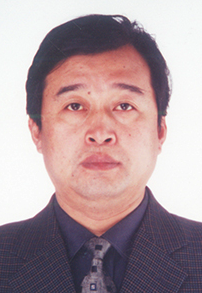 Kunzheng Wang (王坤正)合作论文数The Second Affiliated Hospital of Xi'an Jiaotong University (Xibei Hospital);Xi'an Jiaotong University Health Science Center57
 ZhiChao Tong (同志超)合作论文数13
 Xijing He (贺西京)合作论文数Health Science Center, Xi'an Jiaotong University13Yi Li (李毅)合作论文数11Zhibin Shi合作论文数10LongZhu Zhao (赵龙柱)合作论文数8Qiang Liu (刘强)合作论文数8
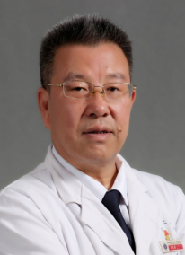 Haopeng Li (李浩鹏)合作论文数The Second Affiliated Hospital of Xi'an Jiaotong University8
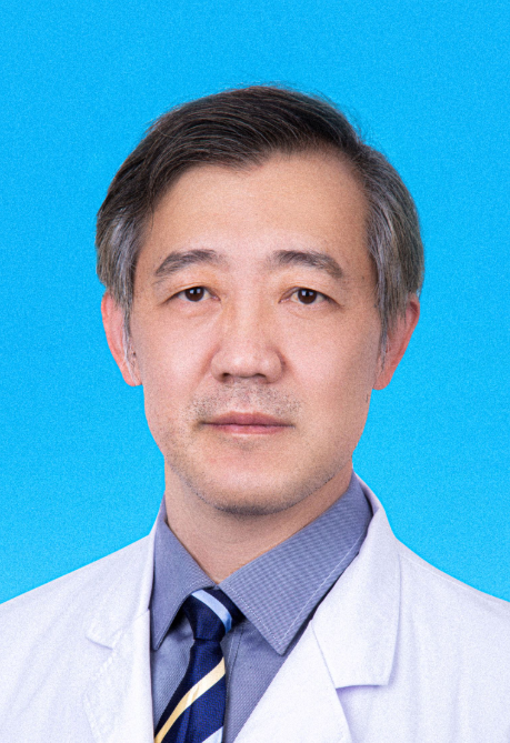 Chunsheng Wang (王春生)合作论文数The Second Affiliated Hospital of Xi'an Jiaotong University8
 Xiaoqian Dang (党晓谦)合作论文数The Second Affiliated Hospital of Xi'an Jiaotong University8GuoLian Yuan (袁国莲)合作论文数7
 Dou Wu (吴斗)合作论文数7zhe yang (杨哲)合作论文数6AnQing Liu (刘安庆)合作论文数6
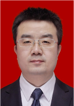 Zongqiang Gao (高宗强)合作论文数The Second Affiliated Hospital of Xi'an Jiaotong University6
 Xiong Guo (郭雄)合作论文数School of Public Health, Xi'an Jiaotong University Health Science Center;Global Health Institute, Xi'an Jiaotong University6
 Dong Wang (王栋)合作论文数The Second Affiliated Hospital of Xi'an Jiaotong University6youfen li (李幼芬)合作论文数5xudong li (李旭东)合作论文数5TuanMin Yang (杨团民)合作论文数5WU Yong-gang (武永刚)合作论文数4
 Weizhuo Wang合作论文数4Xiong GUO (郭雄)合作论文数4HuiRu Lv (吕惠茹)合作论文数4YongGang Wu (武永刚)合作论文数4DaZhi Yang (杨大志)合作论文数4SiYue Xu (徐思越)合作论文数4KaiFang Zhang (张开放)合作论文数4Qiang Gong (龚强)合作论文数4Qi-jun AN (安奇君)合作论文数3Miao Liu合作论文数3Xiangdong Lu合作论文数3WeiJuan Ma (马玮娟)合作论文数3JinHui Song (宋金辉)合作论文数3Hong Xiao (肖虹)合作论文数3Xiaoqiang Han (韩小强)合作论文数3Chen Duan合作论文数3Qi-sheng GU (顾其胜)合作论文数2lvzhen mao (毛履真)合作论文数2wei pang (庞炜)合作论文数2wang bin (王斌)合作论文数2shiqing feng (冯世庆)合作论文数2li zhu (朱利)合作论文数2LIU Qiang (刘强)合作论文数2Min SU (苏敏)合作论文数2hui li (李辉)合作论文数2Liaosha Jin (金辽沙)合作论文数2Xiaoliang Xu合作论文数2




