Objective·To assess the effect of veno-arterial extracorporeal membrane oxygenation(VA-ECMO)treatment on the mortality rate of patients suffering from cardiogenic shock and cardiac arrest in hospital.Methods·A total of 19 patients with cardiogenic shock or cardiac arrest who were treated with VA-ECMO treatment in Suzhou Kowloon Hospital,Shanghai Jiao Tong University School of Medicine from September 2017 to March 2022 were included in the retrospective study.Patients were divided into extracorporeal cardiopulmonary resuscitation(ECPR)group(n=9)and VA-ECMO for cardiogenic shock(E-CS)group(n=10)according to whether cardiac arrest had occurred.The general demographic data,clinical data,Sequential Organ Failure Assessment(SOFA)scores,postoperative complications and prognostic indicators of the two groups of patients were collected.Univariate and multivariate Cox proportional hazard regression analyses were used to evaluate the correlation between each covariate and hospital mortality.Results·Among the included patients,there were 15 males(78.9%),with an average age of 46.5(34.5,61.6)years.The incidence of postoperative complications was as follows:bleeding(47.4%),AKI(36.8%),infection(31.6%),limb ischemia(15.8%)and cerebrovascular accident(5.3%).The duration of VA-ECMO was 4.0(2.0,6.8)days,and the intensive care duration was 11.5(5.8,26.2)days;the ECMO withdrawal success rate was 63.2%,and the hospital mortality was 63.2%.The results of univariate Cox proportional hazard regression analysis showed that AKI(prior to VA-ECMO initiation),postoperative complications of infection and limb ischemia were correlated with the hospital mortality of patients(all P<0.05).The results of multivariate Cox proportional hazard regression analysis showed that AKI(prior to VA-ECMO initiation),postoperative complications of infection and limb ischemia were also independent risk factors for the hospital mortality of patients(all P<0.05).Conclusion·For patients with cardiogenic shock and cardiac arrest treated with VA-ECMO,AKI(prior to VA-ECMO initiation),postoperative infection and limb ischemia are independently associated with higher hospital mortality.
目的:研究外周血管穿刺困难患者予以超声引导技术辅助穿刺的临床应用效果.方法:2021年3月至2023年3月共计98例外周血管穿刺困难者,对临床资料予以回顾性分析,根据穿刺方法分组,常规组按传统盲穿法行血管穿刺,试验组采用超声引导技术下外周血管穿刺,对比两组的穿刺成功率、穿刺时间、疼痛评分等指标,以评估应用效果.结果:①试验组的一次穿刺成功率、穿刺总成功率均高于常规组,x2=9.046,4.170,P均<0.05;②相较而言,试验组的穿刺次数、穿刺时间、疼痛感评分(VAS)均处于更低水平,且试验组的留置时间
目的 分析行局部枸橼酸抗凝连续床边血液净化期间,不同滤器后游离钙浓度的应用效果.方法 以2021年1月-2022年12月医院收治的63例行连续床边血液净化患者为研究对象,根据滤器后游离钙浓度目标值及组间性别、年龄、急性生理与慢性健康(APACHE Ⅱ)评分、既往基础病史等基线资料均衡可比的原则进行分组.对照组(31 例)以 0.36~0.50mmpl/L为目标值,观察组(32 例)目标值为 0.20~0.35mmol/L,对比两组的滤器后游离钙离子浓度、凝血相关指标、滤器使用寿命以及不良反应发生情况.结果 经对应治疗后,观察组患者的滤器后游离钙浓度较对照组更低,且滤器使用寿命更长久,组间差异有统计学意义(P<0.05);两组患者的活化部分凝血活酶时间(APTT)、凝血酶时间(TT)、血肌酐(Scr)、尿素氮(BUN)水平均较治疗前有所改善,但治疗后两组患者上述各项生化指标比较,差异无统计学意义(P>0.05).观察组患者的总不良反应发生率(6.25%)较对照组(29.03%)更低,组间差异有统计学意义(P<0.05).结论 在局部枸橼酸抗凝的连续床边血液净化期间,不同滤器后游离钙浓度均可取得满意效果,且较低滤器后游离钙浓度可延长滤器使用时间,降低不良反应风险.
目的 系统评价艾司洛尔控制心室率对脓毒性休克患者血流动力学及预后的影响.方法 计算机检索PubMed、Cochrane library、EMBASE、MEDLINE、OVID、中国生物医学文献数据库、中国知网、万方数字化期刊全文数据库和维普数据库自建库至2020年6月有关艾司洛尔治疗脓毒症患者临床疗效的随机对照试验,由2名评价者独立选择实验、提取资料和评估方法学质量,采用stata 12.0软件进行Meta分析.结果 共纳入9项随机对照实验研究,共计795例受试者(实验组398例,对照组397例).Meta分析结果提示:与使用常规方案治疗脓毒症病人相比,艾司洛尔够明显降低患者28天的病死率[RR=0.67,95%CI(0.56,0.79),P<0.05],降低患者的心率[SMD=-1.94,95%CI(-2.82,-1.05),P<0.05],降低患者平均动脉压[SMD=-1.56,95%CI(-2.86,-0.25),P=0.049],以及改善患者全心舒张末期血容积指数[SMD=0.81,95%CI(0.37,1.25),P<0.01].结论 现有临床结论显示艾司洛尔可以降低脓毒性休克患者的28天病死率、心率、平均动脉压,改善全心脏舒张末期血容积指数.对中心静脉血氧饱和度、血乳酸、心脏指数、心脏每搏指数等影响不大,仍需长期随访的大样本随机对照试验予以评价.
目的 简要分析临床病例ECPR抢救成功率及并发症发生率.方法 分析2017年12月至2020年12月期间我院重症医学科收治的12例常规ECMO与ECPR的患者的资料.分为常规ECMO组(n=7)和ECPR组(n=5),两组患者常规给与重症超声置管后上机,重症超声实时监测进行器官支持和器官保护等,观察患者行ECMO术时间(置管时血管穿刺至ECMO成功运转的时间)、并发症(消化道出血及血流感染)、生存率(存活及死亡)等指标.结果 两组患者在并发症(消化道出血及血流感染)上,两组无统计学差异(P>0.05),但两组在行ECMO术时间、生存率上有统计学差异,常规ECMO组优于ECPR组(P<0.05).结论 建议在ICU收治重症患者中,需要尽早评估ECMO指征,充分评估后若患者有ECMO指征则尽早进行ECMO术,以便提高患者的抢救成功率.
International Journal of Laboratory HematologyVolume 42, Issue 5 p. e204-e206 LETTER TO THE EDITOR Analysis of hematological indexes of COVID-19 patients from fever clinics in Suzhou, China Jindan Kong, Jindan Kong Department of Hematology, The First Affiliated Hospital of Soochow University, National Clinical Research Center for Hematologic Diseases, Jiangsu Institute of Hematology, Suzhou, China Department of Critical Care Medicine, The First Affiliated Hospital of Soochow University, Suzhou, ChinaSearch for more papers by this authorTing Wang, Ting Wang Department of Nursing, The First Affiliated Hospital of Soochow University, Suzhou, ChinaSearch for more papers by this authorZou Di, Zou Di Department of Critical Care Medicine, The Affiliated Infectious Diseases Hospital of Soochow University, Suzhou, ChinaSearch for more papers by this authorBingyu Shi, Bingyu Shi Department of Hematology, The First Affiliated Hospital of Soochow University, National Clinical Research Center for Hematologic Diseases, Jiangsu Institute of Hematology, Suzhou, ChinaSearch for more papers by this authorXiaoming Yu, Xiaoming Yu Department of Critical Care Medicine, The Sixth People's Hospital of Kunshan, Kunshan, ChinaSearch for more papers by this authorChaofa Huang, Chaofa Huang Department of Critical Care Medicine, Suzhou Kowloon Hospital, Shanghai Jiaotong University School of Medicine, Shanghai, ChinaSearch for more papers by this authorYan Yang, Yan Yang Department of Critical Care Medicine, The First People's Hospital of Kunshan, Kunshan, ChinaSearch for more papers by this authorHaiwei Sun, Haiwei Sun Department of Emergency and Critical Care Medicine, The Second Affiliated Hospital of Soochow University, Suzhou, ChinaSearch for more papers by this authorDejing Yuan, Dejing Yuan Department of Nursing, The First Affiliated Hospital of Soochow University, Suzhou, ChinaSearch for more papers by this authorDepei Wu, Corresponding Author Depei Wu [email protected] Department of Hematology, The First Affiliated Hospital of Soochow University, National Clinical Research Center for Hematologic Diseases, Jiangsu Institute of Hematology, Suzhou, China Correspondence Depei Wu and Jianhong Fu, Department of Hematology, The First Affiliated Hospital of Soochow University, National Clinical Research Center for Hematologic Diseases, Jiangsu Institute of Hematology, Shizi Street 188, Suzhou 215006, China. Emails: [email protected] (DW); [email protected] (JF)Search for more papers by this authorJianhong Fu, Corresponding Author Jianhong Fu [email protected] orcid.org/0000-0002-9044-024X Department of Hematology, The First Affiliated Hospital of Soochow University, National Clinical Research Center for Hematologic Diseases, Jiangsu Institute of Hematology, Suzhou, China Correspondence Depei Wu and Jianhong Fu, Department of Hematology, The First Affiliated Hospital of Soochow University, National Clinical Research Center for Hematologic Diseases, Jiangsu Institute of Hematology, Shizi Street 188, Suzhou 215006, China. Emails: [email protected] (DW); [email protected] (JF)Search for more papers by this author Jindan Kong, Jindan Kong Department of Hematology, The First Affiliated Hospital of Soochow University, National Clinical Research Center for Hematologic Diseases, Jiangsu Institute of Hematology, Suzhou, China Department of Critical Care Medicine, The First Affiliated Hospital of Soochow University, Suzhou, ChinaSearch for more papers by this authorTing Wang, Ting Wang Department of Nursing, The First Affiliated Hospital of Soochow University, Suzhou, ChinaSearch for more papers by this authorZou Di, Zou Di Department of Critical Care Medicine, The Affiliated Infectious Diseases Hospital of Soochow University, Suzhou, ChinaSearch for more papers by this authorBingyu Shi, Bingyu Shi Department of Hematology, The First Affiliated Hospital of Soochow University, National Clinical Research Center for Hematologic Diseases, Jiangsu Institute of Hematology, Suzhou, ChinaSearch for more papers by this authorXiaoming Yu, Xiaoming Yu Department of Critical Care Medicine, The Sixth People's Hospital of Kunshan, Kunshan, ChinaSearch for more papers by this authorChaofa Huang, Chaofa Huang Department of Critical Care Medicine, Suzhou Kowloon Hospital, Shanghai Jiaotong University School of Medicine, Shanghai, ChinaSearch for more papers by this authorYan Yang, Yan Yang Department of Critical Care Medicine, The First People's Hospital of Kunshan, Kunshan, ChinaSearch for more papers by this authorHaiwei Sun, Haiwei Sun Department of Emergency and Critical Care Medicine, The Second Affiliated Hospital of Soochow University, Suzhou, ChinaSearch for more papers by this authorDejing Yuan, Dejing Yuan Department of Nursing, The First Affiliated Hospital of Soochow University, Suzhou, ChinaSearch for more papers by this authorDepei Wu, Corresponding Author Depei Wu [email protected] Department of Hematology, The First Affiliated Hospital of Soochow University, National Clinical Research Center for Hematologic Diseases, Jiangsu Institute of Hematology, Suzhou, China Correspondence Depei Wu and Jianhong Fu, Department of Hematology, The First Affiliated Hospital of Soochow University, National Clinical Research Center for Hematologic Diseases, Jiangsu Institute of Hematology, Shizi Street 188, Suzhou 215006, China. Emails: [email protected] (DW); [email protected] (JF)Search for more papers by this authorJianhong Fu, Corresponding Author Jianhong Fu [email protected] orcid.org/0000-0002-9044-024X Department of Hematology, The First Affiliated Hospital of Soochow University, National Clinical Research Center for Hematologic Diseases, Jiangsu Institute of Hematology, Suzhou, China Correspondence Depei Wu and Jianhong Fu, Department of Hematology, The First Affiliated Hospital of Soochow University, National Clinical Research Center for Hematologic Diseases, Jiangsu Institute of Hematology, Shizi Street 188, Suzhou 215006, China. Emails: [email protected] (DW); [email protected] (JF)Search for more papers by this author First published: 13 July 2020 https://doi.org/10.1111/ijlh.13290Citations: 11 Jindan Kong, Ting Wang and Di Zou contributed equally to this work. Read the full textAboutPDF ToolsRequest permissionExport citationAdd to favoritesTrack citation ShareShare Give accessShare full text accessShare full-text accessPlease review our Terms and Conditions of Use and check box below to share full-text version of article.I have read and accept the Wiley Online Library Terms and Conditions of UseShareable LinkUse the link below to share a full-text version of this article with your friends and colleagues. Learn more.Copy URL Share a linkShare onEmailFacebookTwitterLinkedInRedditWechat No abstract is available for this article. REFERENCES 1Fan BE, Chong V, Chan S, et al. Hematologic parameters in patients with COVID-19 infection. Am J Hematol. 2020; 95(6): E131-E134. 10.1002/ajh.25774 CASPubMedWeb of Science®Google Scholar 2Tang N, Li D, Wang X, et al. Abnormal coagulation parameters are associated with poor prognosis in patients with novel coronavirus pneumonia. J Thromb Haemost. 2020; 18(4): 844-847. 10.1111/jth.14768 CASPubMedWeb of Science®Google Scholar 3Huang C, Wang Y, Li X, et al. Clinical features of patients infected with 2019 novel coronavirus in Wuhan, China. Lancet. 2020; 395(10223): 497-506. 10.1016/S0140-6736(20)30183-5 CASPubMedWeb of Science®Google Scholar 4Yao XH, Li TY, He ZC, et al. A pathological report of three COVID-19 cases by minimally invasive autopsies. Zhonghua Bing Li Xue Za Zhi. 2020; 49: E9. CASPubMedGoogle Scholar 5Russell CD, Parajuli A, Gale HJ, et al. The utility of peripheral blood leucocyte ratios as biomarkers in infectious diseases: a systematic review and meta-analysis. J Infect. 2019; 78(5): 339-348. 10.1016/j.jinf.2019.02.006 PubMedWeb of Science®Google Scholar 6Qin C, Zhou L, Hu Z, et al. Dysregulation of immune response in patients with COVID-19 in Wuhan, China. Clin Infect Dis. 2020. ciaa248. [Epub ahead of print]. 10.1093/cid/ciaa248 Web of Science®Google Scholar 7Wu C, Chen X, Cai Y, et al. Risk factors associated with acute respiratory distress syndrome and death in patients with coronavirus disease 2019 pneumonia in Wuhan, China. JAMA Intern Med. 2020. e200994. [Epub ahead of print]. 10.1001/jamainternmed.2020.0994 Web of Science®Google Scholar 8Bhimraj A, Morgan RL, Shumaker AH, et al. Infectious diseases society of america guidelines on the treatment and management of patients with COVID-19. Clin Infect Dis. 2020. ciaa478. [Epub ahead of print]. 10.1093/cid/ciaa478 PubMedGoogle Scholar 9Zhou F, Yu T, Du R, et al. Clinical course and risk factors for mortality of adult inpatients with COVID-19 in Wuhan, China: a retrospective cohort study. Lancet (London, England). 2020; 395(10229): 1054-1062. 10.1016/S0140-6736(20)30566-3 CASPubMedWeb of Science®Google Scholar Citing Literature Volume42, Issue5October 2020Pages e204-e206 ReferencesRelatedInformation
RATIONALE:Esophagopleural fistula (EPF) is a rare critical life-threatening condition that features high misdiagnosis rate. Although various surgical and conservative techniques have been developed for the treatment of EPF, the mortality rate of EPF remains high.PATIENT CONCERNS:An 81-year-old man with hepatic cirrhosis caused by schistosomiasis was admitted with upper gastrointestinal bleeding.DIAGNOSES:Upper endoscopy revealed bleeding large esophageal varices, and endoscopic injection sclerotherapy (EIS) was performed. Two weeks after the EIS was performed, the patient developed pyrexia, left-sided pleuritic chest pain. Air and pleural effusion were showed in the left pleural cavity by high-resolution computed tomography (HRCT), and a linear fistulous communication was noticed from the distal esophagus. These findings were consistent with hepatic cirrhosis, esophageal varices, upper gastrointestinal bleeding, and esophagopleural fistula.INTERVENTIONS:The patient was intensively treated with endoscopic self-expandable metallic stent (covered-SEMS) implantation and comprehensive treatments (including thoracic closed drainage, antibiotics, nasojejunal nutrition, and antacids).OUTCOMES:The patient was completely cured without recurrence during a 6 months of follow-up by comprehensive conservative treatments.LESSONS:This case indicates that pleural effusion with food residue is a specific finding in EPF. Thorax CT exhibited high sensitivity for the diagnosis of EPF. Endoscopic self-expandable metallic stent implantation and comprehensive conservative treatments may be preferable for the severe liver disease with EPF.
目的 观察血必净用于治疗老年重症肺部感染对患者炎症因子及凝血机制的影响.方法 回顾性分析该院及苏州市立医院2015年3月—2017年3月收治的重症肺部感染的患者76例,其中42例连续使用血必净治疗至少10 d,为血必净组,另外34例未使用血必净治疗,为对照组.对比治疗前及治疗10 d后两组患者凝血功能指标和炎症因子的变化.结果 血必净组治疗第10天PT、APTT、FIB、TT分别为(12.08±4.09)s、(30.06±10.07)s、(2.33±0.87)g/L、(13.37±3.42)s,CRP、PCT、TNF-α水平分别为(18.74±6.92)mg/L、(0.21±0.03)ng/mL、(13.47±3.91)g/L,均较对照组以上指标水平更优,差异有统计学意义(t=3.753、2.543、10.411、10.438、5.894、31.126、9.811,P<0.05).结论 血必净用于治疗老年重症肺部感染可明显改善患者高凝状态,降低炎症因子水平,利于患者病情的好转.
目的 比较血栓弹力图(TEG)与常规凝血试验在重症肺炎中的检测结果,探讨TEG对监测重症肺炎凝血功能障碍的价值,以指导临床对重症肺炎患者病情进一步评估.方法 回顾性分析2015年5月~2016年9月苏州九龙医院重症医学科43例重症肺炎患者的临床资料,并根据急性生理学与慢性健康状况评分系统Ⅱ(APACHEⅡ)评分分为APACHEⅡ评分≤18分组(17例)和APACHEⅡ评分>18分组(26例);另选取同时期本院健康体检人员15例作为对照组.取静脉血检测常规凝血指标,同时进行TEG检测,比较三组间各指标的差异,采用Spearman相关性分析重症肺炎患者APACHEⅡ评分与各项指标的相关性.结果 在常规凝血实验指标中,与对照组[凝血酶原时间(PT)(12.65±0.52)s,活化部分凝血活酶时间(APTT)(34.90±2.79)s,纤维蛋白原(FBG)(3.82±0.60)g/L,D-二聚体(0.63±0.23)mg/L,血小板(PLT)(214.27±52.07)×109/L]相比,APACHEⅡ评分>18分组的PT(14.43±2.65)s、APTT(44.92±12.89)s及D-二聚体(5.71±3.83)mg/L显著升高,FBG(3.24±0.93)g/L及PLT(98.73±27.37)×109/L降低,差异均具有统计学意义(P<0.05);对照组与APACHEⅡ评分≤18分组相比,各常规凝血指标间的差异无统计学意义;提示常规凝血试验能在一定程度上反映病情较重时的低凝状态,但不能很好地反映病情相对较轻的重症肺炎的高凝状态.TEG参数中,与对照组[凝血反应时间(R值)(6.31±0.62)min,血块生成时间(K值)(1.68±0.39)min,最大宽度值(MA值)(58.83±5.09)mm,凝血综合指数(CI值)(0.54±0.49)]相比,R值和K值在重症肺炎APACHEⅡ评分≤18分组R值(5.02±1.43)min,K值(1.37±0.33)min降低,而在APACHEⅡ评分>18分组R值(7.08±2.10)min,K值(3.19±0.54)min则明显升高;而MA值、CI值在APACHEⅡ评分≤18分组MA值(65.03±4.73)mm,CI值(2.73±1.24)升高,而在APACHEⅡ评分>18分组MA值(47.42±4.04)mm,CI值(-2.86±0.83)则明显降低(P<0.05),提示TEG可反应重症肺炎病情中的低凝及高凝改变.相关性分析结果显示,APACHEⅡ评分与PT、APTT、D—二聚体、R值、K值呈正相关(P<0.05),与FBG、PLT、MA值、CI值呈负相关(P<0.05).结论 对比常规凝血功能检测,TEG能更有效地监测重症肺炎患者凝血功能改变,识别高凝、低凝状态,客观评价病情严重程度,指导临床风险评估.
目的:分析脑外伤患者实施APACHE评分和GCS评分的效果.方法:以我院55例脑外伤患者作为研究对象,ICU治疗期间由神经外科医生分别进行APACHE-Ⅱ评分和GCS评分,观察评分和死亡率之间的关系,比较存活患者和死亡患者的评分结果.结果:GCS评分越低、APACHE-Ⅱ评分越高,患者死亡率越高.存活患者的GCS评分高于死亡患者,APACHE-Ⅱ评分低于死亡患者(P<0.05),差异有统计学意义.结论:脑外伤患者在病情评估上,APACHE-Ⅱ评分要优于GCS评分,可以明确患者的病情严重程度,提高预后预测的准确性.
OBJECTIVE To discuss the clinical strategy for treatment of pan drug resistant Acinetobacter baumannii (PDRAB) infection so as to control this infection .METHODS In an open ,parallel experimental design ,86 pa-tients with nosocomial pneumonia caused by PDRAB during Jun .2009 to Jun .2013 were divided into group A (n=32) ,group B (n=33) and group C (n=21) .The patients in group A were treated with cefperazone/sul-bactam (suiperazone ,SPZ) alone by means of lengthening the infusion time .The patients in group B were treated with SPZ combined with meropenem (MEC) .The patients in group C were treated with SPZ combined with tige-cycline (tygacil) .The treatment course lasted for ten days .The three groups were compared for changes in tem-perature and hemogram to evaluate the clinical effect of extended antibiotic infusing time and antibiotic combination for treatment of PDRAB .RESULTS The anal temperature and the blood cell count were significantly decreased at 5 and 10 days after the intensive treatment compared to those before treatment (P<0 .01) .The total effective rate for the three groups was 66 .7% at 5 days and 83 .3% at 10 days after initial treatment .The effective rates of ini-tial treatment and final treatment were 73 .9% and 82 .6% respectively .The effective rate for group C was 95 .2%at 5 days and 100 .0% at 10 days after the intensive treatment ,significantly higher than 59 .4% and 78 .1% for group A and 78 .8% and 87 .9% for group B (P< 0 .05) .CONCLUSION Cefoperazone-sodium(SPZ)by means of lengthening the infusion time can effectively treat PDRAB infection ,and it works quicker when combined with MEC .And the clinical curative effect of SPZ combined with tygacil to treat PDRAB infection is better than with MEC .
目的:探讨ICU脑外伤合并肺部感染患者的致病高危因素分析及其处理措施。方法选取我院2014年1月~2015年1月ICU接收治的脑外伤患者的100例,其中有48例患者合并肺部感染,将其作为观察组,其余无感染的52例患者作为对照组,分析引发肺部感染的危险因素,探讨可行的处理措施。结果将引发肺部感染的危险因素进行对比后发现,观察组患者的年龄、饮酒、吸氧、昏迷、休克、意识障碍、肺部疾病、营养不良、气管切开、使用脱水剂、使用激素、机械通气以及开放性脑外伤的发生率明显高于对照组,对比差异显著(<0.05),具有统计学意义。结论引发ICU脑外伤患者合并肺部感染的危险因素有很多,包括患者自身原因、药物原因、感染等,采取积极有效的措施进行预防,可降低感染的机率。
急性重症心肌炎,起病急,病情重,病死率高。部分患者由于其心肌酶学、心电图改变,以及临床表现等与急性心肌梗死患者非常相似,常导致临床医师误诊。在发病早期给予恰当的支持治疗,可明显改善预后。本文报道了1例经主动脉球囊反搏和心脏临时起搏成功抢救的似急性前壁心肌梗死的重症心肌炎病例。
Poly(lactic acid)-b-poly(ethylene glycol)(MePEG-b-PLA) based nanocapsules containing perfluorohexane were fabricated through a double emulsion-solvent evaporation method.In the preparation,three kinds of hydrophilic polymers,i.e.,poly(vinyl-alcohol)(PVA),chitosan(CS),and carboxymethyl dextran(CMG) were used as emulsifier or co-emulsifier to modify the surface of nanocapsules.Besides,two kinds of MePEG-b-PLA copolymers with different block length ratio were used as main shell composition to fabricate nanocapsules.Nanocapsule size,morphology,zeta potential,water stability of six nanocapsule samples fabricated by different polymers or emulsifiers were characterized by Malvern nanosizer analyzer,transmission electron microscope( TEM) and UV-Visible spectrophotometer( UV-Vis),respectively.The results showed that nanocapsules using 1% PVA + 1% CMG as an emulsifier and MePEG-b-PLA[m( MePEG) ∶ m( PLA) = 1∶ 3] as shell material had monodispersed small size and stable water property.These nanocapsules are not only in nanometer-scale but also possess favorable ultrasonic imaging property.Ultrasonic imaging property of nanocapsules was charaeterized using in vitro ultrasound tester and made a comparison with a commercialized ultrosonic imaging agent SonoVueTM.The results demonstrated a brighter contrast scale and edurable ultrasonic property of MePEG-b-PLA nanocapsules.In addition,the cytotoxicity by incubating MePEG-b-PLA nanocapsules with rat liver cells line BRL-3A was characterized through 3-(4,5-dimethylthiazol-2-yl)-2,5-diphenyltetrazolium bromide(MTT)assay.The results showed that the PLA-based nanocapsules imposed no significant inhibition effect on proliferation of BRL-3A cells.
Objective To evaluate the effect of immunosuppressive therapy for paraquat poisoning. Method Relevant published articles concemed with immunosuppressive therapy for paraquat poisoning both in Chinese and in English were searched,and Meta-analysis was carried out with Stata software. Results Eight non-randomized comparative studies and two randomized controlled trials were enrolled for Meta-analysis according to the inclusion and exclusion criteria. The results indicated the relative risk of immunosuppressive therapy in im-proving mortality of paraquat poisoning were 0. 54(95% CI=0. 44~0. 66)and 0. 76(95% CI=0. 62~0. 94)for non-randomised stud-ies and randomized controlled studies,respectively. Conclusion This anaIysis Suggests that immunosuppressive therapy reduce mortality in paraquat poisoning compared with conventional medical therapy. However,due to significant heterogeneity,a large randomized controlled trial is required to affirm the role of immunosuppression in paraquat poisoning.
目的:研究外源性H2S对心肺复苏(cardiopulmonary resuscitation,CPR)后大鼠记忆障碍的影响,并探究其作用机制.方法:将SD大鼠随机分为5组,对照组、模型组(CPR)、低剂量NaHS治疗组(CPR+ NaHS)、中间剂量NaHS治疗组、高剂量NaHS治疗组.采用Morris水迷宫法测定各组大鼠学习记忆能力;比色法检测大鼠大脑组织中超氧化物歧化酶(superoxide dismutase,SOD)活性及丙二醛(malonaldehyde,MDA)含量;HE染色观察海马组织形态学改变.结果:与对照组比较,模型组大鼠在Morris水迷宫实验中,逃避潜伏期显著延长,在平台所在象限停留时间减少,穿越平台次数减少,SOD活性显著降低,MDA含量显著升高,海马神经元出现变性改变.与模型组比较,NaHS治疗组大鼠逃避潜伏期缩短,在平台所在象限搜索时间延长,穿越平台次数明显增多,SOD活性升高,MDA含量显著降低,海马神经元的变性得到改善.结论:心肺复苏可损害大鼠的学习记忆能力,外源性H2S供体NaHS减轻了CRP对大鼠学习记忆的损害,其作用机制可能与H2S清除自由基和衰减脂质过氧化反应有关.
Objective: To evaluate the early diagnostic and prognosis value of procalcitonin (PCT), interleukin 6 (IL-6), and PCT kinetics for central venous catheter related bloodstream infections (CRBSI) in selected critically ill patients. Methods: Thirty-six patients received central venous catheter, who were suspected of having CRBSI. All the patient blood samples were obtained for detection of PCT, IL-6 levels, and for blood culture on the first day, and catheter were also obtained for culture. PCT levels were measured on day 1, 2, 3, and 7 after infection. The patients were divided into CRBSI group and non-CRBSI group. The CRBSI group was divided into controlled infection group and non-controlled infection group, and PCT kinetics for prognostic value was observed. Results: Fifteen patients were diagnosed as CRBIS (41.67%). Levels of PCT and IL-6 were significantly higher in patients with CRBIS than those without CRBIS on the first infection day (P<0.001 and P<0.005). The ROC curves indicated that the area under the curve (AUC) for PCT and IL-6 was 0.832 and 0.78, respectively. The cutoff points of PCT and IL-6 were set to be 4.3 μg/L and 464.5 ng/L for patients with proven CRBSI, respectively. The corresponding sensitivity, specificity, positive predictive value, and negative predictive value were 80.0% and 77.3%, 95.2% and 66.7%, 92.25% and 61.12%, and 86.95% and 77.76%, respectively. PCT serum levels tended to decrease in patients with controlled CRBSI, whereas in non-controlled patients was proven to remain stably in high level. Conclusion: PCT and IL-6 might be valuable early diagnostic parameters for CRBIS with high specificity and negative predictive value, and PCT kinetics has certain predictive value for the prognosis of CRBSI.
 Changjiang Wu合作论文数13MingLiang Sui (隋明亮)合作论文数13Tuan-jie ZHU (朱团结)合作论文数7YANG Jinhu (杨进虎)合作论文数5Ya-di YANG (杨雅迪)合作论文数5NingJun Li (黎宁君)合作论文数5HU Xianzhu (胡贤著)合作论文数4Weibing Tang合作论文数4Damei Xia合作论文数3Hui-ping LIU (刘会平)合作论文数2Jindan Kong合作论文数1Bingyu Shi (石冰玉)合作论文数1Di Zou合作论文数1Zhong-xia SUN (孙中霞)合作论文数1Haiwei Sun合作论文数1
 Jianhong Fu合作论文数1Hui WANG (汪惠)合作论文数1Lin LIU (刘琳)合作论文数1DeJing Yuan (袁德敬)合作论文数1Yi Jiang (姜毅)合作论文数1Hao Wang (汪浩)合作论文数1
 Ting Wang (王婷)合作论文数Clinical Pediatrics School, Soochow University1
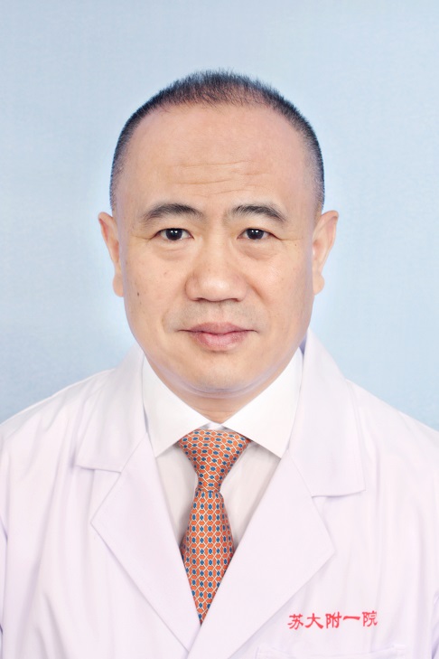 Depei Wu (吴德沛)合作论文数The First Affiliated Hospital of Soochow University;State Key Laboratory of Radiation Medicine and Protection, Soochow University1Li Zhang (张俐)合作论文数1




