目的 比较蓝色半导体激光(蓝激光)和近红外激光对牙龈卟啉单胞菌(Porphyromonas gingivalis,Pg)的杀菌作用和安全性.方法 采用丝线结扎SD大鼠上颌第二磨牙(M2)并在龈沟内涂布Pg菌液的方法构建牙周炎大鼠模型.通过HE染色、CT扫描结合牙槽骨三维模型重建检测建模是否成功.建模成功后,25只模型组大鼠进行实验.10只大鼠左侧M2牙周袋使用1 W蓝激光,右侧M2牙周袋使用1 W近红外激光;另外10只大鼠左侧M2牙周袋使用1.5 W蓝激光,右侧M2牙周袋使用1.5 W近红外激光.其余5只大鼠使用假激光,不开激光电源,左、右侧M2的牙周袋分别使用蓝激光和近红外激光的光纤伸入牙周袋做提拉动作.在第0,3,7天照射后收集龈下菌斑标本,荧光定量PCR检测大鼠龈下菌斑中Pg拷贝数并计算杀菌率.通过0 d和7 d的牙周组织切片HE染色检测蓝激光和近红外激光照射后的组织学变化.结果 模型组大鼠牙周组织出现牙周炎病理改变,牙槽骨吸收,牙周炎大鼠模型构建成功.在第0,3,7天,功率为1 W或1.5 W蓝激光或近红外激光照射牙周袋后Pg拷贝数均较假激光处理后明显减少(P<0.05).蓝激光和近红外激光杀菌率相似.功率为1 W时,杀菌率均在90%左右;功率为1.5 W时,杀菌率均在99%左右.HE染色结果显示在相同功率(1 W或1.5 W),蓝激光均比近红外激光对牙周组织损伤小.结论 蓝激光与近红外激光能对牙周袋内Pg有效杀菌,且杀菌效果相同,而蓝激光安全性更高.
目的 评估Nd:YAG激光辅助龈下刮治术和根面平整术(SRP)在重度慢性牙周炎治疗中的疗效.方法 重度慢性牙周炎纳入患者的牙齿分布在口内4个象限,牙周探诊深度(PD)5~8 mm,附着丧失(AL)≥5 mm,探诊出血(BOP)阳性.符合纳入标准的患者按照就诊顺序随机分为SRP组、SRP+L组(SRP后行Nd:YAG激光治疗)及L+SRP组(Nd:YAG激光治疗后行SRP).记录各组在基线时与治疗8周时临床牙周指标BOP、PD和AL,并进行统计学分析.结果 治疗后8周,各组的BOP、PD和AL较基线均有所改善(P<0.05).SRP+L组和L+SRP组的BOP阳性率较SRP组明显减少(P<0.05).SRP+L组的PD较SRP组和L+SRP组明显变浅(P<0.05),其中基线时PD=7mm的位点,SRP+L组较SRP组和L+SRP组明显变浅(P<0.05).SRP组较SRP+L组和L+SRP组的AL明显减少(P<0.05).结论 Nd:YAG激光辅助SRP治疗重度慢性牙周炎可以更好地减少BOP,使PD变浅,且SRP后进行Nd:YAG激光治疗对于较深的牙周袋PD减少更明显,但对AL的改善无优势.
Objective:To explore the effect of 3D printing periodontal bone defect model of anterior teeth combined with case-based learning (CBL) teaching mode in the standardized residency training of loose teeth fixation.Methods:Sixty general practitioner of stomatology students were equally divided into two groups, with one group receiving CBL teaching (CBL group) and the other group receiving 3D printing model combined CBL teaching (3D+CBL group). After the teaching, the teacher evaluated the overall practice effects, tested the residents' theory and practice scores, and designed a questionnaire to evaluate the teaching effect. SPSS 16.0 software was used for statistical analysis, and t test was used for comparison between the two groups. Results:The theoretical score (89.0±3.2) and the practical score (90.0±2.5) in the 3D+CBL group were significantly higher than the theoretical score (83.3±3.6) and the practical score (84.2±3.5) in the CBL group ( P<0.05). The questionnaire showed that the students of the 3D+CBL group performed significantly better than those in CBL group in the following eight aspects, including mastery of basic periodontal knowledge, professional knowledge of loose teeth fixation, ability to diagnose cases and formulate treatment plans, clinical operation ability, interesting teaching content, willingness to communicate with teachers, awareness of actively receiving patients, and satisfaction of standardized residency training ( P<0.05). Conclusion:The 3D printing periodontal bone defect model of anterior teeth combined with the CBL teaching mode has achieved good results in the teaching of loose teeth fixation, and it provides a reference for the application of 3D printing in clinical teaching for periodontology.
目的:评价3D打印牙周导板辅助松牙固定术完成树脂塑形方法的临床应用效果.方法:选取2018年3月~2019年8月西安交通大学口腔医院牙周科就诊的下前牙慢性重度牙周炎患者40例,金属丝结扎下前牙.其中20例经传统方法手工塑形复合树脂夹板(传统法),20例在3D打印牙周导板辅助下完成复合树脂的塑形(3D打印法).比较两种方法操作时间,患者美观感受,以及复诊3、6、12个月后金属丝暴露,牙齿松动度以及菌斑、牙石指数.结果:传统法松牙固定树脂塑形、光固化及修整总时间(74±10)min,3D打印组(18±5)min,差异有统计学意义(P<0.001).6个月复查时,传统组金属丝暴露5人,3D打印组金属丝暴露2人;12个月复查时,传统组金属丝暴露15人,3D打印组金属丝暴露5人.两种方法制作夹板在复查3、6、12个月时均无松动.在复查3、6、12个月时,菌斑指数、牙石指数差异均无统计学意义(P>0.05).结论:相较于传统法,3D打印牙周导板可以快速、精准引导树脂成型,在松牙固定中有一定的临床应用价值.
The invention relates to a plastifying method for a photosensitive composite material quickly by a template of a 3D printed resin periodontal splint and an application thereof. The method comprises the following steps: importing CBCT data of a patient in a dcm format to Mimics Research 19.0 software, intercepting a tooth position needed to design according to a clinical demand, and copying the shapes of a lip cheek and a tongue palate side tooth face, wherein the gap demand of the splint template and the tongue palate side tooth face is as follows: the width is 0.6 mm, and copying the shapes of a lip cheek and a tongue palate side tooth face; the splint template design demand is as follows: designing a root area and a far middle face sealing area, and designing an adjacent tooth auxiliaryretention position; storing the final design in a format of st1 and inputting the design to a 3D printer to be printed. The method has the characteristics of copying the tooth face shape fully and being uniform in thickness, smooth in surface, primary to form and high in speed, and preventing resin from compressing gum and abducted gap.
Objective :To observe the clinical effects on prevention of alveolar bone absorption by site preservation after tooth extraction due to serious chronic periodontitis .Methods :The fresh sockets were immediately grafted and filled with CGF compounded with Bio‐Oss and CGF membrane after minimally invasive tooth extraction , the clinical effect was observated change by taking X‐ray and CBCT after 6 months .Results :No infection case was found in all patients ,After six months ,alveolar bone height and width in good condition ,gingival was plump .Conclu‐sion :CGF compounded with osteoinduction active material technique can have good effect on guiding bone regenera ‐tion ,and can meet the needs of post‐implanting .
目的:初步探讨半导体激光在牙龈切除术及成形术中的应用,比较半导体激光与高频电刀和传统手术方法在临床疗效之间的差异。方法:收集需进行牙龈切除术及成形术的患者(20例,88颗牙),随机分为3组,分别使用半导体激光(8 例,28颗牙)、高频电刀(6 例,30颗牙)、传统手术刀(6 例,30颗牙)行牙龈切除术及成形术。术后记录患者疼痛情况、创面愈合或感染情况及唇侧/颊侧龈缘正中至牙齿断缘/牙冠切缘/边缘嵴的垂直距离(H 值)。结果:牙龈切除术及成形术术后3组疼痛情况、创面愈合及感染情况、H 值变化情况均无统计学意义(P >0.05),术后 H 值变化在0~0.5mm 范围内。结论:牙龈切除术及成形术术后疼痛情况、牙龈愈合及感染情况、术后牙龈位置不受手术所用仪器影响,半导体激光可成为牙龈切除术及成形术的一种新方法。
牙周病学实验教学是口腔医学生专业理论知识学习和专业技能培养的重要组成部分,也是牙周病临床实习前期训练的重要环节.为提高学生的临床思维能力和操作技能,提高牙周病学实验教学效果,缩短临床实习适应期,结合自身实践经验,根据相关教学大纲编写典型案例,将CBL教学方法应用于牙周病学实验教学,探讨了CBL教学模式在应用于牙周病学实验教学的优势、意义及局限性.
目的探索牙周临床技能考核中标准化病人( SP)的培训模式。方法通过对SP志愿者的选择、SP培训病案的撰写、培训流程和 SP考核评估形式的探讨,促进SP培训的发展与成熟。结果根据临床教学实施情况,结合文献,进行牙周病标准化病人的培训。结论 SP 在教学与评估上扮演极其重要的角色,规范有效的培训方式为将SP应用于牙周临床技能的考核奠定了基础。
OBJECTIVE:To screen a variety of Porphyromonas gingivalis (Pg) common outer membrane proteins with two-dimensional liquid phase fractionation (PF2D) and matrix-assisted laser desorption ionization time-of-flight/time-of-flight mass spectrometry (MALDI-TOF/TOF-MS) and provide candidate target antigen for the design of vaccines with cross protection against a variety of Pg.METHODS:The outer membrane proteins of Pg301, PgATCC33277 and PgW83 were extracted through ultracentrifugation, and then they were separated by ProteomeLab PF2D protein fractionation system. After separation, the outer membrane proteins were obtained through comparison, and the primary structure of the proteins was identified by MALDI-TOF/TOF-MS and database.RESULTS:Ninety-nine protein samples out of 3 strains of Pg were obtained after the high performance chromato focusing (HPCF) separation process. B7 fractions of 3 strains of Pg were separated by the reversed-phase high performance liquid chromatography (RP-HPCL) separation process. After comparison of peak and retention time of chromatogram, the 8 common protein peaks of 3 strains of Pg were confirmed. The protein samples were identified by MALDI-TOF/TOF-MS, and one of them was known protein arg-gingipain A.CONCLUSIONS:PF2D protein fractionation system is of good reproducibility and high resolution. A combination of PF2D and MALDI-TOF/TOF-MS can be used to identify the common outer membrane proteins of Pg.
 Junyi Sun合作论文数7
 Ang Li (李昂)合作论文数Hospital of Stomatology, Xi'an Jiaotong University6
 Jianzhong Gou (苟建重)合作论文数Hospital of Stomatology, Xi'an Jiaotong University4Yue Chen (陈悦)合作论文数3DongHua Gu (谷冬华)合作论文数2JianLing Song (宋健玲)合作论文数2Jin Liu (刘瑾)合作论文数2Shanmei Zhao (赵珊梅)合作论文数2Li Ang合作论文数1
 Dongling Li合作论文数1Xiang-di YANG (杨相笛)合作论文数1Dandan Pei合作论文数1Jian-feng SHI (石建峰)合作论文数1JiangLin Zhang (张江琳)合作论文数1HaoYu Wang (王浩宇)合作论文数1
 Xiaoyi Hu (虎小毅)合作论文数西安交通大学口腔医院口腔颌面外科1
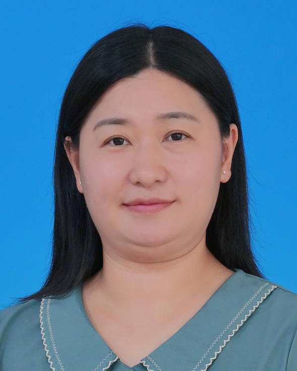 Chunhui Zhu (朱春辉)合作论文数School of Electronic Science and Engineering, Xiamen University1
 Dan Li (李丹)合作论文数School of Public Administration, Sichuan University1Bing Lei (雷冰)合作论文数1
 Sicen Wang (王嗣岑)合作论文数Food & Drug Analysis Center, School of Pharmacy, Xi'an Jiaotong University Health Science Center1
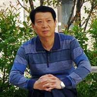 Guozhou Rao (饶国洲)合作论文数Research Center Laboratory, Hospital of Stomatology, Xi'an Jiaotong University1




