肌萎缩侧索硬化症(ALS)是一种累及上、下运动神经元的神经退行性疾病,目前尚无法治愈。针对ALS的临床管理较复杂,需要多学科合作,对症治疗及康复治疗是重要干预手段。运动训练能防止ALS患者肌肉废用性萎缩,同时也有助于患者心血管系统健康,被认为是ALS患者的一种潜在治疗方法,但仍存在一定争议。本文将阐述运动训练治疗ALS的机制、临床应用及对ALS的影响,以期为运动训练治疗ALS患者提供更多参考资料。
Objective:To compare the effects of repetitive transcranial magnetic stimulation (TMS) at three different relatively high frequencies on neuropathic pain so as to find the best frequency.Methods:One hundred Sprague-Dawley rats were randomly divided into a sham operation group, a model group, a TMS group and a control stimulation group. The TMS group was further divided into a 5Hz group, a 10Hz group, and a 20Hz group. The rats in the model, control stimulation and TMS groups received constriction injury of the sciatic nerve, while the rats in the sham group were given a sham version of the operation. On the third day after the operation the rats in the TMS group and the control stimulation group began to receive TMS treatment. Neuropatic pain was evaluated on the day before the operation, and on the 3rd, 5th, 7th, 10th and 12th days after the operation. The evaluations included the paw withdrawal thermal latency (PWTL) and the paw withdrawal mechanical threshold (PWMT).Results:The average PWTLs and PWMTs in all of the TMS groups increased with the TMS treatment. Those of the 10Hz and 20Hz groups were significantly higher than the 5Hz group′s average, while there were no significant differences between the 10Hz group and 20Hz group.Conclusions:High-frequency TMS at different frequencies has different effects on neuropathic pain, at least in rats. The treatment efficacy at 10 and 20Hz is superior to that at 5Hz.
Paired associated stimulation (PAS) has been confirmed to play a role in motor recovery after stroke, but the underlying mechanism has not been fully elucidated. In this study, we employed a comprehensive battery of measurements, including behavioral test, electrophysiology and 1H-NMR approaches, to investigate the therapeutic effects of PAS in rat model of cerebral ischemia and its underlying mechanism. Rats were randomly divided into a transient middle cerebral artery occlusion group (tMCAO group), a tMCAO + PAS group (PAS group), and a sham group. PAS was applied over 7 consecutive days in PAS group. The behavioral function of rats was evaluated by modified Garcia Scores and Rota-rod test. Electrophysiological changes were measured by motor evoked potentials (MEP). Metabolic changes of ischemic penumbra were detected by 1H-NMR. After PAS intervention, the performances on Rota-rod test and Garcia test improved and the amplitude of MEP increased significantly. The gamma-aminobutyric acid (GABA) in penumbra cortex was decreased significantly, whereas the glutamate showed the opposite changes. The results suggested that post-stroke recovery promoted by PAS may be related to the metabolites alteration in ischemic penumbra and also regulate the excitability of motor cortex.
下肢静脉血栓(VTE)是临床常见并发症。神经肌肉电刺激(NMES)作为一种安全、便携的治疗技术,近年来在预防下肢VTE形成方面得到了新的临床应用,但尚未得到充分利用。本综述主要阐述NMES在预防下肢VTE形成中的疗效、作用机制、治疗参数以及存在的问题等,旨在为后期的临床应用和临床研究思路提供参考。
We present a case report of recovery from severe COVID ‐ 19 (novel coronavirus 2019) in a patient with cognitive decline (84‐year old, female), behavioral and psychiatric symptoms, hip arthroplasty, sacroiliac decubital necrosis, cachexia, after comprehensive therapy.
Objective:To investigate the effect of paired associative stimulation (PAS) on the recovery of neurological function after cerebral infarction, and to explore whether any such effect is associated with autophagy in the ischemic penumbra.Methods:Sixty adult male Sprague-Dawley rats were randomly divided into a sham operation group ( n=20) and an experimental group ( n=40). The rats of the experimental group underwent 90 minutes of right middle cerebral artery occlusion (MCAO), while the sham group received a sham operation. The experimental group was subsequently divided into a model group ( n=20) and a PAS group ( n=20). The PAS group received 14 days of paired associative stimulation (PAS) beginning 24 hours after the operation. Neurological dysfunction was evaluated with a modified neurological severity scale (mNSS) and the elevated body swing test (EBST) on the 1st, 7th and 14th day after the MCAO. The rats were then euthanized and the expression of LC3Ⅰ, LC3Ⅱ, Beclin1, and Cathepsin B in the ischemic penumbra were detected using Western blotting, while the distribution of LC3 in neurons was detected using double immunofluorescent staining. Results:Compared with the sham group, the average mNSS scores and EBST values of the model and PAS groups were both higher on the 7th and 14th day after the MCAO, with those of the PAS group significantly lower than those of the model group on those days. The average mNSS score on the 14th day was significantly lower than on the 7th day. Compared with the sham group, the average LC3Ⅱ/Ⅰ values, Beclin 1 and Cathepsin B levels of both the model group and the PAS group were significantly higher on the 7th and 14th day after the MCAO, with the LC3Ⅱ/Ⅰ values of the PAS group significantly lower than those of the model group at both time points. The PAS group also had significantly lower Beclin1 and Cathepsin B levels on day 14. On the 7th and 14th days after the MCAO, the average number of LC3-positive cells and the ratio of LC3-positive neurons to total neurons in the model and PAS groups were significantly greater than the those of sham group, with the PAS group′s values significantly lower than those of the model group at each time point.Conclusion:PAS can significantly promote neurological recovery after stroke. The beneficial effects may involve inhibition of neuronal autophagy in the ischemic penumbra.
目的:观察成对关联刺激对缺血性脑卒中大鼠缺血半暗带区NogoA、NgR、RhoA蛋白表达的影响.方法:90只健康雄性SD大鼠随机分为假手术组、模型组和PAS组,每组又分为7d、14d、28d三个亚组(n=10).模型组和PAS组大鼠采用Longa线栓法制作MCAO模型,假手术组除不插入线栓外,其余操作与模型组和PAS组相同.PAS组于术后第1d开始给予0.05Hz、共90对脉冲的PAS治疗,每日1次,每次持续30min;模型组和假手术组不予任何干预措施,各时间点治疗结束后取缺血半暗带区脑组织,Western Blot和免疫荧光检测缺血半暗带NogoA、NgR、RhoA蛋白的表达和分布情况.结果:术后第7、14、28d,假手术组NogoA、NgR、RhoA蛋白的表达水平均最低,模型组在各时间点NogoA、NgR、RhoA含量均较假手术组明显升高(P<0.05),PAS组术后各时间点NogoA、NgR、RhoA的含量高于假手术组(P<0.05),低于同时间点模型组(P<0.05),PAS组NogoA、NgR、RhoA蛋白在术后28d的含量低于同组第7d、14d(P<0.05).结论:PAS可抑制NogoA/NgR/RhoA信号通路蛋白的表达,可能是PAS促进脑缺血大鼠神经再生,改善脑缺血大鼠的感觉运动功能障碍的内在分子机制之一.
脑卒中后各种功能障碍不仅源于脑部中枢损伤,也源于脊髓中枢继发性病理改变,减轻脑卒中后继发性脊髓病变有望成为脑卒中康复治疗的有利靶点.本文对脑卒中后脊髓病理改变以及相关发生机制及干预措施进行综述,旨在为临床治疗脑卒中提供新的思路.
Paired associative stimulation is a relatively new non-invasive brain stimulation technique that combines transcranial magnetic stimulation and peripheral nerve stimulation. The effects of paired associative stimulation on the excitability of the cerebral cortex can vary according to the time interval between the transcranial magnetic stimulation and peripheral nerve stimulation. We established a model of cerebral ischemia in rats via transient middle cerebral artery occlusion. We administered paired associative stimulation with a frequency of 0.05 Hz 90 times over 4 weeks. We then evaluated spatial learning and memory using the Morris water maze. Changes in the cerebral ultra-structure and synaptic plasticity were assessed via transmission electron microscopy and a 64-channel multi-electrode array. We measured mRNA and protein expression levels of brain-derived neurotrophic factor and N-methyl-D-aspartate receptor 1 in the hippocampus using a real-time polymerase chain reaction and western blot assay. Paired associative stimulation treatment significantly improved learning and memory in rats subjected to cerebral ischemia. The ultra-structures of synapses in the CA1 area of the hippocampus in rats subjected to cerebral ischemia were restored by paired associative stimulation. Long-term potentiation at synapses in the CA3 and CA1 regions of the hippocampus was enhanced as well. The protein and mRNA expression of brain-derived neurotrophic factor and N-methyl-D-aspartate receptor 1 increased after paired associative stimulation treatment. These data indicate that paired associative stimulation can protect cognition after cerebral ischemia. The observed effect may be mediated by increases in the mRNA and protein expression of brain-derived neurotrophic factor and N-methyl-D-aspartate receptor 1, and by enhanced synaptic plasticity in the CA1 area of the hippocampus. The animal experiments were approved by the Animal Ethics Committee of Tongji Medical College, Huazhong University of Science & Technology, China (approval No. TJ-A20151102) on July 11, 2015.
Objective To observe the effects of paired associative stimulation ( PAS) on synaptic ultrastruc-ture, neuron apoptosis and BDNF in rats with cerebral infarct, and explore the possible underlying mechanisms. Methods Forty-five male Sprague-Dawley rats were randomly divided into three groups:a sham operation group, a model group and a PAS group, with 15 rats in each. All the rats underwent a surgical operation for transient middle cerebral artery occlusion ( MCAO) on the right side to model focal cerebral ischemia, with those in the sham opera-tion group left without real occlusion. PAS treatment was given to rats in the PAS group 24h after MCAO model was successfully established, while no special intervention was given to those in the sham operation group and model group. After 28 days of treatment, transmission electron microscopy was used to investigate the ultrastructure of the is-chemic penumbra, TUNEL was used to observe the apoptosis of cortex neurons, and real time-PCR to investigate BDNF mRNA expression. Results It was found that after 28 days treatment:①The synaptic curvature, the synapse length and the post-synaptic density ( PSD) decreased significantly in the rats of model group in contrast to those of the sham control group ( P<0.05) . And compare to model group, the synaptic curvature, the synapse length and the PSD increased significantly in the rats of PAS group ( P<0.05) . ②Compare to sham control group, the apoptosis rate of model group and PAS group increased significantly ( P<0.05) . And the apoptosis rate of PAS group decreased sig-nificantly in contrast to those of model group ( P<0.05) .③Compare to sham control group, BDNF mRNA expression of the PAS group increased significantly( P<0.05) , while BDNF mRNA expression of the model group decreased sig-nificantly( P<0.05) . BDNF mRNA expression of the PAS group increased significantly in contrast to those of model group ( P<0.05) . Conclusion PAS promotes neural plasticity and inhibits apoptosis of cortex neurons of the ische-mic penumbra in rats with ischemic cerebral infarction. One of the underlying mechanisms might be related to the up-regulation of BDNF mRNA expression.
近年来无骨折脱位型脊髓损伤( SCIWORA)备受关注.据报道国内、外SCIWORA流行病学特征存在差异,且致伤原因也不尽相同.在SCIWORA损伤初期若处理不当,可能导致迟发性神经功能障碍加重或复发,颈托外固定、限制高危活动是目前普遍采用的SCIWORA治疗方法,若患者神经功能障碍程度进行性加重或MRI显示脊髓受压、韧带损伤时可考虑手术治疗.目前认为SCIWORA患者远期预后与损伤初期神经功能缺损程度密切相关.
缺血性脑卒中可诱发脑组织内神经元、星形胶质细胞等各种细胞的自噬活性变化.自噬是维持细胞内环境稳态的重要因素之一,与细胞的存活息息相关.探讨缺血性脑卒中后自噬活性的变化情况及其意义、以及相关干预措施,有望为缺血性脑卒中的治疗提供新的思路.
目的:探讨情景模拟互动训练对脑卒中偏瘫患者日常生活自理能力及生活质量的影响.方法:选取我院神经内科确诊为脑卒中,并在康复医学科进行康复治疗的脑卒中患者112例,随机分成对照组和观察组各56例.对照组予以常规治疗及护理,观察组在此基础上增加情景模拟互动训练.于治疗前、治疗后6及12个月,分别对2组患者进行Barthel指数(BI)量表、世界卫生组织生存质量量表(WHOQOL-BREF)评定.结果:治疗12个月后,2组优良率均较治疗前明显提高(均P<0.05),且观察组较对照组更显著提高(P<0.05);治疗6及12个月后,2组同时间点比较,观察组BI评分和WHOQOL-BREF评分均高于治疗前和对照组(均P<0.05);对照组治疗12个月后,BI评分高于治疗前(P<0.05),对照组治疗6及12个月后,WHOQOL-BREF评分均明显高于治疗前(均P<0.05).结论:情景模拟互动训练对提高脑卒中患者日常生活自理能力及生活质量具有明显疗效.
脊髓损伤(SCI)常继发手功能、步行功能、感觉功能等多种功能障碍,严重影响患者的生活质量.虚拟现实(VR)技术作为一项新兴的人工智能技术,越来越广泛地被应用于神经系统疾病的康复治疗.本文主要综述VR技术在SCI康复评估和康复治疗中的应用进展,包括在SCI后运动功能障碍、神经病理性疼痛、心理障碍等评估和治疗中的应用,以及相关作用机制.
BACKGROUND Supernumerary phantom limb(SPL) caused by spinal cord injury(SCI) has previously been reported in several studies. However, the mechanisms and management of SPL in SCI patients are still not fully understood. Herein, we report a rare case of SPL in a patient with incomplete SCI.CASE SUMMARY A 46-year-old man complained of four hands 7 d after SCI. He was diagnosed with SPL complicated with actual limb neuropathic pain. Following a period of treatment with neurotrophic agents and Chinese traditional and analgesic medications, SPL symptoms and actual limb pain did not improve. However, his symptoms gradually lessened after combined treatment with high-frequency repetitive transcranial magnetic stimulation(rTMS), a promising neuromodulation technique, over the M1 cortex and visual feedback. After 7 wk of this treatment, SPL disappeared completely and actual limb pain was significantly relieved.CONCLUSION Cerebral plasticity changes may be a mechanism underlying the occurrence of non-painful SPL in SCI patients, and high-frequency rTMS applied to the M1 cortex could be a promising treatment method for SPL.
Objective To explore the effects and the underlying mechanisms by which paired associative stimulation ( PAS) of tibial nerve electrostimulation and M1 cortex transcranial magnetic stimulation ( TMS) in pro-moting the recovery of forelimb dysfunction in rats with cerebral ischemic stroke. Methods Resting motor thresholds of left extensor carpi radialis muscle ( ECR) were determined 5 min before and 5 min, 30 min, 60 min after PAS,respectively, in 8 male Sprague-Dawley ( SD) rats. Then 48 male SD rats were divided into a sham group ( n=16) subject to sham surgery, an experimental group (n=32) which was further divided into a MCAO group (n=16) and a PAS group (n=16) after cerebral ischemic stroke model was established successfully by occluding the right middle cerebral artery. 24 hours after surgery, PAS consisting of left tibial nerve stimulation and right M1 cortex area TMS was applied to PAS group once daily for 7 consecutive days. The corner tests and grip strength tests were per-formed before and after 7 days of PAS treatment in each group. The RMTs of left ECR were determined, metabolites of the left area tissue of cervical spinal cord were measured by nuclear magnetic resonance spectrum, and expressions of Bcl-2 and Bax of left and right area tissue of cervical spinal cord enlargement were detected by Western Blot tech-nique after 7 days of intervention. Results The average RMTs of left ECR at 5 min, 30 min, 60 min after PAS were significantly lower than those at 5 min before PAS ( P<0.05) . All rats in experimental group showed significant higher turning scores and lower grip strength when compared with sham group (P<0.001 or P<0.01). After PAS interven-tion, PAS group demonstrated lower turning scores, higher grip strength and lower RMT of left ECR as compared with MCAO group ( P<0.05 or P<0.01) . The expression of GABA of left cervical enlargment was significantly decreased in MCAO group when compared with the sham group ( P<0.05) , and there was no significant difference between MCAO group and PAS group. Meanwhile, other metabolites showed no significant difference among the three groups. The av-erage expression level of Bcl-2 and Bax proteins in both sides of cervical spinal cord enlargment showed no significant difference among three groups either. Conclusions Tibial nerve-M1 cortex area PAS may increase the excitability of motor cortical representation of forelimbs in rats, by which PAS promotes the recovery of forelimb dysfunction in rats with cerebral ischemic stroke.
Paired associative stimulation (PAS), combining transcranial magnetic stimulation (TMS) with electrical peripheral nerve stimulation (PNS) in pairs with an optimal interstimulus interval (ISI) in between, has been shown to influence the excitability of the motor cortex (MC) in humans. However, the underlying mechanisms remain unclear. This study was designed to explore an optimal protocol of PAS, which can modulate the excitability of MC in rats, and to investigate the underlying mechanisms. The resting motor thresholds (RMTs) of TMS-elicited motor evoked potentials (MEPs) recorded from the gastrocnemius muscle and the latency of P1 component of somatosensory evoked potentials (SEPs) induced by electrical tibial nerve stimulation were determined in male Sprague-Dawley rats ( n =10). Sixty rats were then randomly divided into 3 groups: a PAS group (further divided into 10 subgroups at various ISIs calculated by using the latency of P1, n =5, respectively), a TMS (only) group ( n =5) and a PNS (only) group ( n =5). Ninety repetitions of PAS, TMS and PNS were administered to the rats in the 3 groups, respectively, at the frequency of 0.05 Hz and the intensity of TMS at 120% RMT and that of PNS at 6 mA. RMTs and motor evoked potentials’ amplitude (MEPamp) were recorded before and immediately after the interventions. It was found that the MEPamp significantly decreased after PAS at ISI of 5 ms ( P <0.05), while the MEPamp significantly increased after PAS at ISI of 15 ms, as compared with those before the intervention ( P <0.05). However, the RMT did not change significantly after PAS at ISI of 5 ms or 15 ms ( P >0.05). PAS at other ISIs as well as the sole use of TMS and PNS induced no remarkable changes in MEPamp and RMT. In conclusion, PAS can influence motor cortex excitability in rats. Neither TMS alone nor PNS alone shows significant effect.
Objective To observe the effect of paired associative stimulation ( PAS) on the recovery of sensorimotor function and to explore the mechanism in terms of neural plasticity. Methods Ninety male adult Sprague-Dawley rats were randomly divided into a sham operation group (Sham group), a model group (Model group) and a paired associative stimulation group ( PAS group) , each of 30. Each group was then subdivided into 7-, 14-and 28-day subgroups with 10 rats in each. A model of focal cerebral ischemia and reperfusion was estab-lished using the Longa suture method in the Model and PAS groups. The rats in the Sham group underwent the same surgical procedure except for the occlusion of the middle cerebral artery. The rats received 30 minutes of paired pe-ripheral nerve stimulation and transcranial magnetic stimulation comprising 90 pairs at 0.05 Hz beginning 24 h after the occlusion. The impulse wave width of the peripheral nerve stimulation was 200 μs and the intensity was 6 mA. The intensity of the transcranial magnetic stimulation was 120% of the resting motor threshold. The other two groups weren't given any intervention. Neurological function was tested using Garcia scores on the 1st, 7th, 14th and 28th day after surgery. The rats were then sacrificed and the expression of MAP-2 and GAP-43 in the ischemic penumbra were detected using western blotting and immunohistochemistry. Results No neurological dysfunction was ob-served in the Sham group at any time. Compared with the Sham group at the same time points, the average Garcia scores of the Model and PAS groups were significantly lower (P≤0.05). However, the average Garcia scores on the 7th, 14th and 28th day were significantly higher in the PAS group compared with the Model group at the same time points ( P≤0.05) . The average Garcia scores of the Model and PAS groups on the 28th day after surgery were significantly higher than those on the 1st day (P≤0.05), but only the PAS group's average Garcia score on the 28th day was significantly higher than that on the 7th day. Compared with the Sham group at the same time points, the expression of MAP-2 and GAP-43 protein in the Model and PAS groups was significantly higher, but with that of the Model group significantly lower than that of the PAS group ( all P≤0.05) . The protein expression of MAP-2 and GAP-43 protein in the PAS group on the 14th day was significantly higher than on the 7th and 28th day ( P≤0.05 for both) . Conclusions PAS can promote the recovery of sensorimotor function after cerebral thrombosis, at least in rats. That may be due to its promoting the expression of the neuroplasticity-associated proteins MAP-2 and GAP-43 in the ischemic penumbra.
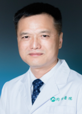 Tiecheng Guo (郭铁成)合作论文数Tongji Hospital, Tongji Medical College, Huazhong University of Science and Technology13
 Song Zhang (张松)合作论文数深圳市光明新区人民医院5
 Xiangyu Zhang合作论文数4siliang ma (马思亮)合作论文数3Xiang-Yu Zhang合作论文数3Yan Hu合作论文数2ShanShan Zhu (朱珊珊)合作论文数2
 Jun Tian (田峻)合作论文数Zhongnan Hospital, Wuhan Universiiv2Sai-hua Wang合作论文数1Yan-fang Sui合作论文数1Shanshan Zhu合作论文数1fengxia zhang (张凤霞)合作论文数1HU Mengting (胡梦婷)合作论文数1Lun Luo合作论文数1yuhui ke (柯于辉)合作论文数1
 Jinfeng Wu合作论文数1Fengxia Zhang合作论文数1
 N. Xia (夏楠)合作论文数Collaborating Ctr Training & Res Rehabil, World Hlth Org1Xiujuan Zhang合作论文数1
 Ze-Jian Chen合作论文数1lifang ke (柯丽芳)合作论文数1Yan Huang (黄燕)合作论文数1
 Jun Hu (胡军)合作论文数College of Life Science and Technology, Huazhong University of Science and Technology1di ye (叶頔)合作论文数1
 Xinhua Ding (丁新华)合作论文数Tongji Hospital, Tongji Medical College, Huazhong University of Science and Technology1
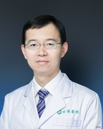 Xiaohua Han (韩肖华)合作论文数Tongji Hospital, Tongji Medical College, Huazhong University of Science and Technology1




