Mesenchymal stem/stromal cells (MSCs) show tremendous potential for regenerative medicine due to their self-renewal, multi-differentiation and immunomodulatory capabilities. Largely studies had indicated conventional tissue-derived MSCs have considerable limited expandability and donor variability which hinders further application. Induced pluripotent stem cell (iPSCs)-derived MSCs (iMSCs) have created exciting source for standardized cellular therapy. However, the cellular and molecular differences between iMSCs and the cognate tissue-derived MSCs remains poorly explored. In this study, we first successfully reprogrammed human umbilical cords-derived mesenchymal stem/stromal cells (UMSCs) into iPSCs by using the cocktails of mRNA. Subsequently, iPSCs were further differentiated into iMSCs in xeno-free induction medium. Then, iMSCs were compared with the donor matched UMSCs by assessing proliferative state, differentiation capability, immunomodulatory potential through immunohistochemical analysis, flow cytometric analysis, transcriptome sequencing analysis, and combine with coculture with immune cell population. The results showed that iMSCs exhibited high expression of MSCs positive-makers CD73, CD90, CD105 and lack expression of negative-maker cocktails CD34, CD45, CD11b, CD19, HLA-DR; also successfully differentiated into osteocytes, chondrocytes and adipocytes. Further, the iMSCs were similar with their parental UMSCs in cell proliferative state detected by the CCK-8 assay, and in cell rejuvenation state assessed by β-Galactosidase staining and telomerase activity related mRNA and protein analysis. However, iMSCs exhibited similarity to resident MSCs in Homeobox (Hox) genes expression profile and presented better neural differentiation potential by activation of NESTIN related pathway. Moreover, iMSCs owned enhanced immunosuppression capacity through downregulation pools of pro-inflammatory factors, including IL6, IL1B etc. and upregulation anti-inflammatory factors NOS1, TGFB etc. signals. In summary, our study provides an attractive cell source for basic research and offers fundamental biological insight of iMSCs-based therapy.
Mesenchymal stem cells (MSC) isolated from different tissue sources exhibit multiple biological effects and have shown promising therapeutic effects in a broad range of diseases. In order to fulfill their clinical applications in context of precision medicine, however, more detailed molecular characterization of diverse subgroups and standardized scalable production of certain functional subgroups would be highly desired. Thus far, the generation of induced pluripotent stem cell (iPSC)-derived MSC (iMSC) seems to provide the unique opportunity to solve most obstacles that currently exist to prevent the broad application of MSC as an advanced medicinal product. The features of iMSC include their single cell clone origins, and defined and controllable cultural conditions for their derivation and proliferation. Still, comprehensive research of the molecular and functional heterogeneity of iMSC, just like MSC from any other tissue types, would be required. Furthered on previous efforts on iMSC differentiation and expansion platform and transcriptomic studies, advantages of single cell multi-omics analysis and other up-to-dated technologies would be taken in order to elucidate the molecular origin and regulation of heterogeneity and to obtain iMSC subgroups homogeneous enough for particular clinical conditions. In this perspective, the current obstacles in MSC applications, the advantages of iMSC over MSC and their implications for biological research and clinical applications will be discussed.
Senile osteoporosis (SOP) is a worldwide age-related disease characterized by the loss of bone mass and decrease in bone strength. Bone mesenchymal stem cells (BMSCs) play an important role in the pathology of senile osteoporosis. Abnormal expression and regulation of non-coding RNA (ncRNA) are involved in a variety of human diseases. In the present study, we aimed to identify differentially expressed mRNAs and ncRNAs in senile osteoporosis patient-derived BMSCs via high-throughput transcriptome sequencing in combination with bioinformatics analysis. As a result, 415 mRNAs, 30 lncRNAs, 6 circRNAs and 27 miRNAs were found to be significantly changed in the senile osteoporosis group. Gene Ontology (GO) and Kyoto Encyclopedia of Genes and Genomes (KEGG) analysis were applied to analyze the function of differentially expressed mRNAs and ncRNAs. The circRNA–miRNA–mRNA regulatory network was constructed using the cytoHubba plugin based on the Cytoscape software. Interestingly, circRNA008876-miR-150-5p-mRNA was the sole predicted circRNA-miRNA-mRNA network. The differential expression profile of this ceRNA network was further verified by qRT-PCR. The biological function of this network was validated by overexpression and knockdown experiments. In conclusion, circRNA008876-miR-150-5p-mRNA could be an important ceRNA network involved in senile osteoporosis, which provides potential biomarkers and therapeutic targets for senile osteoporosis.
Frozen shoulder is a common shoulder disorder characterized by a gradual increase of pain and a limited range of motion. However, its pathophysiologic mechanisms remain unclear and there is no consensus as to the most effective treatment. The purpose of the study was to investigate the effect of transforming growth factor‐β (TGF‐β) on fibrosis and inflammatory response of the shoulder joint of rat models and to explore the therapeutic effect of the peroxisome proliferator‐activated receptor‐γ (PPAR‐γ) agonist. In the study, the effect of PPAR‐γ agonist CDDO‐IM treatment on cell proliferation, migration, and extracellular matrix proteins synthesis (vimentin, α‐smooth muscle actin, collagen I, and collagen III) were tested by cell proliferation test, scratches test, real‐time quantitative polymerase chain reaction, and Western blot analysis. The frozen shoulder was also established on the rat model by injecting adenovirus‐TGF‐β1 into rats' shoulder capsule. Pathological changes of the frozen shoulder tissue of the experimental group and PPAR‐γ agonist treatment group were evaluated. The stiffness of joints of the three groups was tested. Inflammatory mediators' expression including cyclooxygenase‐1, interleukin‐1β, and tumor necrosis factor‐α of the shoulder was tested by enzyme‐linked immunosorbent assay, and the expression of extracellular matrix proteins was evaluated by hematoxylin and eosin staining and immunohistochemistry. The results showed that pathological changes of the frozen shoulder in the rat model include an abnormal proliferation of fibroblasts, infiltration of inflammatory cells, and disorder of fibrous structure, while rosiglitazone reduced the severity of the frozen shoulder in the treatment group. Clinically, PPAR‐γ agonists may be a promising target for the treatment of the frozen shoulder.
背景:冻结肩是一种以肩关节疼痛和活动受限为主要特征的疾病,目前病因不明,治疗效果欠佳.目的:研究转化生长因子-β1(TGF-β1)对大鼠肩关节囊纤维化及炎症反应等病理改变的影响,以及过氧化物酶体增殖剂激活受体-γ(PPAR-γ)激动剂对大鼠冻结肩的治疗作用,为临床治疗冻结肩提供新思路.方法:利用TGF-β1诱导人皮肤成纤维细胞(HSF)异常纤维化,PPAR-γ激动剂三萜类化合物(CDDO-IM)作为治疗药物,检测细胞增殖、迁移情况及细胞外基质蛋白的表达情况.向SD大鼠的肩关节腔注射TGF-β1过表达的腺病毒构建大鼠冻结肩模型.2周后,其中一半大鼠用罗格列酮灌胃饲养2周,作为罗格列酮治疗组.检测大鼠肩关节活动度,利用ELISA检测关节腔炎症因子,HE染色和免疫组织化学法检测大鼠肩关节囊组织细胞外基质蛋白的表达情况.结果:在细胞实验中,TGF-β1组的增殖率为83.6%,TGF-β1+CDDO-IM组增殖率为19.3%(P<0.01);TGF-β1组的迁移率为84.9%,TGF-β1+CDDO-IM组迁移率为44.1%(P<0.05);TGF-β1能够诱导HSF增殖、迁移及异常纤维化.动物实验成功构建了TGF-β1诱导的大鼠冻结肩模型,免疫组织化学结果提示,罗格列酮能够抑制大鼠冻结肩的炎症形成,减少基质蛋白生成,重塑纤维组织结构.结论:冻结肩的病理改变主要是纤维细胞异常增生,炎症细胞浸润,纤维结构紊乱,而PPAR-γ激动剂可以缓解大鼠冻结肩关节僵硬,减弱炎症反应,抑制异常纤维化.
内侧副韧带(MCL)是膝关节内侧维持关节稳定性的最主要韧带,其功能与前交叉韧带(ACL)具有协同作用,共同维持膝关节的稳定性.MCL损伤是膝关节最常见的韧带损伤,其损伤后将对ACL生物力学产生影响,同时增加了ACL损伤的风险.研究MCL损伤后对ACL生物力学的影响可以更加深入理解MCL在维持膝关节稳定性中的重要性,为临床治疗提供更多理论支持.笔者对近年来MCL的解剖、功能、生物力学、损伤后对ACL生物力学的影响及治疗现状进行综述.
Introduction Currently, osteoarthritis (OA) receives global increasing attention because it associates severe joint pain and serious disability. Stem cells intra-articular injection therapy showed a potential therapeutic superiority to reduce OA development and to improve treating outputs. However, the long-term effect of stem cells intra-articular injection on the cartilage regeneration remains unclear. Recently, miR-140-5p was confirmed as a critical positive regulator in chondrogenesis. We hypothesized that hUC-MSCs overexpressing miR-140-5p have better therapeutic effect on osteoarthritis. Materials and methods To enhance stem cell chondrogenic differentiation, we have transfected human umbilical cord mesenchymal stem cells (hUC-MSCs) with miR-140-5p mimics and miR-140-5p lentivirus to overexpress miR-140-5p in a short term or a long term accordingly. Thereafter, MSCs proliferation, chondrogenic genes expression and extracellular matrix were assessed. Destabilization of the medial meniscus (DMM) surgery was performed on the knee joints of SD rats as an OA model, and then intra-articular injection of hUC-MSCs or hUC-MSCs transfected with miR-140-5p lentivirus was carried to evaluate the cartilage healing effect with histological staining and OARSI scores. The localization of hUC-MSCs after intra-articular injection was further confirmed by immunohistochemical staining. Results Significant induction of chondrogenic differentiation in the miR-140-5p-hUC-MSCs (140-MSCs), while its proliferation was not influenced. Interestingly, intra-articular injection of 140-MSCs significantly enhanced articular cartilage self-repairing in comparison to normal hUC-MSCs. Moreover, we noticed that intra-articular injection of high 140-MSCs numbers reinforces cells assembling on the impaired cartilage surface and subsequently differentiated into chondrocytes. Conclusions In conclusion, these results indicate therapeutic superiority of hUC-MSCs overexpressing miR-140-5p to treat OA using intra-articular injection.
目的 探讨过氧化物酶体增殖物激活受体(PPAR)β/δ激动剂(GW0742)对人关节液来源的间充质干细胞(hSF-M SC)成软骨分化及炎性环境下炎症因子释放的影响.方法 抽取膝关节骨关节炎患者患侧关节腔内的关节液并分离培养及鉴定.设置空白对照组(不完全成软骨诱导培养基)、转化生长因子(TGF)-β1组(含浓度为10 ng/mL TGF-β1的成软骨诱导培养基)、TGF-β1+GW0742组(含10 ng/mL TGF-β1、1μmol/L GW0742的成软骨诱导培养基)及GW0742组(含浓度为1μmol/L GW0742的成软骨诱导培养基),采用Pellet法诱导hSF-MSC成软骨分化.通过比较各组成软骨分化相关标志物SOX-9基因表达和蛋白合成以及阿利新蓝、甲苯胺蓝染色等来评估GW0742对hSF-MSC增殖及成软骨分化能力的影响.另设置空白对照组(20% 关节液+80% 完全培养基)、hSF-MSC组(20% 关节液+80% 完全培养基+1×10⁵个/mL hSF-MSC)、hSF-MSC+GW0742组(20% 关节液+80% 完全培养基+1μmol/L GW0742+1×10⁵个/mL hSF-MSC),通过比较第1、3天各组培养液中炎症因子肿瘤坏死因子(TNF)-α表达来评估GW0742+hSF-MSC组免疫抑制能力.结果 应用直接贴壁法成功从炎性关节液中分离培养hSF-MSC.TGF-β1、TGF-β1+GW0742及GW0742对hSF-MSC增殖能力无明显影响;聚合酶链反应(PCR)和Western Blot检测表明,GW0742组hSF-MSC成软骨分化作用最为明显;甲苯胺蓝和阿利新蓝染色显示,GW0742组hSF-MSC能分泌较多的蛋白聚糖,且较TGF-β1+GW0742组细胞分泌的蛋白聚糖更为致密;hSF-MSC+GW0742组炎症因子表达显著低于hSF-MSC组.结论 PPARβ/δ激动剂GW0742不仅能提升hSF-MSC成软骨分化能力,还能增强hSF-MSC抗炎能力.
肩周炎是一种常见的肩关节疾病,患者常出现疼痛及肩关节活动受限,也被称为冻结肩、五十肩.文献报道其地区发病率为2%~5%[1],集中发病年龄在55岁上下,且女性发病率高于男性,左肩发病率高于右肩.肩周炎的主要特征为:①患肢三角肌止点周围存在慢性隐痛;②患肢夜间疼痛明显,影响睡眠;③肩关节上举活动和外旋活动均受限;④正常X线片表现.关于肩周炎的病因和发病机制,尚未明确,但目前研究已经证实肩周炎的病理改变包括盂肱关节囊增厚,伴有部分挛缩,部分患者可伴有关节囊周围韧带组织弥漫性炎症及纤维化,从而导致肩关节主被动活动明显受限[2].笔者就肩周炎不同治疗方式对缓解疼痛、改善肩关节活动度的效果进行综述,报道如下.
过氧化物酶体增殖物激活受体(PPAR)主要由PPAR α、PPARγ和PPAR β/δ 3种亚型组成.PPAR在炎症和机体代谢的调节过程中均起到重要作用,因此,与骨关节炎的发生和发展密切相关.而PPAR作为重要的核受体转录因子,在干细胞和软骨细胞的诸多功能中都起到关键性的调节作用.该文对PPAR在骨关节炎和软骨组织工程中的研究进展作一综述.
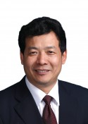 Daping Wang (王大平)合作论文数Shenzhen Second People's Hospital;Guangzhou Medical University7
 Yiyun Geng合作论文数7
 Jinfu CHEN (陈锦富)合作论文数6
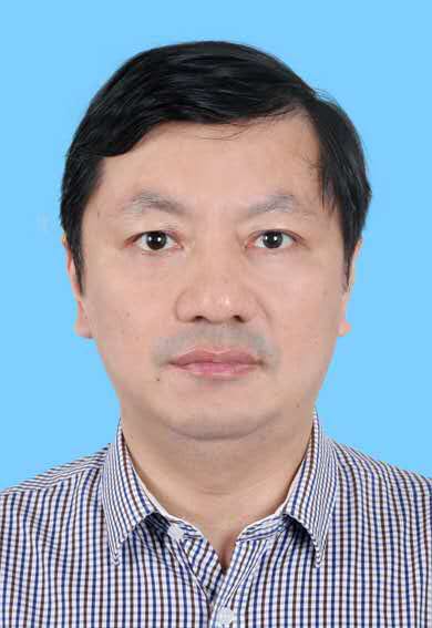 Jianyi Xiong (熊建义)合作论文数Shenzhen Second People's Hospital;Guangzhou Medical University6
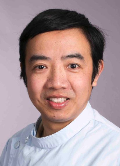 Weimin Zhu (朱伟民)合作论文数Affiliated Stomatological Hospital of Nanchang University6Tianfei Zhu合作论文数4
 Kuokuo Li合作论文数3
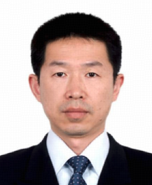 Wei Lu (陆伟)合作论文数Shenzhen Second People's Hospital;Guangzhou Medical University3
 Cui Jiaming合作论文数3
 Manyi Wang (王满宜)合作论文数Beijing Jishuitan Hospital3Luoyong Jiang合作论文数2
 Guangqian Zhou (周光前)合作论文数Medicine School, Shenzhen University2
 Lisha Mou (牟丽莎)合作论文数深圳市第二人民医院2Qiuting Deng合作论文数1Mingzhuang Chen合作论文数1Yanru An合作论文数1Kang Chen合作论文数1Yong Huang (黄勇)合作论文数1
 Liao Jinqi合作论文数1Quanlei Wang合作论文数BGI Shenzhen, Shenzhen 518083, Peoples R China1Juan Zhang合作论文数1Fei Chen合作论文数1Fei CHEN (陈斐)合作论文数1Yong Zhou (周勇)合作论文数1
 Yan Zhou合作论文数Lungene Technologies Co., Ltd.1Yuwei Wang合作论文数1Feilong Ma合作论文数1
 Murad Alahdal合作论文数Shenzhen Key Lab Tissue Engn, Shenzhen Univ1
 Junjun Zhu合作论文数1
 Zhenhan Deng合作论文数1
 Padhiar Arshad Ahmed合作论文数Institute of Glycobiology, Dalian Medical University1
 Xufeng Qi (齐绪峰)合作论文数College of Life Science and Technology, Jinan University;Key Laboratory for Regenerative Medicine, Ministry of Education, China, Jinan University - The Chinese University of Hong Kong1
 Li Duan (段俐)合作论文数Institute of Mechanics, Chinese Academy of Sciences1Kan Ouyang (欧阳侃)合作论文数1
 Dongqing Cai (蔡冬青)合作论文数College of Life Science and Technology, Jinan University1




