Cirrhotic cardiomyopathy and hepatopulmonary syndrome are two quite frequent clinical entities that may complicate the course of liver cirrhosis. The common pathophysiological origin and the same clinical presentation make them difficult to compare. Cirrhotic cardiomyopathy and hepatopulmonary syndrome may start with dyspnea and breathlessness but the former is characterized by a chronic cardiac dysfunction and the latter by a defect of oxygenation due to pulmonary shunts formation. The focus is to differentiate them as soon as possible since the treatment is different until the patient undergoes liver transplant that is the real unique cure for them.
Objectives Information is lacking on the appearance of varices in cirrhotics, either affected or not by portal hypertensive gastropathy (PHG). We assessed whether the absence or presence and the grade of PHG influenced the development of varices in cirrhotics without varices over time. Patients and methods Forty cirrhotics without varices affected or not by PHG at baseline underwent follow-up endoscopy after 5 years. One-tailed t-test and the χ 2-test were used to evaluate variable comparison and the presence of associations. Multivariate logistic regression analysis and the analysis of variance test were carried out to compare the variables and identify predictors of varices. Results The Child–Pugh score at baseline and after 5 years was significantly different (5.72±0.98 vs. 6.25±1.67, P<0.001). After 5 years, 10 (25%) cirrhotics were affected by varices, whereas 30 (75%) patients remained without varices. PHG was associated significantly with varices (P=0.001), proving to be a significant predictive independent factor for their development over time (F=4.765, significant=0.004; analysis of variance test, P<0.001). Conclusion A link between the duration of PHG and the development of varices is likely. An early therapeutic management of PHG might delay the development of varices in cirrhotics.
Multiple mechanisms of cell death exist (apoptosis, necroptosis, pyroptosis) and the subtle balance of several distinct proteins and inhibitors tightly regulates the cell fate toward one or the other pathway. Here, by combining coimmunoprecipitation, enzyme assays, and molecular simulations, we ascribe a new role, within this entangled regulatory network, to the interleukin-1 receptor antagonist (IL-1Ra). Our study enlightens that IL-1Ra, which usually inhibits the inflammatory effects of IL-1α/β by binding to IL-1 receptor, under advanced pathological states prevents apoptosis and/or necroptosis by noncompetitively inhibiting the activity of caspase-8 and -9. Consensus docking, followed by cumulative 10 μs of molecular dynamics simulations unprecedentedly reveal that IL-1Ra binds both caspases at their dimeric interface, preventing, in this manner, the formation of their catalytically/signaling active form. The resulting IL-1Ra/caspase-8(9) adducts are stabilized by hydrophobic and by few key hydrogen bonding interactions, formed by residues fully conserved across distinct caspases (-3, -6, -7, -8, and -9), and closely resemble the binding mode of the caspases inhibitors XIAP (X-linked inhibitor of apoptosis) and c-FLIP (cellular FLICE-like inhibitory protein). Tight regulation of the different forms of cell death has a major impact on distinct human illnesses (i.e., cancer, neurodegeneration, ischemic injury, atherosclerosis, viral/bacterial infections, and immune reaction). Hence, our study, pinpointing IL-1Ra as new actor of the intricate cell death regulatory network and gaining an atomic-scale understanding of its mechanism may open new avenues toward innovative therapeutic strategies to tackle major human diseases.
Portopulmonary hypertension and hepatorenal syndrome are both severe local hypertensive complications of liver cirrhosis and portal hypertension. Both are characterized by vasoconstrictive manifestations regarding pulmonary and renal vascular network, respectively. This review addresses the mechanisms underlying the development of vasoconstriction that leads to local vascular hypertension in the lung and in the kidney with the result of organ dysfunction. Potential therapeutic options are available for the management of these two syndromes as a bridge for liver transplantation; clinical efficacy depends in part on the time and rapidity of intervention and in part on how serious the chain of events is that has triggered the entire vasoconstrictive process.
Patients with advanced liver cirrhosis may develop a clinical syndrome characterized by a blunted contractile responsiveness to stress and/or altered diastolic relaxation, called "cirrhotic cardiomyopathy." This syndrome, which is initially asymptomatic, is often misdiagnosed due to the presence of symptoms that characterize other disorders present in patients with advanced liver cirrhosis, such as exercise intolerance, fatigue, and dyspnea. Stress and other conditions such as liver transplantation and transjugular intrahepatic portosystemic shunt (TIPS) may unmask this syndrome. Liver transplantation in this group of patients results in a clinical improvement and can be a cure for the cardiomyopathy. However, post-transplant prognosis depends on the identification of cirrhotics with cardiomyopathy in the pre-transplant phase; an early diagnosis of cirrhotic cardiomyopathy in the pre-transplant phase may avoid an acute onset or worsening of cardiac failure after liver transplantation. Since a preserved left ventricular ejection fraction may mask the presence of cirrhotic cardiomyopathy, the use of newer noninvasive diagnostic techniques (i.e. tissue Doppler, myocardial strain) is necessary to identify cirrhotics with this syndrome, in the pre-transplant phase. A pre-transplant treatment of heart failure in cirrhotics with cardiomyopathy improves the quality of life in this phase and reduces the complications during and immediately after liver transplantation. Since specific therapies for cirrhotic cardiomyopathy are lacking, due to the absence of a clear understanding of the pathophysiology of the cardiomyopathy, further research in this field is required.
Background: Loss of cardiac myocytes due to apoptosis is a relevant feature of ischemic heart disease. It has been described in infarct and peri-infarct regions of the myocardium in coronary syndromes and in ischemia-linked heart remodeling. Previous studies have provided protection against ischemia-induced cardiomyocyte apoptosis by the anti-inflammatory cytokine interleukin-1 receptor-antagonist (IL-1Ra). Mitochondria triggering of caspases plays a central role in ischemia-induced apoptosis. We examined the production of IL-1Ra in the ischemic heart and, based on dual intra/extracellular function of some other interleukins, we hypothesized that IL-1Ra may also directly inhibit mitochondria-activated caspases and cardiomyocyte apoptosis.Methodology/Principal Findings: Synthesis of IL-1Ra was evidenced in the hearts explanted from patients with ischemic heart disease. In the mouse ischemic heart and in a mouse cardiomyocyte cell line exposed to long-lasting hypoxia, IL-1Ra bound and inhibited mitochondria-activated caspases, whereas inhibition of caspase activation was not observed in the heart of mice lacking IL-1Ra (Il-1ra-/-) or in siRNA to IL-1Ra-interfered cells. An impressive 6-fold increase of hypoxia-induced apoptosis was observed in cells lacking IL-1Ra. IL-1Ra down-regulated cells were not protected against caspase activation and apoptosis by knocking down of the IL-1 receptor, confirming the intracellular, receptor-independent, antiapoptotic function of IL-1Ra. Notably, the inhibitory effect of IL-1Ra was not influenced by enduring ischemic conditions in which previously described physiologic inhibitors of apoptosis are neutralized.Conclusions/Significance: These observations point to intracellular IL-1Ra as a critical mechanism of the cell self-protection against ischemia-induced apoptosis and suggest that this cytokine plays an important role in the remodeling of heart by promoting survival of cardiomyocytes in the ischemic regions.
Complex molecular and cellular mechanisms are involved in the pathway of liver fibrosis. Activation and transformation of hepatic stellate cells (HSCs) are considered the two main reasons for the cause and development of liver fibrosis. The peroxisome proliferator-activated receptors (PPARs) belonging to the family of ligand-activated transcription factors play a key role in liver homeostasis, regulating adipogenesis and inhibiting fibrogenesis in HSCs. Normal transcriptional function of PPARs contributes to maintain HSCs in quiescent phase. A reduced expression of PPARs in HSCs greatly induces a progression of liver fibrosis and an increased production of collagen. Here, we discuss role and function of PPARs and we take into consideration molecular factors able to reduce PPARs activity in HSCs. Finally, although further validations are needed, we illustrate novel strategies available from in vitro and animal studies on how some PPARs-agonists have been proved effective as antifibrotic substances in liver disease.
Background: Healing after acute myocardial infarction (AMI) is characterized by an intense inflammatory response and increased Interleukin-1 (IL-1) tissue activity. Genetically engineered mice lacking the IL-1 receptor (IL-1R1-/-, not responsive to IL-1) or the IL-1 receptor antagonist (IL-1Ra, enhanced response to IL-1) have an altered IL-1/IL-1Ra balance that we hypothesize modulates infarct healing and cardiac remodeling after AMI.Methods: IL-1R1-/- and IL-1Ra-/- male mice and their correspondent wild-types (WT) were subjected to permanent coronary artery ligation or sham surgery. Infarct size (trichrome scar size), apoptotic cell death (TUNEL) and left ventricular (LV) dimensions and function (echocardiography) were measured prior to and 7 days after surgery.Results: When compared with the corresponding WT, IL-1R1-/- mice had significantly smaller infarcts (-25%), less cardiomyocyte apoptosis (-50%), and reduced LV enlargement (LV end-diastolic diameter increase [LVEDD], -20%) and dysfunction (LV ejection fraction [LVEF] decrease, -50%), whereas IL-1Ra-/- mice had significantly larger infarcts (+75%), more apoptosis (5-fold increase), and more severe LV enlargement (LVEDD increase, +30%) and dysfunction (LVEF decrease, +70%)(all P values <0.05).Conclusions: An imbalance in IL-1/IL-1Ra signaling at the IL-1R1 level modulates the severity of cardiac remodeling after AMI in the mouse, with reduced IL-1R1 signaling providing protection and unopposed IL-1R1 signaling providing harm.
BACKGROUND:This study aimed to determine the detection rate and clinical relevance of portosystemic collaterals.METHODS:We studied 326 cirrhotics. Portosystemic collaterals, portal vein diameter, and splenic area were evaluated by color Doppler sonography; esophageal varices were detected by endoscopy.RESULTS:Of the cirrhotics, 130 had portosystemic collaterals (39.9% total, left gastric vein 11%, paraumbilical vein 7.4%, splenorenal shunts 13.8%, and combined shunts 7.7%). Cirrhotics without portosystemic collaterals or with a paraumbilical vein had a significantly narrower portal vein diameter than cirrhotics with a left gastric vein (P < 0.001). Cirrhotics with a paraumbilical vein had a significantly smaller splenic area than cirrhotics with a left gastric vein (P < 0.001), splenorenal shunts (P < 0.001), combined shunts (P < 0.001), or without portosystemic collaterals (P < 0.05). A significant association between portosystemic collaterals and Child's classes or presence and type of esophageal varices was found (P < 0.0001 and P = 0.0004, respectively). The highest prevalence of Child's class C and large (F-3) esophageal varices was found in cirrhotics with a left gastric vein (41.7% and 36.1%, respectively), whereas esophageal varices were absent in 47.4% of cirrhotics without portosystemic collaterals and in 58.3% of cirrhotics with a paraumbilical vein.CONCLUSIONS:The left gastric vein is associated with some sonographic and clinical markers of disease severity, whereas the absence of portosystemic collaterals or the presence of paraumbilical veins seems to identify cirrhotics with markers predictive of a more favorable clinical course.
Endothelin (ET)-1 is a vasoconstrictor involved in cardiovascular diseases. Connective tissue growth factor/CCN2 (CTGF) is a fibrotic mediator overexpressed in human atherosclerotic lesions, myocardial infarction, and hypertension. In different cell types CTGF regulates cell proliferation/apoptosis, migration, and extracellular matrix (ECM) accumulation and plays important roles in angiogenesis, chondrogenesis, osteogenesis, tissue repair, cancer and fibrosis. In the present study, we investigated the ET-1 signaling which triggers CTGF expression in cultured adult mouse atrial-muscle HL-1 cells used as a model system. ET-1 activated the CTGF promoter and induced CTGF expression at both mRNA and protein levels. Real-time PCR analysis revealed CTGF induction also in isolated rat heart preparations perfused with ET-1. Several intracellular signals elicited by ET-1 via ET receptors and even Epidermal Growth Factor Receptor (EGFR) contributed to the up-regulation of CTGF, including ERK activation and induction of the AP-1 components c-fos and c-jun, as also evaluated by ChIP analysis. Moreover, in cells treated with ET-1 the expression of ECM component decorin was abolished by CTGF silencing, indicating that CTGF is involved in ET-1 induced ECM accumulation not only in a direct manner but also through downstream effectors. Collectively, our data indicate that CTGF could be a mediator of the profibrotic effects of ET-1 in cardiomyocytes. CTGF inhibitors should be considered in setting a comprehensive pharmacological approach towards ET-1 induced cardiovascular diseases.
The progress of research on the molecular pathogenesis of liver fibrosis and the consequent discoveries are likely to open new possibilities for therapeutic approaches to the management of this disease in the future. A key step towards this goal is a deeper comprehension of both the complex molecular and cellular mechanisms and the signaling involved in the development of hepatic fibrosis. It is not yet clear, in fact, what role apoptosis, cytokines, oxidants and other molecules play and what relationships exist between them in favouring or delaying the onset of these adverse mechanisms. At present, a unique mechanism is recognized to be the main reason for the cause and development of liver fibrosis: sustained hepatic stellate cell activation and transformation. Therefore, in this review, after considering the cause, development of fibrosis and interrelation between molecular and cellular profibrotic mechanisms, the part played in counteracting both of these actions by some anti-oxidants and anti-fibrotic molecules such as cytokines, prostacyclin and others will be taken into consideration. The gene therapy and the possible therapeutic use of liver stem cells and tissue engineering will also be dealt with briefly. At the moment, however, the efficacy of these novel strategies still needs to be further validated in animal studies and confirmed in clinical trials. Some data that are already available from in vitro and animal studies demonstrating the effectiveness of novel approaches to inhibiting or treating liver fibrosis can only offer moderate hope.
Background— Experimental interleukin-1 receptor antagonist gene overexpression has shown that interleukin-1 receptor antagonist is cardioprotective during global cardiac ischemia. The aim of the present study was to test the impact of an exogenous recombinant human interleukin-1 receptor antagonist (anakinra) in experimental acute myocardial infarction. Methods and Results— Two animal studies were conducted: one of immediate anakinra administration during ischemia in the mouse and one of delayed anakinra administration 24 hours after ischemia in the rat. Seventy-eight Institute of Cancer Research mice and 20 Wistar rats underwent surgical coronary artery ligation (or sham operation) and were treated with either anakinra 1 mg/kg or NaCl 0.9% (saline). Treatment was administered during surgery and then daily for 6 doses in the mice and starting on day 2 daily for 5 doses in the rats. Twenty-eight mice underwent infarct size assessment 24 hours after surgery, 6 saline-treated mice and 22 mice treated with increasing doses of anakinra (1 mg/kg [n=6], 10 mg/kg [n=6], and 100 mg/kg [n=10]); 6 mice were euthanized at 7 days for protein expression analysis. The remaining animals underwent transthoracic echocardiography before surgery and 7 days later just before death. Cardiomyocyte apoptosis was measured in the peri-infarct regions. The antiapoptotic effect of anakinra was tested in a primary rat cardiomyocyte culture during simulated ischemia and in vitro on caspase-1 and -9 activities. At 7 days, 15 of the 16 mice (94%) treated with anakinra were alive versus 11 of the 20 mice (55%) treated with saline ( P =0.013). No differences in infarct size at 24 hours compared with saline were observed with the 1- and 10-mg/kg doses, whereas a 13% reduction in infarct size was found with the 100-mg/kg dose ( P =0.015). Treatment with anakinra was associated with a significant reduction in cardiomyocyte apoptosis in both the immediate and delayed treatment groups (3.1±0.2% versus 0.5±0.3% [ P <0.001] and 4.2±0.4% versus 1.1±0.2% [ P <0.001], respectively). Compared with saline-treated animals, anakinra-treated mice and rats showed signs of more favorable ventricular remodeling. In vitro, anakinra significantly prevented apoptosis induced by simulated ischemia and inhibited caspase-1 and -9 activities. Conclusions— Administration of anakinra within 24 hours of acute myocardial infarction significantly ameliorates the remodeling process by inhibiting cardiomyocyte apoptosis in 2 different experimental animal models of AMI. This may open the door for using anakinra to prevent postischemic cardiac remodeling and heart failure.
Background: Doppler ultrasonography (US) of portal blood flow and portal flow volume (PFV) are useful to definechangesinportalhemodynamicsofpatientswithchronic liverdiseases.ThemealtestwithpostmealPFVmeasurements is generally accepted as a reproducible noninvasive test to evaluate the severity of portal hypertension. The aim of this study was to evaluate whether monitoring PFV changes after ingestion of a standard meal would be useful to characterize patientswithchronichepatitisorlivercirrhosisinthepresence or absence of hyperdynamic syndrome (HS) characterized by elevated PFV, splenomegaly, systemic hypotension and/or increased cardiac output. Patients and Methods: Thirty-seven patients (22 men and 15 women, median age 53 years) with hepatitis C virus infection and 20 healthy age- and sex- matchedvolunteers(Controls)wereenrolledinthestudy.There were19(51.4%)patientswithchronichepatitis(GroupA)and 18 (48.6%) with ultrasonographic evidence of liver cirrhosis (Child-Pugh class B), 9 of whom had an HS (Group B) while theremainder(GroupC)didnot.Eachpatientunderwentliver color Doppler US and the test was repeated 30, 60 and 90 minutesafteradministrationofastandardmeal(300kcalfluid mealcontaining12gofproteins,11.6goflipidsand36.8gof carbohydrates). Results: The baseline PFV did not differ (p=NS) between Controls and both GroupsA and C, while the PFV of Group B patients was significantly (p<0.01) higher. After30minutes,thePFVincreased(p<0.01)bothinControls andGroupApatients,whilethedifferenceswerenotsignificant in cirrhotic patients (Groups B and C). Our study confirmed that the postmeal PFV increases in both healthy individuals and in patients with chronic hepatitis, while in cirrhotic patients no significant changes occur. In conclusion, monitoring the portal blood flow in cirrhotic patients before andafteradministrationofastandardmealmightbeasuitable test to evaluate potential disturbances of the flow itself. Moreover, the test could be useful to determine optimal pharmacological or surgical interventions aimed at restoring a better flow to the liver by reducing or favouring the occurrenceofspontaneousmesenteric-systemicvenousshunts.
Background: T-lymphocyte activation within atherosclerotic plaque, and widespread to the myocardium, has been shown in patients with acute coronary syndromes. Objective: To investigate the presence of T-lymphocyte infiltrate at different stages of acute coronary syndromes by studying patients with sudden coronary death, acute myocardial infarction (AMI) and healed infarction, in comparison with patients with myocarditis and patients with non-ischaemic heart failure. Methods: 72 cases were studied at autopsy: 12 dying of sudden coronary death (group 1), 12 dying <4 weeks (group 2) and 12 dying >4 months after AMI (group 3), 12 with active lymphocytic myocarditis (group 4), 12 with hypertensive heart disease (group 5), and 12 control subjects (group 6). Light microscopy was performed to measure the number of activated T-lymphocytes (CD3+/DR+) in the myocardium and coronary artery wall, and intercellular adhesion molecule-1 (ICAM-1) expression in the myocardium. Results: Activated T-lymphocyte infiltrates and ICAM-1 myocardial expression in both remote and peri-infarction regions and activated T-lymphocytes within the epicardial coronary artery wall of both the infarct- and non-infarct-related arteries were found in groups 1, 2 and 3, whereas myocardial, but not coronary, infiltrates were found in groups 4 (p<0.001 vs groups 1, 2 and 3 for coronary infiltrates). Groups 5 and 6 had no evidence of myocardial or coronary inflammation (p<0.001 vs groups 1, 2 and 3). Conclusions: The study shows the presence of a lymphocytic infiltrate in both coronary arteries and myocardium and a proinflammatory phenotype shift in the myocardium associated with acute coronary thrombosis in patients dying suddenly, shortly, or even late after coronary thrombosis.
The aim was to evaluate the predictability of portal diameter (PD) in the diagnosis of esophageal varices (EV) and of large size EV (F3EV) in a large series of patients with cirrhosis. Two-hundred sixty-six persons with cirrhosis (M:F = 153:113; mean age 65.4 +/- 10 y) were studied by abdominal sonography and upper endoscopy. Portal hypertensive gastropathy (PHG) was found in 16.1% and EV was found in 60.9% of patients. Only Child's class (B vs. A: OR 3.4, p < 0.0001; C vs. A: OR 10.3, p < 0.0001; C vs. B: OR 3.1, p = 0.01) and age (OR 1.04, p = 0.03) were independent predictors of EV, whereas PD was not (p = 0.4). Child's class and age were also the only independent predictors of F3EV. Mean PD showed a slight and not significant increase in PHG patients compared with patients with negative endoscopy, a reduction in F1EV patients and then a progressive increase in F2EV and F3EV patients. Patients with PD <12 mm showed a significantly higher prevalence of F1-F2EV (p < 0.05) and a near-significant lower prevalence of endoscopies negative for EV (p = 0.06) than patients with 12 < or = PD < or = 13 mm. PD was not able to predict EV or F3EV in a large series of patients with cirrhosis. The oscillatory trend of PD, proceeding from patients with negative endoscopy to F3EV patients, seems to indicate that EV may unload portal pressure in the initial phases of portal hypertension.
According to current literature, infective processes greatly modify both vascular hemodynamics and anti-oxidant properties of affected tissues, causing a change in homeostasis that regulates the correct functioning of all cells responsible for the physiological and metabolic balance of various organs. As a consequence, the response to the infection that has caused the change is also likely to be weaker and, in the case of septic shock, ineffective. In this review, we will take into consideration these mechanisms and then focus on a group of vasodilator drugs (prostacyclin and its analogs) which, though have been used for over 20 years mainly to treat obstructive vascular diseases, have such hemodynamic and anti-inflammatory properties which prevent homeostatic changes. It is obvious that prostacyclin does not definitively have anti-infective characteristics; however, in association with anti-infective drugs (antibiotics, etc.), the effectiveness of the latter appears improved, at least in some circumstances. Similarly, the fact that prostacyclin and its analogs have a cytoprotective effect on the liver and reduce the ischemia-reperfusion damage following liver transplant is not a novelty and evidence that they improve hepatic hemodynamics suggests their use in those pathologies characterized by possible reduced perfusion or ascertained ischemia of the liver.
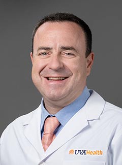 Antonio Abbate合作论文数Advanced Heart Failure and Transplant Center, University of Virginia;Heart and Vascular Center Fontaine, University of Virginia24
 Enrico Zardi合作论文数Department of Clinical Medicine, "Campus Bio-Medico" University20
 E Vecile合作论文数13Antonella Afeltra合作论文数Department of Clinical Medicine, Immunology and Rheumatology, University Campus Bio-Medico of Rome11Elena Vecile合作论文数10Giuseppe Patti合作论文数9
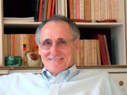 Germano Di Sciascio合作论文数Department of Cardiovascular Sciences, Campus Bio-Medico University9
 Alfonso Baldi合作论文数8Ermanno Nardon合作论文数8
 Andrea D'Ambrosio合作论文数Department of Cardiovascular Sciences, Campus Bio-Medico University7
 Antonio Picardi合作论文数Università di Roma7Vito Michele Fazio合作论文数Laboratory of Molecular Medicine and Biotechnology, Campus Bio-Medico University of Rome7
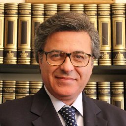 Giovanni Ambrosino合作论文数University of Padua School of General Surgery Padua Italy7
 Mads Bergholt合作论文数Faculty of Dentistry, Oral & Craniofacial Sciences, King’s College London6
 John Marshall Harlan合作论文数Department of Medicine, University of Washington;Department of Hematology, University of Washington6Rossana Bussani合作论文数6Furio Silvestri合作论文数6Luigi M. Biasucci合作论文数6Gianfranco Guarnieri合作论文数Clinica Medica, University of Trieste6
 Fadi N Salloum合作论文数VCU Pauley Heart Center, Virginia Commonwealth University6M Pausa合作论文数5F Baldi合作论文数5Giordano Dicuonzo合作论文数5T M Carlos合作论文数4Manzoli A合作论文数4
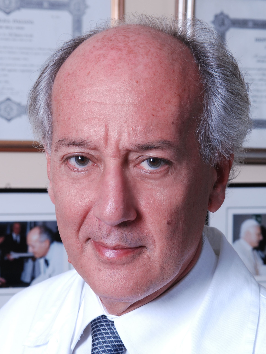 Filippo Crea合作论文数Dipartimento di Scienze Cardiovascolari e Pneumologiche, Università Cattolica del Sacro Cuore;Dipartimento Universitario di Scienze Cardiovascolari e Pneumologiche, Università Cattolica di Roma4
 Benjamin Van Tassell合作论文数Department of Pharmacotherapy and Outcomes Science, School of Pharmacy, Virginia Commonwealth University4Pierluigi Patriarca合作论文数4Domenico Maria Zardi合作论文数4Nicola Fiotti合作论文数4C Giansante合作论文数3Ggl Biondi-Zoccai合作论文数3Valentina Uwechie合作论文数3
 Anindita Das合作论文数3Nicholas N. Hoke合作论文数3
 Alberto Mantovani合作论文数Istituto Clinico Humanitas, Humanitas University;William Harvey Research Institute, Queen Mary University3Marina Cinco合作论文数3
 Oscar Burrone合作论文数Molecular Immunology Laboratory, International Centre for Genetic Engineering and Biotechnology3D Camilot合作论文数3
 Carolina Gravina合作论文数3
 George W. Vetrovec合作论文数Ambulatory Care Center Virginia Commonwealth University3Vittorio Laghi合作论文数3Rakesh C. Kukreja合作论文数3T Zacchi合作论文数3Giorgio Stanta合作论文数3
 Fabio Cacciapaglia合作论文数Department of Toxicology, University of Cagliari3D. Seripa合作论文数2D. M. Zardi合作论文数2M G Matera合作论文数2




