Infection by influenza A virus (IAV) and other viruses causes disease exacerbations in chronic obstructive pulmonary disease (COPD). Immune responses are blunted in COPD, a deficit compounded by current standard-of-care glucocorticosteroids (GCS) to further predispose patients to life-threatening infections. The immunosuppressive effects of elevated transforming growth factor-β (TGF-β) in COPD may amplify lung inflammation during infections while advancing fibrosis. In the present study, we investigated potential repurposing of pirfenidone, currently used as an antifibrotic for idiopathic pulmonary fibrosis, as a nonsteroidal treatment for viral exacerbations of COPD. Murine models of lung-specific TGF-β overexpression or chronic cigarette smoke exposure with IAV infection were used. Pirfenidone was administered daily by oral gavage commencing pre- or postinfection, and inhaled pirfenidone and GCS treatment preinfection were also compared. Tissue and BAL were assessed for viral replication, inflammation, and immune responses. Overexpression of TGF-β enhanced the severity of IAV infection, contributing to unrestrained airway inflammation. Mechanistically, TGF-β reduced innate immune responses to IAV by blunting IFN-regulated gene expression and suppressing production of antiviral proteins. Prophylactic pirfenidone administration opposed these actions of TGF-β, curbing IAV infection and airway inflammation associated with TGF-β overexpression and cigarette smoke-induced COPD. Notably, inhaled pirfenidone caused greater inhibition of viral loads and inflammation than inhaled GCS. These proof-of-concept studies demonstrate that repurposing pirfenidone and employing a preventative strategy may yield substantial benefit over antiinflammatory GCS in COPD. Pirfenidone can mitigate damaging viral exacerbations without attendant immunosuppressive actions and merits further investigation, particularly as an inhaled formulation.
Transforming growth factor β1 (TGFβ1) is a pleiotropic cytokine implicated in the pathophysiology of chronic lung diseases such as asthma and chronic obstructive pulmonary disease. Epithelial TGFβ1 is released in response to injury, inflammatory stimuli, and during bronchoconstriction to induce fibrosis. We hypothesized that elevated expression of endogenous TGFβ1, localized to the lung, would elicit autocrine effects to alter airway responsiveness. We utilized a transgenic mouse model of doxycycline (Dox)-induced, lung-specific overexpression of active TGFβ1 by giving Dox (0.25 mg/mL in drinking water, 8 wk), or normal water as a control. Comparing Dox with control groups, levels of TGFβ1 were ∼30-fold higher in bronchoalveolar lavage fluid (BALF), but not in serum, as measured by ELISA. BALF cells, predominantly macrophages, were ∼3.5-fold higher, with no evidence of tissue inflammation in hematoxylin and eosin (H&E)-stained sections from Dox mice. Higher collagen deposition was evident around the airways in Masson's trichrome-stained sections [subepithelial thickness (µm): control 10.4 ± 10.9, n = 9; Dox 25.8 ± 1.5, n = 13, P < 0.0001]. TGFβ1 overexpression increased baseline airway resistance and induced airway hyperresponsiveness (AHR) to methacholine (MCh) in vivo, as measured using in vivo plethysmography. Comparing precision-cut lung slices (PCLS) from separate Dox-treated and control mice, maximum contraction of intrapulmonary airways to MCh was increased ex vivo. Overall, elevated lung TGFβ1 levels resulted in localized airway fibrosis associated with increased airway contraction to MCh. These autocrine effects of endogenous TGFβ1 implicate its potential contribution to AHR, suggesting that targeting TGFβ1 may provide a novel approach to oppose excessive airway contraction in chronic lung diseases.NEW & NOTEWORTHY TGFβ upregulation is common in respiratory diseases. Here, the authors have utilized for the first time a mouse model of lung-specific overexpression of active TGFβ to demonstrate the dual role of TGFβ1 in structural remodeling and dysregulation of airway contractility. Given these pathologies are common to asthma and COPD, this model provides a unique opportunity to identify essential novel therapeutics for the treatment of chronic lung diseases.
Severe acute respiratory syndrome coronavirus 2 (SARS-CoV-2) binds to angiotensin-converting enzyme 2 (ACE2) on host cells, via its spike protein, and transmembrane protease, serine 2 (TMPRSS2) cleaves the spike-ACE2 complex to facilitate virus entry. As rate-limiting steps for virus entry, modulation of ACE2 and/or TMPRSS2 may decrease SARS-CoV-2 infectivity and COVID-19 severity. In silico modeling suggested the natural bioactive flavonoid quercetin can bind to ACE2 and a recent randomized clinical trial demonstrated that oral supplementation with quercetin increased COVID-19 recovery. A range of cultured human cells were assessed for co-expression of ACE2 and TMPRSS2. Immortalized Calu-3 lung cells, cultured and matured at an air-liquid interface (Calu-3-ALIs), were established as the most appropriate. Primary bronchial epithelial cells (PBECs) were obtained from healthy adult males (N = 6) and cultured under submerged conditions to corroborate the outcomes. Upon maturation or reaching 80% confluence, respectively, the Calu-3-ALIs and PBECs were treated with quercetin, and mRNA and protein expression were assessed by droplet digital PCR and ELISA, respectively. SARS-CoV-2 infectivity, and the effects of pre- and co-treatment with quercetin, was assessed by median tissue culture infectious dose assay. Quercetin dose-dependently decreased ACE2 and TMPRSS2 mRNA and protein in both Calu-3-ALIs and PBECs after 4 h, while TMPRSS2 remained suppressed in response to prolonged treatment with lower doses (twice daily for 3 days). Quercetin also acutely decreased ADAM17 mRNA, but not ACE, in Calu-3-ALIs, and this warrants further investigation. Calu-3-ALIs, but not PBECs, were successfully infected with SARS-CoV-2; however, quercetin had no antiviral effect, neither directly nor indirectly through downregulation of ACE2 and TMPRSS2. Calu-3-ALIs were reaffirmed to be an optimal cell model for research into the regulation of ACE2 and TMPRSS2, without the need for prior genetic modification, and will prove valuable in future coronavirus and respiratory infectious disease work. However, our data demonstrate that a significant decrease in the expression of ACE2 and TMPRSS2 by a promising prophylactic candidate may not translate to infection prevention.
1. The increased susceptibility to and severity of viral infections in patients with respiratory diseases may be due to the immunosuppressive actions of elevated lung transforming growth factor-beta (TGFb), prophylactic inhaled glucocorticosteroids (GCS) and oral GCS for exacerbations. The oral anti-fibrotic and anti-inflammatory drug pirfenidone (PFD) may offer a novel, non-immunosuppressive alternative (Thomas et al, Respirology, 2021). 2. Compare the efficacy of inhaled and oral PFD to standard GCS treatment in the context of viral-induced exacerbations. 3. Transgenic TGFb-overexpressing mice were treated daily for 2 days prior to infection with IAV (102 PFU, HKx31) then 3 days post-infection. Groups (n=4-6): intranasal prior to infection, then oral, vehicle (i/oVeh), i/oPFD (13.3mg/kg, 100mg/kg), o/oPFD (100mg/kg) or i/oGCS (1mg/kg). Lung viral loads and inflammation in bronchoalveolar lavage fluid were measured. Effects of treatments on IAV-induced impairment of dilator responses to salbutamol (SALB) were measured using precision cut lung slices (PCLS) from separate mice (Donovan et al, Clin Sci, 2016). 4. Treatment with o/oPFD, but not i/oPFD, reduced viral load, while both viral load and weight loss were increased with i/oGCS (p<0.01). KC was reduced by o/oPFD but not i/oGCS. In airways precontracted with methacholine, relaxation to SALB (1mM) was only 20% in PCLS from i/oVeh post-infection mice but 45-70% in PCLS from both PFD- and GCS-treated mice. 5. Treatment with PFD may offer greater protection against TGFβ-enhanced viral infection severity and impaired dilator responses than GCS. Further investigation is warranted into the repurposing of PFD for viral-induced exacerbations of respiratory diseases.
TANK-binding kinase 1 (TBK1) is a key signalling component in the production of type-I interferons, which have essential antiviral activities, including against SARS-CoV-2. TBK1, and its homologue IκB kinase-ε (IKKε), can also induce pro-inflammatory responses that contribute to pathogen clearance. While initially protective, sustained engagement of type-I interferons is associated with damaging hyper-inflammation found in severe COVID-19 patients. The contribution of TBK1/IKKε signalling to these responses is unknown. Here we find that the small molecule idronoxil inhibits TBK1/IKKε signalling through destabilisation of TBK1/IKKε protein complexes. Treatment with idronoxil, or the small molecule inhibitor MRT67307, suppresses TBK1/IKKε signalling and attenuates cellular and molecular lung inflammation in SARS-CoV-2-challenged mice. Our findings additionally demonstrate that engagement of STING is not the major driver of these inflammatory responses and establish a critical role for TBK1/IKKε signalling in SARS-CoV-2 hyper-inflammation.
Cytoplasmic detection of DNA by cyclic GMP-AMP (cGAMP) synthase (cGAS) is an essential component of antiviral responses. Upon synthesis, cGAMP binds to the stimulator of interferon (IFN) genes (STING) in infected and adjacent cells through intercellular transfer by connexins forming gap-junctions, eliciting a strong IFN-β-driven antiviral response. We demonstrate here that Genistein, a flavonoid compound naturally occurring in soy-based foods, inhibits cGAS-STING antiviral signaling at two levels. First, Genistein pretreatment of cGAMP-producing cells inhibited gap-junction intercellular communication, resulting in reduced STING responses in adjacent cells. In addition, Genistein directly blocked STING activation by the murine agonist DMXAA, by decreasing the interaction of STING with TBK1 and IKKε. As a result, Genistein attenuated STING signaling in human and mouse cells, dampening antiviral activity against Semliki Forest Virus infection. Collectively, our findings identify a previously unrecognized proviral activity of Genistein mediated via its inhibitory effects at two levels of cGAS-STING signaling. IMPORTANCE Several reports suggest that Genistein exhibits antiviral activities against DNA viruses. Our work uncovers a previously unrecognized proviral effect of Genistein, through inhibition of the cGAS-STING pathway at the level of cGAMP transfer and its sensing by STING. This suggests that the use of Genistein as an antiviral should be taken with caution as it may reduce the protective antiviral effects elicited by host STING activation.
1. Precision cut lung slices (PCLS) offer a unique integrated experimental platform for investigating both inflammatory and immune responses to viral infection. 2. This study aimed to develop a protocol for robust infection of PCLS ex vivo with influenza A virus (IAV) with a view to its application for assessment of antiviral agents. 3. PCLS were prepared using agarose-inflated lungs from naïve C57Bl6 mice and infected with IAV (HKx31 mouse strain). Matched slices from each mouse at 3 IAV concentrations (1x10⁴-1x10⁶ PFU) were assessed 24 and 48 hr post infection (n=4-8). Viral loads were assessed by plaque assay of homogenised PCLS. Conditioned media was used to measure both cell death via LDH assay and inflammatory responses to infection via TNFα ELISA. 4. A dose-dependent increase in viral load was observed at 24 hr, with a further 3-fold increase in plaque numbers at 48 hr at the lowest IAV concentration only (1x10⁴PFU, p<0.05, paired t-test). LDH was increased at 48 hr, irrespective of viral load. TNFα levels in conditioned media were increased 5-fold between 24 and 48 hr with 1x10⁴PFU IAV (57±11, 243± 56 pg/ml, p<0.01). 5. Treatment of mouse PCLS with IAV ex vivo elicits dose- and time-dependent infection and release of inflammatory cytokines. Further studies assessing immune responses and sensitivity to viral treatment are required. PCLS may be a viable screening tool for pre-clinical assessment of mechanisms of infection and validation of new therapeutic targets.
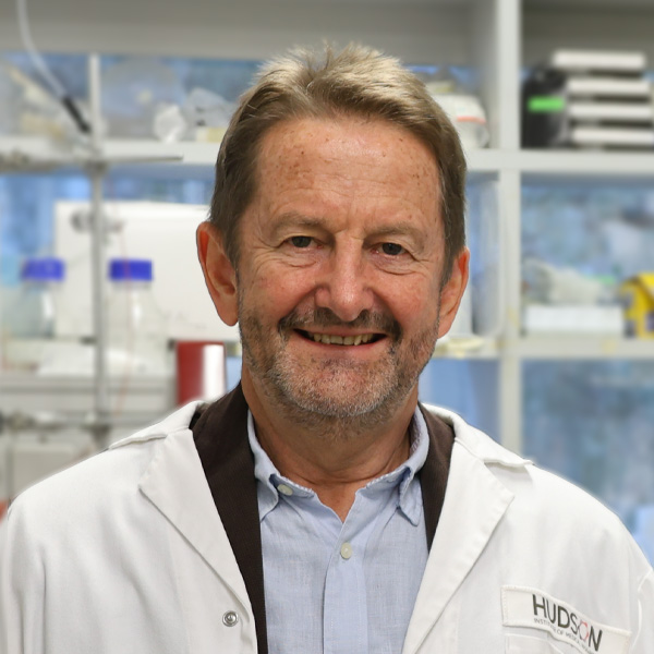 Philip Bardin合作论文数Centre for Innate Immunity and Infectious Diseases, Hudson Institute of Medical Research37
 Jane Bourke合作论文数Department of Pharmacology, School of Biomedical Sciences, Monash University16
 J. Chitty合作论文数13
 Jack A Elias合作论文数Department of Molecular Microbiology and Immunology, Brown University;Department of Molecular Biology, Cell Biology and Biochemistry, Brown University8
 Paul Robert King合作论文数8
 Saleela Ruwanpura合作论文数Monash Health8
 Lam Maggie M合作论文数7
 Michael Gantier合作论文数Centre for Innate Immunity and Infectious Diseases, Hudson Institute of Medical Research7
 Paul Hertzog合作论文数Hudson Institute of Medical Research;Monash University5K. Kan-o合作论文数5
 Phillip Nagley合作论文数Department of Biochemistry and Molecular Biology, Monash University4
 Ashley Mansell合作论文数Department of Microbiology, Anatomy, Physiology & Pharmacology, La Trobe University4C. Burnie合作论文数3Kathryn Hjerrild合作论文数3Rebecca Piganis合作论文数3Lovisa Dousha合作论文数3Geneviève Pépin合作论文数3L. Dousha合作论文数3Sharon Byers合作论文数3Seow Huei Jiunn合作论文数3Kan-O Keiko合作论文数3Lo Camden Y合作论文数3
 Bryan Williams合作论文数Centre for Cancer Research, Hudson Institute of Medical Research;Department of Molecular and Translational Science, Monash University3
 Kristie Jenkins合作论文数3
 Simon Royce合作论文数Victorian Heart Institute, Monash University3
 Ross Vlahos合作论文数Department of Pharmacology;University of Melbourne;Department of Pharmacology, University of Melbourne3
 Reena Ghildyal合作论文数Ctr Res Therapeut Solut, Univ Canberra3Jodee Gould合作论文数3
 Suresh Mahalingam合作论文数University of Canberra3
 Michelle Tate合作论文数Centre for Innate Immunity and Infectious Diseases, Hudson Institute of Medical Research3Ainslie L.K. Roberts合作论文数2Charlotte Nejad合作论文数2Judy Callaghan合作论文数2
 Barton R. Jennings合作论文数2Rebecca L. Ambrose合作论文数2Roleen Sharma合作论文数2
 Katherine R Balka合作论文数2Martin MacDonald合作论文数2
 Tomalika Rahmat Ullah合作论文数Dept Mol & Translat Sci, Monash Univ2Ian Simpson合作论文数2
 Kim O'Sullivan合作论文数Centre for Inflammatory Disease Monash Health, Monash University;Department of Medicine, Monash University2
 Penny A Rudd合作论文数2Jaffar Jade合作论文数2
 Philip M. Hansbro合作论文数The Priority Research Centre for Healthy Lungs, Hunter Medical Research Institute, The University of Newcastle2
 Chihiro Sasakawa (笹川千尋)合作论文数Medical Mycology Research Center, Chiba University2Steven Lim合作论文数2
 Lorinda Turner合作论文数2Ferrand Jonathan合作论文数MIMR-PHI Institute of Medical Research, Monash University2
 Dominic De Nardo合作论文数Department of Anatomy and Developmental Biology, School of Biomedical Sciences, Faculty of Medicine, Nursing and Health Sciences, Monash University2




