1. The increased susceptibility to and severity of viral infections in patients with respiratory diseases may be due to the immunosuppressive actions of elevated lung transforming growth factor-beta (TGFb), prophylactic inhaled glucocorticosteroids (GCS) and oral GCS for exacerbations. The oral anti-fibrotic and anti-inflammatory drug pirfenidone (PFD) may offer a novel, non-immunosuppressive alternative (Thomas et al, Respirology, 2021). 2. Compare the efficacy of inhaled and oral PFD to standard GCS treatment in the context of viral-induced exacerbations. 3. Transgenic TGFb-overexpressing mice were treated daily for 2 days prior to infection with IAV (102 PFU, HKx31) then 3 days post-infection. Groups (n=4-6): intranasal prior to infection, then oral, vehicle (i/oVeh), i/oPFD (13.3mg/kg, 100mg/kg), o/oPFD (100mg/kg) or i/oGCS (1mg/kg). Lung viral loads and inflammation in bronchoalveolar lavage fluid were measured. Effects of treatments on IAV-induced impairment of dilator responses to salbutamol (SALB) were measured using precision cut lung slices (PCLS) from separate mice (Donovan et al, Clin Sci, 2016). 4. Treatment with o/oPFD, but not i/oPFD, reduced viral load, while both viral load and weight loss were increased with i/oGCS (p<0.01). KC was reduced by o/oPFD but not i/oGCS. In airways precontracted with methacholine, relaxation to SALB (1mM) was only 20% in PCLS from i/oVeh post-infection mice but 45-70% in PCLS from both PFD- and GCS-treated mice. 5. Treatment with PFD may offer greater protection against TGFβ-enhanced viral infection severity and impaired dilator responses than GCS. Further investigation is warranted into the repurposing of PFD for viral-induced exacerbations of respiratory diseases.
1. Precision cut lung slices (PCLS) offer a unique integrated experimental platform for investigating both inflammatory and immune responses to viral infection. 2. This study aimed to develop a protocol for robust infection of PCLS ex vivo with influenza A virus (IAV) with a view to its application for assessment of antiviral agents. 3. PCLS were prepared using agarose-inflated lungs from naïve C57Bl6 mice and infected with IAV (HKx31 mouse strain). Matched slices from each mouse at 3 IAV concentrations (1x10⁴-1x10⁶ PFU) were assessed 24 and 48 hr post infection (n=4-8). Viral loads were assessed by plaque assay of homogenised PCLS. Conditioned media was used to measure both cell death via LDH assay and inflammatory responses to infection via TNFα ELISA. 4. A dose-dependent increase in viral load was observed at 24 hr, with a further 3-fold increase in plaque numbers at 48 hr at the lowest IAV concentration only (1x10⁴PFU, p<0.05, paired t-test). LDH was increased at 48 hr, irrespective of viral load. TNFα levels in conditioned media were increased 5-fold between 24 and 48 hr with 1x10⁴PFU IAV (57±11, 243± 56 pg/ml, p<0.01). 5. Treatment of mouse PCLS with IAV ex vivo elicits dose- and time-dependent infection and release of inflammatory cytokines. Further studies assessing immune responses and sensitivity to viral treatment are required. PCLS may be a viable screening tool for pre-clinical assessment of mechanisms of infection and validation of new therapeutic targets.
Introduction: Novel dilators are required to overcome the reduced efficacy of β2-agonists in inflamed remodelled airways in severe asthma. Relaxin (RLX), a Relaxin Family Peptide Receptor 1 (RXFP1) agonist, reverses established allergen-induced fibrosis and in vivo AHR (Royce, Endocrinology, 2009) and also elicits acute in vitro bronchodilation (Lam, Pharm & Ther, 2018). Aim: To assess if RLX potentiates relaxation to salbutamol (SALB) in inflamed, fibrotic mouse airways or human airways. Methods: Airway responses to SALB +/- RLX (100 nM) were visualised in PCLS from mice (control, 4 day house dust-mite (HDM), 8 weeks dox-induced TGFβ overexpression, n=6-16) or human lungs (unused donors, n=3). Inflammation, fibrosis and RXFP1 expression were assessed. Results: HDM increased BAL neutrophils (x104 cells/ml: control 0.9±0.1; HDM19.0±4.3) and eosinophils (control <1; HDM 4.1±0.8) and reduced maximum relaxation to SALB (control 50±4%; HDM 27±10%, P<0.05). With TGFβ overexpression, peribronchial fibrosis increased by 40%, but relaxation to SALB was not impaired. In both models, airway RXFP1 was maintained and RLX increased relaxation to SALB by ~30%. In human non-asthmatic airways, RLX or SALB alone induced near-complete relaxation and RLX increased SALB potency ~3-fold. RXFP1 expression was not decreased in asthmatic airways. Conclusion: RLX potentiates relaxation to SALB in mouse airways under inflammatory and fibrotic conditions, and also has efficacy in human airways. RLX may oppose both acute and chronic aspects of airway pathology to improve outcomes in poorly controlled asthma.
A patient with myocardial disease and pulsus alternans was found to have cyclic alternation in the intensity of a summation gallop sound recorded from the left ventricle. This sound was of greater amplitude immediately preceding the weaker beats. A reasonable explanation for the alternate waxing and waning of these vibrations makes use of the concept of Straub and the observations of more recent investigators that myocardial relaxation may be incomplete preceding the weaker beats of ventricular alternation.
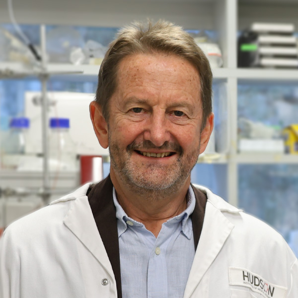 Philip Bardin合作论文数Centre for Innate Immunity and Infectious Diseases, Hudson Institute of Medical Research15
 Jane Bourke合作论文数Department of Pharmacology, School of Biomedical Sciences, Monash University15
 Belinda Thomas合作论文数Department of Genetic Medicine, Women's and Children's Hospital;Department of Respiratory and Sleep Medicine, Monash Medical Centre;Monash Medical Centre, Women's and Children's Hospital13Lam Maggie M合作论文数5C. Burnie合作论文数3
 Simon Royce合作论文数Victorian Heart Institute, Monash University3Seow Huei Jiunn合作论文数2
 Jack A Elias合作论文数Department of Molecular Microbiology and Immunology, Brown University;Department of Molecular Biology, Cell Biology and Biochemistry, Brown University2
 Paul Robert King合作论文数2
 Saleela Ruwanpura合作论文数Monash Health2K. Kan-o合作论文数2Lovisa Dousha合作论文数1Cathrin Maksdi合作论文数1WR MEADOWS合作论文数1Jaffar Jade合作论文数1John T. Sharp合作论文数1
 Ross Vlahos合作论文数Department of Pharmacology;University of Melbourne;Department of Pharmacology, University of Melbourne1Milton L. Fort合作论文数1Glen P. Westall合作论文数1Ghazi Jawad Kanber合作论文数1




