Membrane contact sites (MCSs) establish organelle interactomes in cells to enable communication and exchange of materials. Volume EM (vEM) is ideally suited for MCS analyses, but semantic segmentation of large vEM datasets remains challenging. Recent adoption of artificial intelligence (AI) for segmentation has greatly enhanced our analysis capabilities. However, we show that organelle boundaries, which are important for defining MCS, are the least confident predictions made by AI. We outline a segmentation strategy termed AI-directed voxel extraction (AIVE), which refines segmentation results and boundary predictions derived from any AI-based method by combining those results with electron signal values. We demonstrate the precision conferred by AIVE by applying it to the quantitative analysis of organelle interactomes from multiple FIB-SEM datasets. Through AIVE, we discover a previously unknown category of mitochondrial contact that we term the mitochondrial intrusion. We hypothesize that intrusions serve as anchors that stabilize MCS and promote organelle communication.
The structures and functions of organelles in cells depend on each other but have not been systematically explored. We established stable knockout cell lines of peroxisomal, Golgi and endoplasmic reticulum genes identified in a whole-genome CRISPR knockout screen for inducers of mitochondrial biogenesis stress, showing that defects in peroxisome, Golgi and endoplasmic reticulum metabolism disrupt mitochondrial structure and function. Our quantitative total-organelle profiling approach for focussed ion beam scanning electron microscopy revealed in unprecedented detail that specific organelle dysfunctions precipitate multi-organelle biogenesis defects, impair mitochondrial morphology and reduce respiration. Multi-omics profiling showed a unified proteome response and global shifts in lipid and glycoprotein homeostasis that are elicited when organelle biogenesis is compromised, and that the resulting mitochondrial dysfunction can be rescued with precursors for ether-glycerophospholipid metabolic pathways. This work defines metabolic and morphological interactions between organelles and how their perturbation can cause disease.
Mitophagy preserves overall mitochondrial fitness by selectively targeting damaged mitochondria for degradation. The regulatory mechanisms that prevent PTEN-induced putative kinase 1 (PINK1) and E3 ubiquitin ligase Parkin (PINK1/Parkin)-dependent mitophagy and other selective autophagy pathways from overreacting while ensuring swift progression once initiated are largely elusive. Here, we demonstrate how the TBK1 (TANK-binding kinase 1) adaptors NAP1 (NAK-associated protein 1) and SINTBAD (similar to NAP1 TBK1 adaptor) restrict the initiation of OPTN (optineurin)-driven mitophagy by competing with OPTN for TBK1. Conversely, they promote the progression of nuclear dot protein 52 (NDP52)-driven mitophagy by recruiting TBK1 to NDP52 and stabilizing its interaction with FIP200. Notably, OPTN emerges as the primary recruiter of TBK1 during mitophagy initiation, which in return boosts NDP52-mediated mitophagy. Our results thus define NAP1 and SINTBAD as cargo receptor rheostats, elevating the threshold for mitophagy initiation by OPTN while promoting the progression of the pathway once set in motion by supporting NDP52. These findings shed light on the cellular strategy to prevent pathway hyperactivity while still ensuring efficient progression. Mitophagy is an important quality control pathway. Here, the authors identify the mechanisms enabling the TBK1 adaptors NAP1 and SINTBAD to prevent hyperactivation of PINK1/Parkin mitophagy while promoting the pathway once set in motion.
Cargo sequestration is a fundamental step of selective autophagy in which cells generate a double membrane structure termed an autophagosome on the surface of cargoes. NDP52, TAX1BP1 and p62 bind FIP200 which recruits the ULK1/2 complex to initiate autophagosome formation on cargoes. How OPTN initiates autophagosome formation during selective autophagy remains unknown despite its importance in neurodegeneration. Here, we uncover an unconventional path of PINK1/Parkin mitophagy initiation by OPTN that does not begin with FIP200 binding nor require the ULK1/2 kinases. Using gene-edited cell lines and in vitro reconstitutions, we show that OPTN utilizes the kinase TBK1 which binds directly to the class III phosphatidylinositol 3-kinase complex I to initiate mitophagy. During NDP52 mitophagy initiation, TBK1 is functionally redundant with ULK1/2, classifying TBK1’s role as a selective autophagy initiating kinase. Overall, this work reveals that OPTN mitophagy initiation is mechanistically distinct and highlights the mechanistic plasticity of selective autophagy pathways.
Mitochondrial diseases are a group of inherited diseases with highly varied and complex clinical presentations. Here, we report four individuals, including two siblings, affected by a progressive mitochondrial encephalopathy with biallelic variants in the cardiolipin biosynthesis gene CRLS1. Three affected individuals had a similar infantile presentation comprising progressive encephalopathy, bull’s eye maculopathy, auditory neuropathy, diabetes insipidus, autonomic instability, cardiac defects and early death. The fourth affected individual presented with chronic encephalopathy with neurodevelopmental regression, congenital nystagmus with decreased vision, sensorineural hearing loss, failure to thrive and acquired microcephaly. Using patient-derived fibroblasts, we characterized cardiolipin synthase 1 (CRLS1) dysfunction that impaired mitochondrial morphology and biogenesis, providing functional evidence that the CRLS1 variants cause mitochondrial disease. Lipid profiling in fibroblasts from two patients further confirmed the functional defect demonstrating reduced cardiolipin levels, altered acyl-chain composition and significantly increased levels of phosphatidylglycerol, the substrate of CRLS1. Proteomic profiling of patient cells and mouse Crls1 knockout cell lines identified both endoplasmic reticular and mitochondrial stress responses, and key features that distinguish between varying degrees of cardiolipin insufficiency. These findings support that deleterious variants in CRLS1 cause an autosomal recessive mitochondrial disease, presenting as a severe encephalopathy with multi-systemic involvement. Furthermore, we identify key signatures in cardiolipin and proteome profiles across various degrees of cardiolipin loss, facilitating the use of omics technologies to guide future diagnosis of mitochondrial diseases.
Interfaces within biological tissues not only connect different regions but also contribute to the overall functionality of the tissue. This is especially true in the case of the aortic heart valve. Here, melt electrowriting (MEW) is used to engineer complex, user-defined, interfaces for heart valve scaffolds. First, a multi-modal imaging investigation into the interfacial regions of the valve reveals differences in collagen orientation, density, and recruitment in previously unexplored regions including the commissure and inter-leaflet triangle. Overlapping, suturing, and continuous printing methods for interfacing MEW scaffolds are then investigated for their morphological, tensile, and flexural properties, demonstrating the superior performance of continuous interfaces. G-codes for MEW scaffolds with complex interfaces are designed and generated using a novel software and graphical user interface. Finally, a singular MEW scaffold for the interfacial region of the aortic heart valve is presented incorporating continuous interfaces, gradient porosities, variable layer numbers across regions, and tailored fiber orientations inspired by the collagen distribution and orientation from the multi-modal imaging study. The scaffold exhibits similar yield strain, hysteresis, and relaxation behavior to porcine heart valves. This work demonstrates the ability of a bioinspired approach for MEW scaffold design to address the functional complexity of biological tissues.
Damaged, dysfunctional, or excess mitochondria are removed from cells via a selective form of macroautophagy termed mitophagy. The clearance of mitochondria during mitophagy is mediated by double-membrane vesicles called autophagosomes, which encapsulate mitochondria that have been tagged for mitophagic removal before delivering them to lysosomes for degradation. A variety of different mitophagy pathways exist that differ in their mechanisms of initiation but share a common pathway of autophagosome formation. Autophagosome biogenesis is regulated by a number of autophagy factors which translocate from the cytosol to spatially defined focal points (foci) on the mitochondrial surface after mitophagy has been initiated. The functional analysis of autophagosome biogenesis requires the use of microscopy-based techniques which assess the recruitment of autophagy factors to mitophagic foci representing autophagosome formation sites. Here, we describe a routine method for the quantitative 3D analysis of mitophagic foci in PINK1/Parkin mitophagy immunofluorescence samples through the application of object-based image analysis (OBIA) to 3D confocal imaging datasets. The approach enables unbiased high-throughput characterisation of autophagosome biogenesis during mitophagy.
Introduction: The increasing integration of high-throughput genomic strategies, particularly whole exome (WES) and genome sequencing (WGS) has revolutionised mitochondrial diagnostics with the discovery of a growing list of novel disease genes. Application of 'multi-omics' technologies including proteomics, lipidomics and transcriptomics will improve the diagnostic gap. Case report: We present two individuals with progressive encephalopathy and biallelic variants identified on WES in the cardiolipin synthase 1 (CRLS1) gene. Additionally, the female infant had hypertrophic cardiomyopathy, bull's eye maculopathy, auditory neuropathy, diabetes insipidus and died at 3.5 months. The 19-year-old male presented with neurodevelopmental regression, congenital nystagmus, sensorineural hearing loss and acquired microcephaly. Results: Respiratory chain enzyme analysis of skeletal muscle showed low Complex IV activity in the first patient. Mitochondrial morphology and biogenesis were impaired in her fibroblasts. Expression of wild-type CRLS1 restored her abnormal fibroblast morphology. Fibroblast proteomic profiling and mouse Crls1 knockout cell lines identified endoplasmic reticular, mitochondrial stress responses, and varying degrees of cardiolipin insufficiency. Lipid profiling in both demonstrated reduced cardiolipin levels, altered acyl-chain composition and significantly increased levels of phosphatidylglycerol. Collectively, these findings support that deleterious CRLS1 variants cause a novel mitochondrial disorder with multisystemic involvement. Conclusion: Innovative multi-omics approaches help establish novel diagnoses and provide insights into disease mechanisms.
The sole proteases of the macroautophagy/autophagy machinery, the ATG4s, contribute to autophagosome formation by cleaving Atg8-family protein members (LC3/GABARAPs) which enables Atg8-family protein lipidation and de-lipidation. Our recent work reveals that ATG4s can also promote phagophore growth independently of their protease activity and of Atg8-family proteins. ATG4s and their proximity partners including ARFIP2 and LRBA function to promote trafficking of ATG9A to mitochondria during PINK1-PRKN mitophagy. Through the development of a 3D electron microscopy framework utilizing FIB-SEM and artificial intelligence (termed AIVE: Artificial Intelligence-directed Voxel Extraction), we show that ATG4s promote ER-phagophore contacts during the lipid-transfer phase of autophagosome biogenesis, which requires ATG2B and ATG9A to support phagophore growth. We also discovered that ATG4s are not essential for removal of Atg8-family proteins from autolysosomes, but they can function as deubiquitinase-like enzymes to counteract the conjugation of Atg8-family proteins to other proteins, a process that we have termed ATG8ylation (also known as LC3ylation). These discoveries demonstrate the duality of the ATG4 family in driving autophagosome formation by functioning as both autophagy proteases and trafficking factors, while simultaneously raising questions about the putative roles of ATG8ylation in cell biology.
The sequestration of damaged mitochondria within double-membrane structures termed autophagosomes is a key step of PINK1/Parkin mitophagy. The ATG4 family of proteases are thought to regulate autophagosome formation exclusively by processing the ubiquitin-like ATG8 family (LC3/GABARAPs). We discover that human ATG4s promote autophagosome formation independently of their protease activity and of ATG8 family processing. ATG4 proximity networks reveal a role for ATG4s and their proximity partners, including the immune-disease protein LRBA, in ATG9A vesicle trafficking to mitochondria. Artificial intelligence-directed 3D electron microscopy of phagophores shows that ATG4s promote phagophore-ER contacts during the lipid-transfer phase of autophagosome formation. We also show that ATG8 removal during autophagosome maturation does not depend on ATG4 activity. Instead, ATG4s can disassemble ATG8-protein conjugates, revealing a role for ATG4s as deubiquitinating-like enzymes. These findings establish non-canonical roles of the ATG4 family beyond the ATG8 lipidation axis and provide an AI-driven framework for rapid 3D electron microscopy.
Human hepatitis A virus 3C protease (3Cpro) has recently been shown to cause in vitro caspase-independent cell death, accompanied by previously undescribed cytoplasmic vacuolization with abnormal fusion of various types of endosomal-lysosomal organelles. Yeasts are unicellular eukaryotes widely used as a biological model to study fundamental cellular processes. In particular, yeast models of cell death are useful due to the similarity of regulated cell death mechanisms between yeast and humans. This, together with the functional similarity of yeast vacuoles and the lysosomal/endosomal compartment of mammalian cells, suggests yeasts to be a promising model to study molecular mechanisms of 3Cpro action. Here, we expressed 3Cpro and its mutant inactivated form (3Cmut) in Saccharomyces cerevisiae using pYES2 vector. It was shown that expression of 3Cpro, but not 3Cmut, caused cells to lose the ability to proliferate, but maintained viability, thus indicating the enzyme exerts a cytostatic effect. In addition, using vacuolar dyes and light and electron microscopy, it was found that 3Cpro causes vacuole distortion and fragmentation, accompanied by the abnormal vacuolar staining. In summary, the effects of the 3Cpro on yeast cells resemble the response of mammalian cells. Therefore, the experimental system based on S. cerevisiae can be used for further studies of 3Cpro action.
Following the detection of cytosolic double-stranded DNA from viral or bacterial infection in mammalian cells, cyclic dinucleotide activation of STING induces interferon β expression to initiate innate immune defenses. STING activation also induces LC3B lipidation, a classical but equivocal marker of autophagy, that promotes a cell-autonomous antiviral response that arose before evolution of the interferon pathway. We report that STING activation induces LC3B lipidation onto single-membrane perinuclear vesicles mediated by ATG16L1 via its WD40 domain, bypassing the requirement of canonical upstream autophagy machinery. This process is blocked by bafilomycin A1 that binds and inhibits the vacuolar ATPase (V-ATPase) and by SopF, a bacterial effector that catalytically modifies the V-ATPase to inhibit LC3B lipidation via ATG16L1. These results indicate that activation of the cGAS-STING pathway induces V-ATPase–dependent LC3B lipidation that may mediate cell-autonomous host defense, an unanticipated mechanism that is distinct from LC3B lipidation onto double-membrane autophagosomes.
Current models of selective autophagy dictate that autophagy receptors, including Optineurin and NDP52, link cargo to autophagosomal membranes. This is thought to occur via autophagy receptor binding to Atg8 homologs (LC3/GABARAPs) through an LC3 interacting region (LIR). The LIR motif within autophagy receptors is therefore widely recognised as being essential for selective sequestration of cargo. Here we show that the LIR motif within OPTN and NDP52 is dispensable for Atg8 recruitment and selectivity during PINK1/Parkin mitophagy. Instead, Atg8s play a critical role in mediating ubiquitin-independent recruitment of OPTN and NDP52 to growing phagophore membranes via the LIR motif. The additional recruitment of OPTN and NDP52 amplifies mitophagy through an Atg8-dependent positive feedback loop. Rather than functioning in selectivity, our discovery of a role for the LIR motif in mitophagy amplification points toward a general mechanism by which Atg8s can recruit autophagy factors to drive autophagosome growth and amplify selective autophagy.
Volume 8, no. 6, e01874-17, 2017, [https://doi.org/10.1128/mBio.01874-17][1]. The following funding information and acknowledgments should be added: This work, including the efforts of Benjamin S. Padman and Michael Lazarou, was funded by an Australian Research Council (ARC) Future Fellowship (
Mitochondrial apoptosis is mediated by BAK and BAX, two proteins that induce mitochondrial outer membrane permeabilization, leading to cytochrome c release and activation of apoptotic caspases. In the absence of active caspases, mitochondrial DNA (mtDNA) triggers the innate immune cGAS/STING pathway, causing dying cells to secrete type I interferon. How cGAS gains access to mtDNA remains unclear. We used live-cell lattice light-sheet microscopy to examine the mitochondrial network in mouse embryonic fibroblasts. We found that after BAK/BAX activation and cytochrome c loss, the mitochondrial network broke down and large BAK/BAX pores appeared in the outer membrane. These BAK/BAX macropores allowed the inner mitochondrial membrane to herniate into the cytosol, carrying with it mitochondrial matrix components, including the mitochondrial genome. Apoptotic caspases did not prevent herniation but dismantled the dying cell to suppress mtDNA-induced innate immune signaling.
Parkinson's disease is diagnosed upon the presentation of motor symptoms, resulting from substantial degeneration of dopaminergic neurons in the midbrain. Prior to diagnosis, there is a lengthy prodromal stage in which non-motor symptoms, including olfactory deficits (hyposmia), develop. There is limited information about non-motor impairments and there is a need for directed research into these early pathogenic cellular pathways that precede extensive dopaminergic death in the midbrain. The protein tau has been identified as a genetic risk factor in the development of sporadic PD. Tau knockout mice have been reported as an age-dependent model of PD, and this study has demonstrated that they develop motor deficits at 15-months-old. We have shown that at 7-month-old tau knockout mice present with an overt hyposmic phenotype. This olfactory deficit correlates with an accumulation of α-synuclein, as well as autophagic impairment, in the olfactory bulb. This pathological feature becomes apparent in the striatum and substantia nigra of 15-month-old tau knockout mice, suggesting the potential for a spread of disease. Initial primary cell culture experiments have demonstrated that ablation of tau results in the release of α-synuclein enriched exosomes, providing a potential mechanism for disease spread. These alterations in α-synuclein level as well as a marked autophagy impairment in the tau knockout primary cells recapitulate results seen in the animal model. These data implicate a pathological role for tau in early Parkinson's disease.
Mitochondria are double-membraned organelles fundamental to all mammalian life. Despite their importance, mammalian mitochondria are also a major biochemical hazard. Damaged mitochondria generate excessive quantities of reactive oxygen species (ROS), which can damage intracellular content and organelles. Without maintenance of mitochondrial health, ROSinduced mitochondrial damage can escalate into a catastrophic cascade of damage & dysfunction. This process has been implicated in the development of numerous diseases with poor clinical outcomes (Alzheimer's disease, amyotrophic lateral sclerosis, cancer metastasis, MELAS, etc.). Many of these diseases are incurable, but their therapeutic prevention may be possible through maintenance of mitochondrial health. Understanding the regulatory mechanisms of mitochondrial quality control is essential to this objective. My doctoral thesis aimed to characterise the signalling pathways that regulate the degradation of damaged mitochondria via mitophagy. Mitophagy is a mitochondrially targeted variant of macroautophagy, by which cytoplasmic content is sequestered within membranes and delivered to the lysosomes for degradation. Mitophagy is known to exist in mammalian cells, yet the underlying biochemistry has never been linked to an established mechanism of macroautophagy. To characterize the link between mitophagy and macroautophagy, I developed a suite of new genetically encoded mitochondrial probes and a procedure for live-cell correlative light electron microscopy (CLEM). These characterization techniques soon revealed fundamental discrepancies in the experimental basis of two leading mitophagy models, and led to the discovery of a new mechanism regulating the recovery of damaged mitochondria. This new mechanism represents an entirely new paradigm in the context of mitochondrial quality control. The work presented here describes the development of novel characterization techniques, and the unanticipated results yielded by those techniques.
Bacterial viruses are among the most numerous biological entities within the human body. These viruses are found within regions of the body that have conventionally been considered sterile, including the blood, lymph, and organs. However, the primary mechanism that bacterial viruses use to bypass epithelial cell layers and access the body remains unknown. Here, we used in vitro studies to demonstrate the rapid and directional transcytosis of diverse bacteriophages across confluent cell layers originating from the gut, lung, liver, kidney, and brain. Bacteriophage transcytosis across cell layers had a significant preferential directionality for apical-to-basolateral transport, with approximately 0.1% of total bacteriophages applied being transcytosed over a 2-h period. Bacteriophages were capable of crossing the epithelial cell layer within 10 min with transport not significantly affected by the presence of bacterial endotoxins. Microscopy and cellular assays revealed that bacteriophages accessed both the vesicular and cytosolic compartments of the eukaryotic cell, with phage transcytosis suggested to traffic through the Golgi apparatus via the endomembrane system. Extrapolating from these results, we estimated that 31 billion bacteriophage particles are transcytosed across the epithelial cell layers of the gut into the average human body each day. The transcytosis of bacteriophages is a natural and ubiquitous process that provides a mechanistic explanation for the occurrence of phages within the body. IMPORTANCE Bacteriophages (phages) are viruses that infect bacteria. They cannot infect eukaryotic cells but can penetrate epithelial cell layers and spread throughout sterile regions of our bodies, including the blood, lymph, organs, and even the brain. Yet how phages cross these eukaryotic cell layers and gain access to the body remains unknown. In this work, epithelial cells were observed to take up and transport phages across the cell, releasing active phages on the opposite cell surface. Based on these results, we posit that the human body is continually absorbing phages from the gut and transporting them throughout the cell structure and subsequently the body. These results reveal that phages interact directly with the cells and organs of our bodies, likely contributing to human health and immunity.
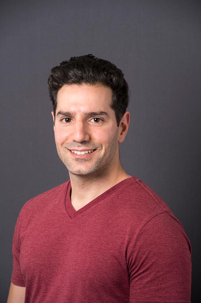 Michael Lazarou合作论文数Department of Biochemistry and Molecular Biology, School of Biomedical Sciences, Faculty of Medicine, Nursing and Health Sciences, Monash University16
 Nguyen Thanh N合作论文数9
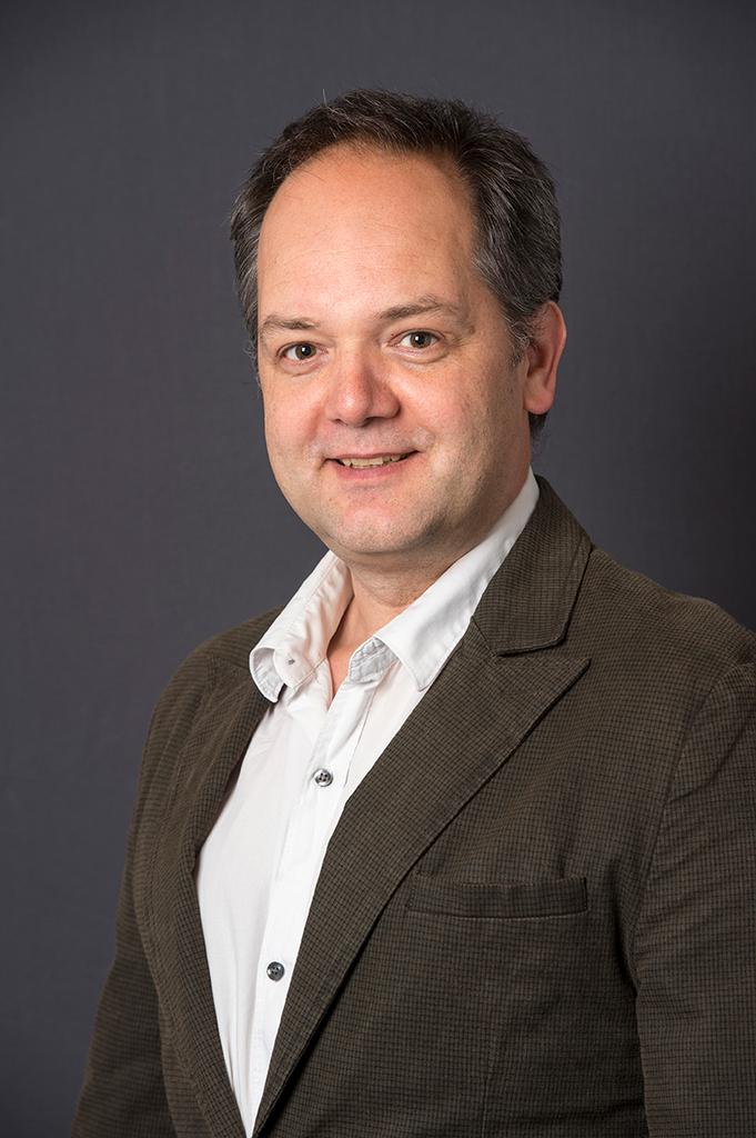 Georg Ramm合作论文数Department of Biochemistry and Molecular Biology, School of Biomedical Sciences, Faculty of Medicine, Nursing and Health Sciences, Monash University7
 Marvin Skulsuppaisarn合作论文数4Richard G. Lee合作论文数Univ Western Australia, Harry Perkins Inst Med Res, Ctr Med Res, QEII Med Ctr, Nedlands, WA 6009, Australia4
 Aleksandra Filipovska合作论文数Centre for Child Health Research, University of Western Australia4
 Gavin E. Reid合作论文数School of Biomedical Sciences, University of Melbourne4Grace Khuu合作论文数3Maike Stentenbach合作论文数3
 Runa Lindblom合作论文数3Yupeng Liu合作论文数3
 Esteban Marcellin Saldana合作论文数Australian Institute for Bioengineering and Nanotechnology, The University of Queensland3Fletcher Janice合作论文数3Geraghty Michael T合作论文数Children's Hospital, Eastern Ontario Research Institute3
 Shanti Balasubramaniam合作论文数Metabolic Unit, Princess Margaret Hospital3
 Kym Boycott合作论文数Department of Pediatrics, University of Ottawa;Children's Hospital of Eastern Ontario Research Institute, University of Ottawa3Markus Bach合作论文数3
 Oliver Rackham合作论文数Corpus Christi College3
 Frederic M. Vaz合作论文数Academic Medical Center;University of Amsterdam;Laboratory Genetic Meta - bolic Diseases;Department of Clinical Chemistry;Academic Medical Center, University of Amsterdam3Rebecca Walsh合作论文数3Tony Roscioli合作论文数Univeristy of New South Wales3Ken Peacock合作论文数2Stephen Shoebridge合作论文数2Tom Kralj合作论文数2Kyle Schlosser合作论文数2Madison R Nuske合作论文数2Ruzeen Patwa合作论文数2
 Louise Uoselis合作论文数Biomedicine Discovery Institute, Monash University;Walter and Eliza Hall Institute of Medical Research2Thomas A Weston合作论文数2Wai Kit Lam合作论文数2Tim McCubbin合作论文数Univ Queensland, Australian Inst Bioengn & Nanotechnol, Brisbane, Qld 4072, Australia2Philip Roberts合作论文数2
 Gabrielle Lemire合作论文数Regional Genetics Program, Children's Hospital of Eastern Ontario Ottawa2
 Elias Adriaenssens合作论文数Univ Antwerp, Inst Born Bunge, Dept Biomed Sci, Peripheral Neuropathy Res Grp, Antwerp, Belgium2
 Antoni Luque合作论文数2
 Richard D. Webster合作论文数Division of Chemistry and Biological Chemistry, School of Physical and Mathematical Sciences, College of Science, Nanyang Technological University2
 Forest Rohwer合作论文数Department of Biology, San Diego State University;Coastal and Marine Institue, San Diego State University2
 Trevor Lithgow合作论文数Centre to Impact AMR, Monash University;School of Biomedical Sciences, Faculty of Medicine, Nursing and Health Sciences, Monash University2
 Rhys A Dunstan合作论文数Department of Microbiology, Monash University2
 David Thorburn合作论文数Department of Paediatrics, University of Melbourne;Murdoch Children's Research Institute;Victorian Clinical Genetics Services Mitochondrial Diagnostic Laboratory2van Klinken Jan Bert合作论文数2Priyadarshi Archana合作论文数2Sawa-Makarska Justyna合作论文数2Yoko Ito合作论文数2
 Richard S Blumberg合作论文数Department of Medicine, Brigham and Women's Hospital;Dana-Farber / Harvard Cancer Center;Harvard Medical School2
 Sascha Martens合作论文数Institute for Genetics, University of Cologne2
 Jeremy Barr合作论文数School of Biological Sciences, Monash University2Kristi Baker合作论文数Department of Pathology, McGill University2Sacha Ferdinandusse合作论文数2




