HISTORY: An 18yo male presented to the sports medicine clinic with 10 days of left thumb pain that began several hours after high school football practice. He denied any acute injury during practice, but he was involved in tackling drills. He is an offensive and defensive lineman. His pain was mostly at the 1st metacarpophalangeal (MCP) joint with associated swelling, but also at the 1st carpometacarpal (CMC) joint with associated popping. He had pain with thumb motion including grasping and pinching. He denied paresthesias or bruising. Xrays done by his primary physician were negative for fracture and he was initially treated with a thumb spica splint then referred for further evaluation. PHYSICAL EXAMINATION: Mild MCP joint swelling, no gross deformity. Weakness with pincer grasp and thumb adduction, otherwise normal thumb flexion and extension strength. Tenderness of the MCP joint and 1st CMC joint. Laxity with thumb ulnar collateral ligament (UCL) stress but good endpoint. Pain with CMC grind, no ligamentous instability. DIFFERENTIAL DIAGNOSIS: Thumb UCL sprain, Thumb fracture, Contusion, Flexor tendon rupture. TEST AND RESULTS: Thumb X-rays: No fractures, No 1st MCP gapping with stress. Ultrasound: Partial tearing of thumb UCL, no Stener lesion. Normal CMC joint. MRI: Nondisplaced intraarticular 1st metacarpal base fracture with effusion. Partial tearing of thumb UCL.FINAL WORKING DIAGNOSIS: Occult 1st Metacarpal base fracture, Partial thumb UCL tear. TREATMENT AND OUTCOMES: The patient was initially treated with thumb spica splinting for 1 month, but was changed to thumb spica casting due to continued pain. After 1 month of casting his pain had largely resolved and the cast was removed. He began home strengthening. His pain resolved at his final visit. This is a unique injury involving both the proximal and distal 1st metacarpal (fracture and ligament sprain), especially given the lack of a clear injury.
PURPOSE:We tested the hypothesis that higher-intensity interval training (HIIT) could be deployed into a standard cardiac rehabilitation (CR) setting and would result in a greater increase in cardiorespiratory fitness (ie, peak oxygen uptake, ) versus moderate-intensity continuous training (MCT).METHODS:Thirty-nine patients participating in a standard phase 2 CR program were randomized to HIIT or MCT; 15 patients and 13 patients in the HIIT and MCT groups, respectively, completed CR and baseline and followup cardiopulmonary exercise testing.RESULTS:No patients in either study group experienced an event that required hospitalization during or within 3 hours after exercise. The changes in resting heart rate and blood pressure at followup testing were similar for both HIIT and MCT. at ventilatory-derived anaerobic threshold increased more (P < .05) with HIIT (3.0 +/- 2.8 mL center dot kg center dot(-1)min(-1)) versus MCT (0.7 +/- 2.2 mL center dot kg center dot(-1)min(-1)). During followup testing, submaximal heart rate at the end of stage 2 of the exercise test was significantly lower within both the HIIT and MCT groups, with no difference noted between groups. Peak Vo2 improved more after CR in patients in HIIT versus MCT (3.6 +/- 3.1 mL center dot kg(.-1)center dot min(-1) vs 1.7 +/- 1.7 mL center dot kg(.-1)center dot min(-1); P < .05).CONCLUSIONS:Among patients with stable coronary heart disease on evidence-based therapy, HIIT was successfully integrated into a standard CR setting and, when compared to MCT, resulted in greater improvement in peak exercise capacity and submaximal endurance.
OBJECTIVES:This study examined the effects of a cardiac rehabilitation (CR) program on functional capacity and health status (HS) in patients with newly implanted left ventricular assist devices (LVADs). BACKGROUND:Reduced functional capacity and HS are independent predictors of mortality in patients with heart failure. CR improves both, and is related to improved outcomes in patients with heart failure; however, there is a paucity of data that describe the effects of CR in patients with LVADs. METHODS:Enrolled subjects (n = 26; 7 women; age 55 ± 13 years; ejection fraction 21 ± 8%) completed a symptom-limited cardiopulmonary exercise test, the Kansas City Cardiomyopathy Questionnaire (KCCQ), a 6-min walk test (6MW), and single-leg isokinetic strength test before 2:1 randomization to CR versus usual care. Subjects in the CR group underwent 18 visits of aerobic exercise at 60% to 80% of heart rate reserve. Within-group changes from baseline to follow-up were analyzed with a paired t-test, whereas an independent t-test was used to determine differences in the change between groups. RESULTS:Within-group improvements were observed in the CR group for peak oxygen uptake (10%), treadmill time (3.1 min), KCCQ score (14.4 points), 6MW distance (52.3 m), and leg strength (17%). Significant differences among groups were observed for KCCQ, leg strength, and total treadmill time. CONCLUSIONS:Indicators of functional capacity and HS are improved in patients with continuous-flow LVADs who attend CR. Future trials should examine the mechanisms responsible for these improvements, and if such improvements translate into improved clinical outcomes. (Cardiac Rehabilitation in Patients With Continuous Flow Left Ventricular Assist Devices:Rehab VAD Trial [RehabVAD]; NCT01584895).
INTRODUCTION: Patient health status (PHS) and peak oxygen uptake ((V) over dotO(2)) are important predictors of clinical outcomes in individuals with heart failure. Preliminary studies of individuals with left ventricular assist devices (LVADs) show improvements in both PHS and peak (V) over dotO(2). However, the relationship between peak (V) over dotO(2) and PHS in this population is not well described. Likewise, data regarding muscular strength are also lacking in this population. We sought to describe the association between peak (V) over dotO(2), muscular strength, and PHS in patients with continuous-flow LVADs.METHODS: Subjects (n = 26; 7 women) completed a symptom-limited graded exercise test within an average of 82 days (range, 33-167 days) of LVAD implant. In addition, subjects underwent a 6-Minute Walk Test and an isokinetic knee extension strength test and completed the Kansas City Cardiomyopathy Questionnaire (KCCQ). Spearman correlation coefficients were performed, adjusting for body weight and gender, to examine relationships between variables.RESULTS: Muscular strength, as measured by peak torque, and peak <(V) over dot>O-2 were both moderately associated with the KCCQ (r = 0.58, P = .006; r = 0.51, P = .019). A subanalysis revealed that muscular strength and peak (V) over dotO(2) were related to different domains within the KCCQ.CONCLUSIONS: Leg muscle strength and peak (V) over dotO(2) appear to be important factors related to PHS in patients with continuous-flow LVADs. This is likely partially a result of deconditioning due to recent hospitalization, as well as persistent heart failure-related peripheral maladaptations in skeletal muscle. Incorporating both a cardiovascular as well as strength training program before and after LVAD implant surgery may be beneficial. V.
Objective: Continuous-flow left ventricular assist devices (LVAD) show improved patient reported health status (HS) and six-minute walk distance (6MW) within the first 1 to 6 months following impla...
Exercise capacity (EC), typically measured as peak oxygen consumption (pVO2), is dependent on both central (e.g. cardiac output) and peripheral factors (e.g. muscular strength, oxidative capacity). In patients with heart failure, EC is reduced as much as 40-60% compared to normal. The EC of patients with continuous flow left ventricular assist devices (LVADs) are not well described, nor is the association between EC and muscular strength (MS). We hypothesize an association between EC and MS in patients with continuous flow LVADs. Subjects (n = 16, 6 females; mean ± SD; age 56 ± 14 years; %EF 21.0 ± 8) completed a symptom-limited graded exercise test with gas-exchange within an average of 79 days (range 33-167 days) of LVAD implant. Additionally, subjects underwent a six minute walk test, an isokinetic knee extension strength test for quadriceps muscle strength (Biodex), and completed the Kansas City Cardiomyopathy Quality of Life Questionnaire (KCCQ). Pearson correlation coefficients were performed to examine relationships between variables. Peak VO2 was very low (13.1 ± 3.8 mL·kg−1·min−1) and was moderately associated with both days after implant (r = 0.560; p = 0.030) and quadriceps MS (r = 0.582; p = 0.029). Additionally, quadriceps MS was related with quality of life score (r = 0.652; p = 0.008). Factors not significantly associated with either FC or MS included: %EF, 6-min walk, age, or BMI. Leg muscle strength appears to be an important factor for both EC and quality of life measures in patient with continuous flow LVADs. This is likely partially a result of deconditioning due to recent hospitalization, as well as persistent heart failure related peripheral maladaptations in skeletal muscle. Incorporating an exercise program to maintain leg strength prior to and after surgery may be beneficial.
While the response of many physiologic measures (e.g. HR, oxygen uptake) to incremental exercise has been reported previously in patients with continuous flow LVADS, the response of the LVAD flow rate (L • min−1) during maximal exercise and its relationship to metabolic demand has not been studied. We hypothesize that an increase in measured LVAD flow during incremental exercise will be positively correlated to oxygen uptake (VO2). Subjects (n = 16, 6 females; mean ±SD; age 56 ± 14 yr; %EF 21.0 ± 8) completed a symptom-limited graded exercise treadmill test. Metabolic demand was assessed by gas exchange analysis and LVAD flow rates were recorded manually from the device output at the end of each stage. A one-way ANOVA with repeated measures was used to determine differences for VO2, LVAD flow, and other parameters for each stage. Pearson correlation coefficients were performed to examine the relationship between change in VO2 and change in LVAD flow. From rest to peak exercise, mean VO2, O2 pulse, HR, and LVAD flow increased by 229% (+746±403 mL/min), 132% (+ 5±2 mL/beat), 41% (+34±23 bpm), and 37% (+1.7±0.9 L/min), respectively (All p<0.05, Figure 1). While the change in VO2 and LVAD flow was weakly correlated from rest to the first stage of exercise (r=0.508, p = 0.044), the overall change in VO2 from rest to peak was not significantly related to the change in VAD flow. Also, change in heart rate was not significantly related to VAD flow, but was strongly associated with change in VO2 (r=0.70, p = 0.002). Discordance between change in VO2 and change in LVAD flow suggests that other factors (i.e., cardiac output of native heart and/or a-VO2 difference) also increase to meet the increased metabolic demands of exercise in patients with LVADs. Because the magnitude of change in a-VO2 difference during exercise would far exceed known physiologic limits of a-VO2 difference alone, our data suggest that an increase in cardiac output of the native heart also occurs during maximal exercise in these patients.
 Dennis J. Kerrigan合作论文数8
 Jonathan Ehrman合作论文数Department of Internal Medicine, Henry Ford Hospital8
 Steven J Keteyian合作论文数7
 John R. Schairer合作论文数7
 Matthew Saval合作论文数5Celeste T. Williams合作论文数5Meghan Swaffer合作论文数4
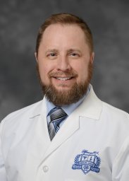 Clinton A. Brawner合作论文数Henry Ford Hospital4Cristina Tita合作论文数3Yelena Selektor合作论文数3Robert Baker合作论文数2Smith Cheryl合作论文数2Velez Mauricio合作论文数2Hibner Brooks A合作论文数2
 David E. Lanfear合作论文数Center for Individualized and Genomic Medicine Research, Henry Ford Health;School of Medicine, Wayne State University;College of Human Medicine, Michigan State University2Steve Keteyian合作论文数1Lisa M. Reasons合作论文数1david e lafear合作论文数1Charles R. C. Marks合作论文数1McCulloch Derek合作论文数1Hill Jason合作论文数1Thompson Tracey M S合作论文数1Saval Mathew A合作论文数1Heather Aldred合作论文数1




