Time-domain diffuse optical tomography (TD-DOT) is a non-invasive technique that utilizes near-infrared light to visualize the optical properties in tissues, showing promising applications in tumor diagnosis and functional brain imaging. The integration of advanced SPAD arrays delivers superior data quality characterized by picosecond timing precision and increased detection channels, enabling high-fidelity reconstruction of unmixed absorption and scattering properties. These gains, however, come with longer acquisition and computation times. We introduce a data-driven view-optimization strategy that exploits the statistical richness of time-resolved measurements to reduce the number of views without sacrificing image quality. We developed a noncontact TD-DOT combining a pulsed laser, a SPAD array, and a rotational stage for view selection. Simulations and phantom experiments show that the proposed method accelerates TD-DOT while maintaining robust reconstruction, comparable to those from full-view acquisition. The accelerated TD-DOT is valuable for monitoring biological dynamics, such as cerebral hemodynamics.
Significance:Cerebral autoregulation (CA) reflects the dynamic coupling among cerebral blood flow (CBF), intracranial pressure (ICP), and arterial blood pressure (ABP); its failure contributes to secondary brain injury. Existing bedside methods rely on indirect or spatially limited CBF surrogates and cannot resolve microvascular flow dynamics across space, depth, and time. Aim:To develop, optimize, and apply a scalable, noncontact time-resolved laser speckle contrast imaging (TR-LSCI) platform for depth-sensitive, high-speed, wide-field CBF imaging during controlled ICP perturbations. Approach:TR-LSCI synchronizes a 20-MHz pulsed laser with a time-gated, single-photon avalanche diode (SPAD) camera (512 × 512 pixels) to detect diffuse photons at varying path lengths, enabling depth-resolved microvascular CBF imaging. Benchtop and mobile TR-LSCI systems were applied in adult rats and a neonatal piglet with synchronized invasive ICP and ABP measurements. Results:TR-LSCI captured spatially heterogeneous, pulsatile CBF dynamics at up to 52 Hz over large cortical fields of view, with heart rate estimates statistically equivalent to those from ICP and ABP. Multivariable analysis identified reproducible, phase-dependent CA transitions encompassing preserved autoregulation, ABP-driven compensation, and ICP-constrained CBF suppression; notably, CBF alone exhibited distinct phase signatures. Conclusions:TR-LSCI enables dynamic, physiology-informed neurovascular monitoring and supports future bedside CA assessment.
In the next generation of experiments in high energy particle physics a large increase in beam interaction density will necessitate upgrades of particle detectors. Examples are the Ring imaging Cherenkov detectors (RICH) in the planned upgrades of the LHCb, Belle II and ALICE 3 experiments. The upgraded RICH detectors will need photodetectors capable of detecting rings of Cherenkov photons at high rates of true and background events as well as large background radiation. Silicon photomultipliers (SiPMs) are an attractive photodetector candidate, with the main remaining technological challenge being the resistance to neutron radiation damage - during the whole experiment run time, the photodetectors are expected to receive a fluence of a few 1013 1-MeV neutron equivalent/cm2. To achieve the targeted radiation tolerance, as well as other RICH detector requirements, dedicated developments and a combination of radiation damage reduction and mitigation techniques, such as cryogenic cooling, are needed. The spadRICH project is developing a CMOS single-photon avalanche diode (SPAD) based photodetector optimized for the application of the planned RICH detectors, with SPADs designed specifically for radiation hardness and cryogenic operation. In this work, we report on dark count rate measurements of SPADs designed by the AQUA Lab in 55 nm BCD technology and 110 nm CMOS image sensor technology, performed down to liquid nitrogen temperatures and for neutron irradiation up to 1012 1-MeV neutron equivalent/cm2.
Diffuse correlation spectroscopy (DCS) is an emerging optical technique for non-invasive cerebral blood flow monitoring. Extraction of the DCS blood flow index typically involves calculating the temporal autocorrelation of the measured light intensity and then fitting its decay to a solution of the correlation diffusion equation. It is well-known that the experimental autocorrelation is a biased estimator of the true autocorrelation. This work explores this phenomenon as it relates to DCS, in particular implementations with single photon avalanche diode arrays (SPAD arrays). After deriving a first-order expression for the bias in DCS, we then quantify its impact as a function of sampling time in both simulation and experiment using SPAD array detection. We then present and explore two bias correction strategies to correct for its impact at fast sampling times (20-200 Hz) and in low-photon regimes.
Precise monitoring of cardiac electrophysiology in vitro is crucial to understanding heart function and cardiac disease. However, high-throughput, contact-free methods for directly measuring excitation-contraction coupling remain limited. Here, we introduce a paradigm for quantitative electrophysiological imaging that combines fluorescence lifetime and intensity information to capture dynamic cardiac signals with high fidelity. We show that lifetime measurements are intrinsically decoupled from motion artifacts and provide calibrated calcium concentration and membrane potential estimates across wide fields of view. Using a gated single-photon avalanche diode camera, we acquire fluorescence lifetime images at up to 200 frames per second with sufficient signal-to-noise ratio such that each frame contains meaningful lifetime information without temporal averaging. This approach yields spatially resolved maps of voltage and calcium values across contracting cardiomyocyte monolayers, revealing heterogeneous cell behaviors within individual assays and uncovering previously unreported dynamics during late-phase repolarization for real-time analysis of excitation-contraction coupling.
Neutrino detectors, particle calorimeters and some dark matter detectors require dense and massive active materials. An extremely fine segmentation is desirable to achieve precise three-dimensional particle tracking. However, such systems introduce significant challenges in construction and demand a large number of readout electronics channels, leading to extremely high costs. In this article, we propose an alternative approach to elementary particle detection that enables ultrafast three-dimensional high-resolution imaging in large volumes of unsegmented scintillator. Enabling technologies are plenoptic systems and time-resolving single-photon avalanche diode array imaging sensors. Together, they enabled us, using a plenoptic camera, to reconstruct the origin of single photons in the scintillator. A case study focused on neutrino detection demonstrates full event reconstruction with a spatial resolution of two hundred micrometres. This work paves the way for a class of particle detectors whose capabilities should be further enhanced through future developments and expanded to Cherenkov light detection, medical imaging and neutron detection.
Purely digital or hybrid digital-analog silicon photomultipliers, based on CMOS single-photon avalanche diode (SPAD) photodetectors, have been developed for a range of high-energy and nuclear physics applications, including at EPFL's AQUA Laboratory in collaboration with external partners. The requirements can widely differ, such as high timing precision and detection at low event rates for time-of-flight PET, very high data rates, spatial granularity, and low light level operation in ring imaging Cherenkov counters (RICH) at colliders, or single-SPAD spatial granularity over large overall areas at low duty cycle when employed as active neutrino targets coupled to scintillating fibers. Emphasis is often on high photon detection efficiency in the visible, coupled to tiling capabilities. Operation can likely take place at low or even cryogenic temperatures and/or high radiation levels. We will highlight how these challenges are being tackled with bespoke devices and sensor architectures, possibly in 3D-stacked implementations.
Fluorescence lifetime imaging (FLIM) offers a powerful approach for assessing drug delivery and target engagement in targeted therapies. However, monitoring drug response in 3D at the mesoscopic scale has been largely constrained by tissue scattering and prolonged acquisition times. In this study, we present the application of our near-infrared to shortwave-infrared (NIR-SWIR) light-sheet mesoscope for rapid 3D probe biodistribution and Forster Resonance Energy Transfer (FRET) mapping in HER2-positive tumor spheroid models.
A room-temperature 3D-stacked flash LiDAR sensor is presented for the short-wave infrared (SWIR). The 96×96 InGaAs-InP SPAD array in the top tier is biased by a circuit at the bottom tier that implements a complementary cascoded gating at the pixel level to control noise and afterpulsing. The bottom-tier chip is fabricated in a 110-nm CMOS technology. The sensor is tested with a 1550nm laser operating at 100μW to 3.1mW average power. The SPADs are gated with 3ns pulses with 500ps skew. Intensity images and depth maps are shown both indoors and outdoors at 10m in 120 klux background light with telemetry up to 100m, having better than 2% accuracy.
We introduce a single-shot fluorescence lifetime imaging method that provides real-time molecular contrast for fluorescence-guided surgery. By delivering rapid, high-fidelity lifetime contrast, it supports accurate tumor delineation and seamless integration into intraoperative clinical workflows.
Diffuse correlation spectroscopy (DCS) is a promising technique for noninvasive measurement of blood flow, especially for cerebral blood flow where other noninvasive techniques have shortcomings. Conventional DCS often requires multiple simultaneous measurements to enhance the signal-to-noise ratio (SNR) especially when probing deep into the brain with large source-detector separations where photons are scarce. However, this limits scalability when using discrete optical detectors. This study demonstrates the application of the 500 x 500 single-photon avalanche diode (SPAD) array, SwissSPAD3, coupled with a custom field-programmable gate array (FPGA) design, which enables significant increases in SNR compared to conventional DCS systems. We validate the fiber-coupled SPAD camera system against a lab-standard CW-DCS system in two-layer liquid phantoms and in human measurements, and demonstrate robust blood-flow tracking at source-detector separations up to 3.25 cm. These results support SPAD-based parallel detection as a scalable route to improved deep-tissue DCS performance in humans.
Significance:Fluorescence-guided surgery (FGS) utilizes molecular contrast agents to highlight critical structures or pathological tissues in real time. The premise of FGS is to enable precise surgical decision-making through accurate visualization and quantitative assessment of fluorophore distribution. However, strong effects of diffusion and absorption of fluorescent light in tissue confound fluorescence images, preventing accurate quantitative assessment of the concentration and distribution of fluorescent markers. These optical artifacts may lead to misinterpretation of tissue boundaries and compromised surgical precision, thereby diminishing the capabilities of FGS. Resolving topological depth maps of fluorophore distribution at the millimeter scale is an important first step in performing quantitative sub-surface fluorescence imaging. Aim:In this study, we present a spatiotemporal deep learning architecture that utilizes picosecond single-photon avalanche diode (SPAD) sensor images to rapidly recover the depth topology of a fluorophore distribution embedded in diffuse media. The network is designed to work with wide-field, epi-illumination geometry and millimeter spatial resolution. Approach:A ConvLSTM-UNet deep learning network was developed for picosecond time-resolved image analysis. This network was trained on 5000 spatiotemporal maps simulated by the optical Monte Carlo method and convolved with the instrument response function (IRF) of the imaging system. The experimental setup utilized a SwissSPAD2 sensor synchronized with a 635 nm picosecond laser diode. Using only 10 selected temporal gates as input, the network could recover depth maps. Reconstruction accuracy was evaluated using mean error metrics across various depths and background concentrations of a fluorophore with a simulated decay time of 100 ps. Results:A total of 75 different test fluorescence video data were evaluated. This set encompassed 15 unique inclusion shapes at five different depths. The network successfully reconstructed fluorescence topography up to 15 mm with a mean absolute error of less than 0.6 mm and mean depth variances below 0.5 mm. The inference time was ∼ 30 ms . Conclusions:Integrating temporal and spatial deep learning networks enabled depth mapping from time-resolved fluorescence data. Utilizing real IRF proved the applicability of SPAD sensors for sub-surface fluorescence mapping.
In this paper, we present a reliability risk of using a virtual guard ring (VGR) in single-photon avalanche diodes (SPADs) and propose an optimized SPAD to mitigate this risk and consequently improve the device performance. Utilizing a lower-doped deep junction to implement a SPAD is an appropriate approach to prevent band-to-band and trap-assisted tunneling and improve the near-infrared efficiency in advanced CMOS technology. To realize the deep-junction-based SPAD, a VGR based on the retrograde doping profile is typically applied to prevent premature edge breakdown. However, because the VGR does not include a physical guard-ring structure at the side of the junction, carriers generated close to the cathode region can be more susceptible to triggering avalanche events in the VGR region, thereby degrading device stability and reliability. To investigate this issue, we fabricated two different VGR-based SPADs based on TCAD simulations and performed comparative analysis in terms of I-V characteristics, dark count rates, waveforms, and lifetimes. The default SPAD operates for only about 1 hour or less at the excess bias voltage of 2 V under dark and illuminated conditions, whereas the optimized SPAD operates stably for over 24 hours at the excess bias voltage of 5 V regardless of illumination. These results clearly reveal the previously underexplored reliability limitation of VGR-based SPADs and demonstrate that cathode-region optimization is an effective strategy for robust and high-performance deep-junction SPADs and high-density SPAD arrays.
Objective.Time-domain diffuse optical imaging (DOI) requires accurate forward models for photon propagation in scattering media. However, existing simulators lack comprehensive experimental validation, especially for non-contact configurations with oblique illumination. This study rigorously evaluates three widely used open-source simulators, including MMC (Mesh-based Monte Carlo), NIRFASTer, and Toast++ (the latter two are finite-element method (FEM)-based), using time-resolved experimental data.Approach.All simulations employed a unified mesh and point-source illumination. Virtual source approximation was applied to FEM solvers to reconcile the directional oblique beam with the isotropic diffusion equation. A time-resolved DOI system with a 32 × 32 single-photon avalanche diode (SPAD) array acquired transmission-mode data from 16 standardized phantoms certified under the BIP protocol with varying absorption coefficientμaand reduced scattering coefficientμs'. The simulation results were quantified across five metrics: spatial-domain (SD) accuracy, time-domain (TD) accuracy, oblique beam accuracy, computational speed, and mesh-density independence. To further quantify the simulators' practical applicability, optical property recovery was conducted using grid-search optimization.Results.Among the three simulators, MMC achieves superior accuracy in SD and TD metrics (SD MSE: 0.072 ± 0.053; TD MSE: 0.179 ± 0.080), and shows robustness across all optical properties. NIRFASTer and Toast++ demonstrate comparable overall performance. In general, MMC is optimal for accuracy-critical TD-DOI applications, while NIRFASTer and Toast++ suit scenarios prioritizing speed (e.g. image reconstruction) with sufficiently largeμs'. Besides, virtual source approximation is essential for non-contact FEM modeling, which reduced average errors by > 34% in large-angle scenarios.Significance.This work suggests a simplified framework for balancing simulation fidelity and computational speed through optimized solver selection and configuration, facilitating fast prototyping of novel TD-DOI systems. Our work represents the first study to systematically validate TD simulators against SPAD array-based data under clinically relevant non-contact conditions, bridging a critical gap in biomedical optical simulation standards.
We present a free-space time-domain diffuse optical tomography (TD-DOT) system that integrates SPAD array detection with geometric calibration for accurate, non-invasive tumor imaging. By combining structured-light surface extraction with calibration of source positions and detector responses, the system enables precise modeling of photon transport in air. Phantom studies demonstrate improved reconstruction accuracy across diverse shapes, while longitudinal in vivo imaging of mice with dorsal tumor reveals consistent changes in tumor volume and optical properties. These results demonstrate that our system enables high-quality, quantitative monitoring of tumor progression, underscoring its utility in preclinical research.
Detectors deployed in high-resolution neutrino experiments, particle calorimetry, or dark matter candidate searches require dense and massive active materials and, in some cases, extremely fine segmentation. This is essential for achieving precise three-dimensional tracking of the interaction products and enabling accurate particle-flow reconstruction. Organic scintillator detectors, for example, in the form of scintillating fibres, offer sub-millimetre spatial and sub-nanosecond temporal resolution. However, such systems introduce significant challenges in construction and demand a large number of readout electronics channels, leading to extremely high costs that are difficult to mitigate. In this article, we propose a paradigm shift in the detection of elementary particles that leads to ultrafast three-dimensional high-resolution imaging in large volumes of unsegmented scintillator. The key enabling technologies are plenoptic systems and time-resolving single-photon avalanche diode (SPAD) array imaging sensors. Together, they allow us, for the first time ever with a plenoptic camera, the reconstruction of the origin of single photons in the scintillator, thereby facilitating an event-by-event analysis. A case study focused on neutrino detection demonstrates the unique potential of this approach, achieving full event reconstruction with a spatial resolution on the order of two hundred micrometres. This work paves the way for a new class of particle scintillator-based detectors, whose capabilities should be further enhanced through future developments and expanded to Cherenkov light detection and calorimetry at collider neutrino experiments, searches for neutrinoless double beta decay, as well as applications such as medical imaging and fast neutron detection.
Fluorescence lifetime imaging microscopy (FLIM) is a powerful tool to discriminate fluorescent molecules or probe their nanoscale environment. Traditionally, FLIM uses time-correlated single-photon counting (TCSPC), which is precise but intrinsically low-throughput due to its dependence on point detectors. Although time-gated cameras have demonstrated the potential for high-throughput FLIM in bright samples with dense labeling, their use in single-molecule microscopy has not been explored extensively. Here, we report fast and accurate single-molecule FLIM with a commercial time-gated single-photon camera. Our optimized acquisition scheme achieves single-molecule lifetime measurements with a precision only about three times less than TCSPC, while imaging with a large number of pixels (512 × 512) allowing for the spatial multiplexing of over 3000 molecules. With this approach, we demonstrate parallelized lifetime measurements of large numbers of labeled pore-forming proteins on supported lipid bilayers, and temporal single-molecule Förster resonance energy transfer measurements at 5-25 Hz. This method holds considerable promise for the advancement of multi-target single-molecule localization microscopy and biopolymer sequencing.
 Edoardo Charbon合作论文数Quantum & Computer Engineering Department, Faculty of Electrical Engineering, Mathematics and Computer Science, Delft University of Technology234
 Arin C. Ulku合作论文数EPFL, Switzerland52
 Samuel Burri合作论文数48
 Ivan Michel Antolovic合作论文数École Polytechnique Fédérale de Lausanne44
 Xavier Michalet合作论文数28
 Francesco Gramuglia合作论文数Advanced Quantum Architecture Laboratory, Ecole polytechnique fédérale de Lausanne (EPFL),23michael a wayne合作论文数18L. Malferrari合作论文数18
 Shimon Weiss合作论文数Department of Chemistry and Biochemisty, University of California, Los Angeles18M. Passaseo合作论文数18
 Xavier Intes合作论文数Department of Biomedical Engineering, Rensselaer Polytechnic Institute17
 Myung-Jae Lee合作论文数Department of Electrical and Electronic Engineering, Yonsei University17Paul Mos合作论文数Advanced Quantum Architecture Laboratory, École polytechnique fédérale de Lausanne16Andrei Ardelean合作论文数16
 Giovanni Darbo合作论文数Istituto Nazionale di Fisica Nucleare, Sezione di Genova16P. Mazzanti合作论文数16G. Ciapetti合作论文数14Tenne Ron合作论文数Dept Phys Complex Syst, Weizmann Inst Sci14D. Barberis合作论文数13Gur Lubin合作论文数Weizmann Institute of Science13V. Casanova合作论文数13
 Dan Oron合作论文数Nanophotonics Laboratory, Department of Molecular Chemistry and Materials Science, Weizmann Institute of Science13C. Bacci合作论文数12D. Barney合作论文数12j batten合作论文数12
 Ming-Lo Wu合作论文数12
 Marco Adinolfi合作论文数School of Physics, University of Bristol12Anatoly I. Frenkel合作论文数sapienza university of rome12Lucia Zanello合作论文数Istituto Nazionale di Fisica Nucleare12Werner Beusch合作论文数12
 Giovanni Carboni合作论文数Università degli Studi di Roma Tor Vergata12Roberto Cardarelli合作论文数INFN Sezione di Roma Tor Vergata, Roma, Italy12M. Dameri合作论文数12
 Anna Di Ciaccio合作论文数University of Rome Tor Vergata11A. Quareni合作论文数11b r french合作论文数11A. Forino合作论文数11M. Adamovich合作论文数11Yu. Alexandrov合作论文数11Jg Mcewen合作论文数11
 Ekin Kizilkan合作论文数Advanced Quantum Architecture Laboratory (AQUA),, École Polytechnique Fédérale de Lausanne (EPFL),11a duane合作论文数11Paolo Martinengo合作论文数CERN11
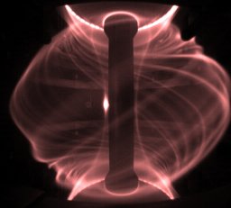 a kirk合作论文数Culham Sci Ctr, CCFE11
 Chockalingam Veerappan合作论文数11G. Martellotti合作论文数11C. Salvo合作论文数11Domizia Orestano合作论文数Roma Tre University and INFN, Rome, Italy11Vincenzo Flaminio合作论文数11




