Cancer therapy–related cardiac dysfunction (CTRCD) is a frequent and clinically relevant complication in patients with lymphoma treated with anthracyclines. Early identification of subclinical myocardial injury is critical to prevent progression to overt heart failure and to avoid chemotherapy dose reductions that may compromise oncologic outcomes. We aimed to evaluate the role of cardiac magnetic resonance (CMR) with T1 mapping and feature-tracking myocardial strain, in comparison with transthoracic echocardiography (TTE) and circulating biomarkers, for the early detection of CTRCD in lymphoma patients receiving anthracycline-based chemotherapy. In this prospective, single-center observational study, forty-nine adult patients with lymphoma scheduled for anthracycline-based chemotherapy underwent serial assessment of high-sensitivity cardiac troponin T (hs-cTnT), NT-proBNP, TTE, and CMR at baseline (visit 0), after the third chemotherapy cycle (visit 1), and at chemotherapy completion (visit 2). TTE was repeated at 12-month follow-up (visit 3). CMR included cine imaging, T1 mapping with extracellular volume (ECV) quantification, late gadolinium enhancement, and feature-tracking strain analysis (global longitudinal [GLS], circumferential [GCS], and radial [GRS] strain). CTRCD was defined and graded according to the 2022 ESC Cardio-Oncology Guidelines. The median age of patients was 46 (29.5–62) years; 53.1
Background: Chagas cardiomyopathy remains a major cause of heart failure (HF) in endemic regions and is increasingly recognized globally, yet data on in-hospital outcomes are limited. Objective: To assess whether Chagas disease is associated with higher in-hospital mortality among patients hospitalized with HF. Methods: We analyzed a nationwide administrative database from the Brazilian Unified Health System (DATASUS/SIHSUS), including adults hospitalized with HF between April 2017 and August 2021. HF was identified using ICD10 code I50.x and Chagas disease using B57.x. The primary outcome was in hospital mortality, evaluated using multivariable Cox models. Results: Among 910,128 HF hospitalizations, 1,082 (0.12%) were associated with Chagas disease. Patients with Chagas were younger but had a more complex clinical profile and higher resource use. In-hospital mortality was higher in the Chagas group (25% vs 12%; p<0.001). After adjustment, Chagas disease remained independently associated with mortality (HR 1.54; 95% CI 1.35, 1.75; p<0.001). Conclusions: In this large real world cohort, Chagas disease was associated with higher in-hospital mortality and greater healthcare utilization. These findings reinforce the high risk nature of Chagas cardiomyopathy and point to the need for more targeted treatment strategies. ### Competing Interest Statement The authors have declared no competing interest. ### Funding Statement No specific funding was received for this work. ### Author Declarations I confirm all relevant ethical guidelines have been followed, and any necessary IRB and/or ethics committee approvals have been obtained. Yes The details of the IRB/oversight body that provided approval or exemption for the research described are given below: The data that support the findings of this study are derived from the Brazilian Unified Health System (DATASUS/SIHSUS, https://datasus.saude.gov.br), which is publicly available. I confirm that all necessary patient/participant consent has been obtained and the appropriate institutional forms have been archived, and that any patient/participant/sample identifiers included were not known to anyone (e.g., hospital staff, patients or participants themselves) outside the research group so cannot be used to identify individuals. Yes I understand that all clinical trials and any other prospective interventional studies must be registered with an ICMJE-approved registry, such as ClinicalTrials.gov. I confirm that any such study reported in the manuscript has been registered and the trial registration ID is provided (note: if posting a prospective study registered retrospectively, please provide a statement in the trial ID field explaining why the study was not registered in advance). Yes I have followed all appropriate research reporting guidelines, such as any relevant EQUATOR Network research reporting checklist(s) and other pertinent material, if applicable. Yes The data that support the findings of this study are derived from the Brazilian Unified Health System (DATASUS/SIHSUS, https://datasus.saude.gov.br), which is publicly available. Processed data and analytic code may be made available upon reasonable request to the corresponding author.
Background: The effects of endovascular therapeutic hypothermia (ETH) in ST-elevation myocardial infarction (STEMI) regional contractility are unknown, and its impact on segmental contractility has still not been evaluated. We sought to evaluate segmental myocardial strain after ETH adjuvant to percutaneous coronary intervention (PCI) in STEMI. Methods: We included patients who underwent 1.5T cardiovascular magnetic resonance exams 5 and 30 days after acute anterior or inferior STEMI in a previous randomized trial. Left ventricle (LV) strain was evaluated on infarcted, adjacent, and remote myocardium. Segmental circumferential (CS) and radial strains (RS) were measured using feature-tracking imaging. Repeated measures of analysis of variance were used for comparisons within time and treatment. Results: Forty patients were divided into hypothermia (ETH, n = 29) and control (n = 11) groups, with 5210 LV segments. In ETH infarcted areas, RS (11.2 +/- 16 vs 14.8 +/- 15.2, p = 0.001) and CS (-5.4 +/- 11.1 vs -8 +/- 11.1, p = 0.001) showed recovery from 5-30 days compared to controls (11.4 +/- 14 vs 13.1 +/- 1 6.8, p = 0.09; -6.5 +/- 10.6 vs -6.4 +/- 12.5, p = 0.94). In control remote areas, RS (28 +/- 18 vs 31.7 +/- 18.5, p = 0.001) and CS (-15.5 +/- 10.7 vs -17.1 +/- 9, p = 0.001) improved from 5-30 days compared to ETH (28.6 +/- 18.6 vs 29 +/- 20, p = 0.44; -15.2 +/- 10.4 vs -15.3 +/- 10.6, p = 0.82). Transmural infarcted areas in ETH improved RS (11.8 +/- 13.2 vs 8.17 +/- 14.7, p = 0.001) and CS (-6.1 +/- 10.9 vs.-3.1 +/- 11.3, p = 0.001) compared to controls, with better contractility at 30 days. Conclusion: In anterior or inferior STEMI patients, ETH adjuvant to PCI is associated with significant improvement in RS and CS of infarcted areas, including transmural segments, but not in remote area. This might further increase our pathophysiological knowledge on early LV remodeling and ultimately suggest potential clinical value.
AI-QCT provided incremental prognostic information compared with CAD-RADS 2.0, CACS, and the modified Duke Index for the prediction of MACE as well as the secondary endpoint of death or nonfatal MI.
Introdução: O distúrbio mineral ósseo da doença renal crônica (DMO-DRC) é caracterizado por uma tríade que envolve calcificação vascular (CV). A CV tem sido associada a piores desfechos clínicos nos pacientes com DRC. A avaliação da presença de CV em diferentes sítios anatômicos, assim como sua gravidade ainda, é pouco estudada, e sua relação com diferentes comorbidades não foi estabelecida em pacientes com DRC na fase pré dialítica. Objetivo: Avaliar a presença de CV em diferentes sítios anatômicos e os fatores associados à sua forma grave em pacientes com DRC e perda de massa óssea. Métodos: Foram analisados dados do início do estudo OSTEO4CKD, de 27 pacientes com DRC 1-4, baixa massa óssea (definida por densitometria com T-score ≤-1) e com interligadores C-terminais do colágeno tipo 1 não suprimidos. Os participantes foram submetidos à realização de tomografia computadorizada sem contraste com cálculo do escore de cálcio em artérias coronárias, carótidas, ilíacas e, aorta torácica e abdominal. CV grave foi definida pela presença de escore coronariano > 400 UA e maior que a mediana da somatória dos escores dos demais sítios. Resultados: A idade dos pacientes foi de 68 (61–74) anos, sendo 14 (52%) do sexo feminino e 9 (33%) autodeclarados negros. Dez pacientes (37%) tinham diabetes mellitus, sendo 6 (60%) deles insulino-dependente, 26 (96%) hipertensão arterial sistêmica, 23 (85%) dislipidemia e 19 (70%) sobrepeso/obesidade. INCLUIR QTOS. PACIENTES TINHAM DCV PRÉVIA XX(YY%). A calcificação coronariana foi identificada em 15 pacientes (55%), sendo grave em 7 (46%). Calcificações nas artérias carótidas foram observadas em 11 pacientes (40%), na aorta torácica em 21 (77%), na aorta abdominal em 22 (81%) e nas artérias ilíacas em 21 (77%). Ao comparar os indivíduos com e sem CV grave, observou-se que os primeiros apresentavam idade significativamente mais elevada [71 (67–80) vs. 61 (55–67) anos; p = 0,012], além de uma tendência a maior prevalência de DRC em estágio 4 (57% vs. 31%; p = 0,063). Não houve diferença em relação aos demais dados laboratoriais. Discussão e Conclusões: A presença de CV foi frequente em todos os sítios avaliados, com predomínio na aorta abdominal. A gravidade das lesões esteve relacionada predominantemente à idade mais avançada.
 Roberto Kalil Filho合作论文数Centro de Cardiologia, Hospital Sirio Libanês107
 Joao A. C. Lima合作论文数Department of Epidemiology, Bloomberg School of Public Health, Johns Hopkins University;School of Medicine, Johns Hopkins University59
 Cesar Nomura合作论文数57
 Wilson Mathias合作论文数São Paulo49José Rodrigues Parga Filho合作论文数48
 Pedro Alves Lemos Neto合作论文数Hospital Israelita Albert Einstein;Heart Institute, Medical School, University of Sao Paulo46Charles Mady合作论文数Heart Institute (InCor),, University of São Paulo Medical School45
 Raul D. Santos合作论文数Hospital Israelita Albert Einstein;Faculdade de Medicina, Universidade de São Paulo;Heart Institute, University of São Paulo;Instituto do Coração - HCFMUSP44
 Armin Arbab-Zadeh合作论文数School of Medicine, Johns Hopkins University;Heart and Vascular Institute, Johns Hopkins University;Division of Cardiology, Johns Hopkins Hospital39Marc Dewey合作论文数39
 Flavio Tarasoutchi合作论文数Heart Institute (InCor), University of São Paulo Medical School38Fábio Fernandes合作论文数Centro de Química-Física Molecular, Instituto Superior Técnico38
 Clerio F. Azevedo合作论文数Department of Medicine;Division of Cardiology;Johns Hopkins Hospital;Department of Medicine, Johns Hopkins Hospital37Hiroyuki Niinuma合作论文数37Ludhmila Abrahão Hajjar合作论文数36Afonso Akio Shiozaki合作论文数36
 Jose Carlos Nicolau合作论文数Fac Med, Univ Sao Paulo35
 Christopher Cox合作论文数Department of Epidemiology, Bloomberg School of Public Health, Johns Hopkins University32
 Melvin Eugene Clouse合作论文数Beth Israel Deaconess Medical Center, Harvard University31
 Magalhaes Tiago A合作论文数Department of Medicine, Johns Hopkins University School of Medicine30Andrea L. Vavere合作论文数29
 David A Bluemke合作论文数Department of Radiology, School of Medicine and Public Health, University of Wisconsin- Madison;Department of Medical Physics, School of Medicine and Public Health, University of Wisconsin- Madison28
 Jose E Krieger合作论文数28Julie M. Miller合作论文数28Jac Lima合作论文数27Lf Avila合作论文数27
 Marcelo L C Vieira合作论文数University of Sao Paulo27
 Jeane Mike Tsutsui合作论文数Heart Institute (InCor),, University of São Paulo Medical School26
 Otavio R Coelho-Filho合作论文数Brigham and Women's Hospital, Boston, MA, USA26Felix José Alvarez Ramires合作论文数Inst Coracao, Univ Sao Paulo24
 Jose A F Ramires合作论文数Medical School, University of São Paulo24
 Múcio Tavares de Oliveira Junior合作论文数Unidade Clínica de Emergência, University of São Paulo24
 Marcelo Nacif合作论文数24
 Luis Henrique Wolff Gowdak合作论文数23
 Roney Sampaio合作论文数23
 Luís Alberto Oliveira Dallan合作论文数Faculdade de Medicina, Universidade de São Paulo;Departamento de Cardio-Pneumologia, Universidade de São Paulo;Hospital Sírio-Libanês23
 Gláucia Maria Moraes De Oliveira合作论文数Departamento de Clínica Médica, Faculdade de Medicina, Universidade Federal do Rio de Janeiro23
 Ibraim Masciarelli Pinto合作论文数Serviço De Métodos Complementares, Instituto Dante Pazzanese de Cardiologia;Universidade de São Paulo22
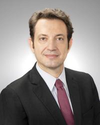 Marcio Sommer Bittencourt合作论文数Department of Medicine, School of Medicine, University of Pittsburgh;University of Pittsburgh Medical Center21
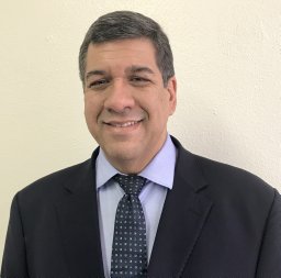 Claudio Mesquita合作论文数21
 Whady Armindo Hueb合作论文数Faculdade de Medicina, University of São Paulo;Instituto do Coração, University of São Paulo21
 Juliano L. Fernandes合作论文数Unicamp, University of Campinas21
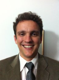 Cerci Rodrigo J合作论文数Division of Cardiology and the Bloomberg School of Public Health, Johns Hopkins University20Matheson Matthew B合作论文数20
 José Antonio Franchini Ramires合作论文数Hospital das Clínicas da Faculdade de Medicina da Universidade de São Paulo20
 Carlos Vicente Serrano Junior合作论文数Faculdade de Medicina, Universidade de São Paulo20Barbara Maria Ianni合作论文数Heart Institute, Clinical Hospital, Faculty of Medicine, University of São Paulo;Departamento de Cardiopneumologia, Faculty of Medicine, University of São Paulo20
 Silvio Henrique Barberato合作论文数20Richard T. George合作论文数Department of Medicine, Johns Hopkins University School of Medicine20




