BACKGROUND:Controversy exists about the impact of ischemia on renal function. We evaluated the creatinine clearance of patients having undergone laparoscopic renal extirpative and ablative surgery.STUDY DESIGN:The records of patients undergoing laparoscopic procedures for renal masses from February 2000 to March 2004 were examined. Creatinine clearance (CrCl) for each patient was determined using the Cockcroft-Gault equation and ideal body weight. We compared CrCl changes of patients undergoing laparoscopic partial nephrectomy (without renal ischemia [LPN-none], with warm ischemia [LPN-warm], and with cold ischemia [LPN-cold]) with patients undergoing laparoscopic radical nephrectomy (LRN) and laparoscopic cryoablation. Patients predisposed to medical renal disease were substratified and evaluated.RESULTS:All patients who underwent LRN or LPN-warm sustained a significant drop in CrCl on the first postoperative day, compared with patients who had LPN without ischemia or cryoablation (p < 0.01). The CrCl decrease correlated directly with warm ischemia time. Six months postoperatively, CrCl changes were no longer significant. Patients with medical renal disease risk factors were more likely to sustain longterm (1 year postoperatively) renal damage if they had renal ischemia, trending toward statistical significance.CONCLUSIONS:Ischemia causes acute renal damage, which is apparently reversible in patients without evidence of medical renal disease. Patients with known medical renal disease have substantial longterm changes in renal function associated with unilateral renal ischemia. Considering the insensitivity of creatinine-based renal function metrics, only eliminating ischemic time will realize the goal of maximal nephron preservation, particularly in patients with preexisting medical renal disease.
OBJECTIVE To evaluate the association of variables of body habitus with direct measurements of ureteric length, as the correct choice of ureteric stent length might help to prevent complications and improve stent tolerance, and to date there are limited data correlating height or other body variables with ureteric length.PATIENTS AND METHODS We prospectively measured pelvi-ureteric junction to vesico-ureteric junction length in 100 patients by placing a ruled 5 F ureteric catheter. Ureteric length was then correlated with patient height, weight, body mass index, and distance from the shoulder (acromium process) to the wrist (head of the ulna; S-W), the elbow (olecranon process) to the wrist (head of the ulna), xyphoid process to umbilicus, xyphoid process to pubis (X-P), umbilicus to pubis, and anterior iliac spine to anterior iliac spine. Patients with pathology affecting the ureteric length were excluded. The results were analysed statistically using a multiple linear regression model with stepwise selection of variables, and a paired t-test.RESULTS The mean right and left ureteric lengths were similar (P = 0.61); height (P < 0.01), weight (P = 0.02), X-P (P = 0.01), and S-W (P = 0.02) distances all correlated with ureteric length. On multivariate regression analysis, weight, height and male gender were associated with mean ureteric length. From these data a formula was constructed to predict ureteric length.CONCLUSIONS It is a challenge to predict ureteric length from body habitus, but height, X-P distance and S-W distance can be used to predict ureteric length.
Purpose: We analyzed the smoke plume produced by various energy-based laparoscopic instruments and determined its effect on laparoscopic visibility.Materials and Methods: The Bipolar Macroforceps, Harmonic Scalpel, Floating Ball, and Monopolar Shears were applied in vitro to porcine psoas muscle. An Aerodynamic Particle Sizer and Electrostatic Classifier provided a size distribution of the plume for particles >500 nm and <500 nm, and a geometric mean particle size was calculated. A Condensation Particle Counter provided the total particle-number concentration. Electron microscopy was used to characterize particle size and shape further. Visibility was calculated using the measured-size distribution data and the Rayleigh and Mie light-scattering theories.Results: The real-time instruments were successful in measuring aerosolized particle size distributions in two size ranges. Electron microscopy revealed smaller, homogeneous, spherical particles and larger, irregular particles consistent with cellular components. The aerosol produced by the Bipolar Macroforceps obscured visibility the least (relative visibility 0.887) among the instruments tested. Particles from the Harmonic Scalpel resulted in a relative visibility of 0.801. Monopolar-based instruments produced plumes responsible for the poorest relative visibility (Floating Ball 0.252; Monopolar Shears 0.026).Conclusions: Surgical smoke is composed of two distinct particle populations caused by the nucleation of vapors as they cool (the small particles) and the entrainment of tissue secondary to mechanical aspects (the large particles). High concentrations of small particles are most responsible for the deterioration in laparoscopic vision. Bipolar and ultrasonic instruments generate a surgical plume that causes the least deterioration of visibility among the instruments tested.
OBJECTIVES:To report our experience with laparoscopic renal cryoablation for patients who have completed a minimum of 3 years of follow-up.METHODS:From July 2000 to March 2005, 81 patients underwent laparoscopic renal cryoablation for renal masses. Of these 81 patients, 31 (38%) underwent laparoscopic renal cryoablation for 36 tumors and have completed a minimal follow-up of 3 years (mean 45.7 months). The postoperative follow-up protocol consisted of serial contrast-enhanced computed tomography or magnetic resonance imaging at 1 day, 1, 3, 6, and 12 months, and yearly thereafter.RESULTS:Twenty-seven tumors were partially exophytic, five were totally endophytic, and four were hilar tumors. The mean operative time was 2.9 hours, with a mean estimated blood loss of 97 mL. The mean renal tumor size was 2.1 cm. In early follow-up, the ablation zone was larger than the tumor but subsequently diminished to the original tumor size 6 months postoperatively. Thereafter, the ablation zone size decreased. The biopsy results revealed that 22 tumors (61%) were malignant and 14 (39%) were benign. The renal tumor 3-year cancer-specific survival rate was 100%, and no patient developed metastatic disease. One patient demonstrated return of abnormal enhancement within the cryolesion during follow-up, suggesting tumor recurrence. One patient had a hemorrhage and urinary leak after cryoablation of an endophytic tumor and was treated conservatively.CONCLUSIONS:Renal cryoablation is safe and offers a minimally invasive nephron-sparing alternative. The oncologic adequacy of renal cryoablation requires long-term follow-up data, but the intermediate-term data seem equivalent to that achieved with extirpative therapy.
Objectives. To evaluate the efficacy of transcutaneous electrical nerve stimulation (TENS) for its effectiveness in eliminating or minimizing discomfort during office-based flexible cystoscopy.Methods. A total of 148 patients were prospectively randomized into one of three groups: flexible cystoscopy with no analgesics, a placebo TENS, or an activated TENS. The patient data collected included patient parameters, number of previous flexible cystoscopies, visual analog pain scores during and after the procedure, surgeon's difficulty rating of procedure, and International Prostate Symptom Score before and 24 hours after the procedure.Results. No statistically significant difference was found among the three groups regarding patient parameters. The visual analog pain scores were similar before and after the procedure. At 30 seconds, the mean visual analog scale score for the control group, placebo group, and TENS study group was 3.73, 3.65, and 3.52, respectively (control versus placebo, control versus active, and placebo versus active: P = 0.97, 0.29, and 0.53, respectively). At 1 and 5 minutes, the corresponding scores were 3.44, 4.37, and 3.50 (P = 0.88, P = 0.99, and P = 0.99) and 0.86, 1.23, and 0.88 (P = 0.97, P = 0.35, and P = 0.56), respectively. The surgeon's mean difficulty rating for the control procedures was 1.08 and for the placebo group was 2.30 (P = 0.02).Conclusions. All patients undergoing flexible cystoscopy in the office setting experienced discomfort. The TENS device provided no significant benefit for pain. The trend toward greater pain scores in the control group could be attributed to the greater degree of difficulty.
PURPOSE:We characterized the size and shape of the ablated area, and the surrounding indeterminate zone associated with renal cryolesions produced by single and multiple cryoablation probe configurations in a porcine model. MATERIALS AND METHODS:Laparoscopic cryoablation, consisting of 2, 10-minute double freeze cycles followed by a 3-minute active thaw, was performed in 18 pigs. Three groups of 6 pigs each were studied, including a single IceRod, 3 rods arranged in a triangular configuration 2 cm apart from each other and 4 rods arranged in a quadratic configuration 2 cm from each other. Cryoablated kidneys were harvested after 14 days for histopathological evaluation. RESULTS:The mean diameter of the area of complete ablation produced by a 1, 3 and 4 rod configuration 1 cm from the tip of the rod was 2.0, 4.4 and 4.9 cm, respectively. Cryolesions were elliptical with 1 cryoablation probe and in the 3 and 4 rod configurations they conformed to triangular and quadratic cross-sectional shapes, respectively. In the zone, ablation a single small focus of peri-arteriolar renal cell viability was noted less than 1 mm from the edge of the cryolesion in 1 of the 18 cryolesions. Otherwise no viable renal cells were detected. CONCLUSIONS:Multirod configurations can be used with templates to space cryoablation probes 2 cm apart from each other with reliable and complete ablation overlap between the rods. However, around larger blood vessels the ice ball must be extended to a larger margin to ensure complete tissue ablation.
Objectives. To evaluate a novel prototype self-anchoring suture (SAS) material that incorporates unidirectional "barbs" designed to grip and approximate tissue without the need for suture ligation for urinary tract reconstructive procedures.Methods. The in vitro failure strength of various porcine tissue approximations with SAS was compared with the strength of similar approximations with standard ligated suture. For in vivo analysis, 3 pigs underwent laparoscopic pyeloplasty with SAS on one side and 2-0 Vicryl suture on the contralateral side. Nine pigs underwent bladder neck anastomoses (BNA) performed with SAS (n = 3) and standard ligated interrupted suturing (n = 3) or standard ligated running suturing (n = 3). The operative times were compared. One week after surgery, the integrity of each anastomosis was evaluated radiographically and histopathologically.Results. The in vitro analysis revealed no statistically different (P = 0.22) failure forces between the approximations performed with SAS (13.2 +/- 2.6 N) and standard suture (14.1 +/- 3.1 N). The in vitro analysis revealed no difference in operative times for the pyeloplasties (P = 0.72) or BNA (P = 0.31). None of the 1-week postoperative radiographic studies revealed extravasation. The histopathologic analysis revealed more fibrotic reaction associated with the SAS BNA procedures (P < 0.01).Conclusions. Self-anchoring suture secures tissue approximations at loads equivalent to tissue approximations with standard tied suture. Self-anchoring suture obviates the need for knot tying and provides a watertight anastomosis. With laparoscopic knot tying experience, anastomotic time with SAS and standard suture do not differ. Self-anchoring suture might induce more fibrosis. Long-term follow-up evaluation will be required before clinical application.
PURPOSE:We performed a randomized, prospective, multi-institutional study evaluating the durability of commercially available flexible ureteroscopes. MATERIALS AND METHODS:A total of 192 patients were randomized to the use of 7 less than 9Fr flexible ureteroscopes, including the Storz 11274AA and Flex-X, the ACMI DUR-8 and DUR-8 Elite, Wolf models 7330.170 and 7325.172, and the Olympus URF-P3. Information about total and lower pole use time, the number and method of ureteroscope insertion, and they type and duration of accessory instrumentation was recorded. Surgeons were asked to rate the visibility and maneuverability of the instrument on a scale of 0-poor to 10-excellent. RESULTS:The indication for ureteroscopy was upper tract calculi in 87% of cases. Of ureteroscope insertions 97% were performed through an access sheath. The average of number of cases before repair ranged from 3.25 for the Wolf 7325 to 14.4 for the ACMI DUR-8 Elite. Average ureteroscope operative time was statistically longer for the DUR-8 Elite (494 minutes) than for the Flex-X (p = 0.047), and the Wolf 7325 and 7330 (p = 0.001 and 0.001, respectively). Duration of use before repair for the URF-P3 (373 minutes) was statistically longer than for the Wolf 7325 and 7330 (p = 0.016 and 0.017, respectively). Minutes of use with an instrument in the working channel were significantly more with the DUR-8 Elite and the URF-P3 than the Wolf 7330 (p = 0.017 and 0.008) and 7325 (p = 0.012 and 0.005, respectively). The ureteroscope that experienced the greatest average duration of lower pole use was the URF-P3, while the shortest was the Wolf 7325 (103 vs 20 minutes, p = 0.005). Average minutes of laser use before breakage was significantly longer for the DUR-8 Elite than for the Wolf 7325 (110 vs 21 minutes, p = 0.021) and 7330 (24 minutes, p = 0.025). CONCLUSIONS:Currently available less than 9Fr flexible ureteroscopes remain fragile instruments. The DUR-8 Elite and Olympus URF-P3 proved to be the most durable devices.
BACKGROUND AND PURPOSE Intraluminal application of pharmacologic agents for acute ureteral dilation may facilitate difficult ureteroscopy. We characterized the in-vivo effects of intraluminal application of verapamil and theophylline on ureteral peristalsis and diameter in a porcine model. MATERIALS AND METHODS Twenty-four female domestic pigs (35-40 kg) were incorporated into the study. We deployed a giant magneto resistive (GMR) sensor and electromagnetic (EMG) electrodes laparoscopically onto the ureteral surface for simultaneous measurement of the mechanical and electrical signals of ureteral peristalsis, respectively. The ureteral-luminal diameter was measured at three levels by digital retrograde pyelography and standardized to a 10-mm laparoscope. The results were calculated as change in peristalsis and ureteral diameter from baseline during the first hour after drug injection. We tested two smooth-muscle relaxants, verapamil (2 mg/kg) and theophylline (70 mg/kg), with saline and dimethylsulfoxide (DMSO; solvent) as controls. Six pigs were studied for each of the four groups. Hydration, anesthesia, and intra-abdominal pressure were standardized. The serum concentrations of the drugs were measured to determine systemic absorption. RESULTS During the first 10 minutes after intraluminal drug injection, theophylline caused a significant decrease in ureteral peristalsis (6.75 waves/10 minutes) compared with the control group (1.00/10 minutes; P = 0.02). This trend persisted for the next hour. However, there were no changes from baseline in ureteral width. Ureteral peristalsis and dilation remained similar after the saline and DMSO injections. Verapamil increased the diameter of the proximal ureter compared with the controls throughout the hour after drug injection. Fifteen minutes after the drug injection, the change in the ureteral diameter with verapamil was 1.38 mm (4.14F), while the control group showed a change of 0.27 mm (P = 0.03). At 1 hour, the width of the proximal ureter in the verapamil group had increased by 1.72 mm (5.16F), while the control group had changed by 0.55 mm (P = 0.03). There were no statistically significant changes in the widths of the mid or distal ureter. No ureteral dilation was observed in the other groups. CONCLUSIONS In the porcine model, intraluminal application of pharmacologic agents produced independent effects on ureteral dilation and peristalsis. Theophylline inhibited ureteral peristalsis, and verapamil produced acute proximal-ureteral dilation. The ability to alter ureteral diameter or peristaltic activity acutely may facilitate ureteroscopy.
Objectives. To compare the mechanical and clinical performance of new and reprocessed harmonic scalpels (HS).Methods. A total of 89 reprocessed and 90 new HS (laparoscopic coagulating shears with a curved blade and 5-mm-diameter shaft) were subjected to visual inspection, destructive testing, and nondestructive mechanical testing. Subsequently, new HS, randomly selected reprocessed HS, and selected reprocessed HS with known abnormalities were graded on clinical performance by 14 surgeons in a porcine model.Results. Visual inspection of the HS discriminated between the new and reprocessed instruments in 1 1 of the 12 visual criteria (P <= 0.02). In vitro mechanical testing revealed greater clamp arm dislodge forces for the new HS (P < 0.01) and greater midshaft temperatures for reprocessed HS (P < 0.01). Overall, 65 (73%) of 89 reprocessed and 7 (7.8%) of 90 new HS had gross abnormalities noted on inspection and mechanical evaluation (P < 0.01). The surgeons' evaluation of the instruments during in vivo testing demonstrated that the new instruments manifested significantly greater hemostatic control compared with randomly selected reprocessed HS (P = 0.01) and were significantly better for tissue sticking (P = 0.01), tissue transection (P = 0.02), tissue dissection (P < 0.01), grasping (P < 0.01), and hemostatic control (P = 0.04) compared with reprocessed HS with obvious defects.Conclusions. In vitro and in vivo data demonstrated significantly greater performance for new HS compared with reprocessed HS. Mechanical testing of reprocessed HS could not adequately distinguish which HS would result in decreased performance. These findings raise important issues of clinical safety in the contemporary quest for cost-effectiveness.
BACKGROUND AND PURPOSE Optimal placement allows intuitive laparoscope positioning between two working trocars (0 degrees angle). However, this configuration may require the assistant to operate in an awkward position. We evaluated the effect of alteration of laparoscope position on surgeon performance and correlated this with surgical experience. SUBJECTS AND METHODS Participants were stratified by laparoscopic experience. Group 1 (N = 10) was naïve (no surgical experience), group 2 (N = 7) had moderate laparoscopic experience (1-100 cases), and group 3 (N = 6) was laparoscopically experienced (>100 cases). Participants were timed performing a simple laparoscopic task three times in a trainer with camera angles randomized along the horizontal plane: 0 degrees , 45 degrees , 90 degrees , 135 degrees , and 180 degrees . RESULTS All participants showed progressive deterioration in performance as the angle deviated from baseline. The mean time required to complete the tasks was significantly higher for group 1 v groups 2 and 3 at 135 degrees (158 v 77 and 73 seconds) and 180 degrees (153 v 89 and 86 seconds). Performance curves for each group revealed more pronounced deterioration of performance with alteration in the angle of vision in group 1 than in groups 2 and 3 (P < 0.01). There was no difference between groups 2 and 3 (P = 0.19). CONCLUSIONS Even modest alteration in laparoscopic perspective results in deterioration of performance for all levels of surgical experience. Experienced laparoscopists adapt more quickly to complexities presented by alteration in camera angles. Novice surgeons should focus on trocar positioning to maintain intuitive surgical perspective and should refrain from working with alterations in camera angles until significant laparoscopic experience has been gained.
You have accessJournal of UrologyDiscussed Poster, Tuesday, May 24, 2005, 1:00 - 5:00 pm1 Apr 20051169: Evaluation of Gas Embolization During Laparoscopic Partial Nephrectomy Kyle J. Weld, Jaime Landman, Caroline D. Ames, Kevin Morrissey, Greg Hruby, Mohamad E. Allaf, and Sam B. Bhayani Kyle J. WeldKyle J. Weld More articles by this author , Jaime LandmanJaime Landman More articles by this author , Caroline D. AmesCaroline D. Ames More articles by this author , Kevin MorrisseyKevin Morrissey More articles by this author , Greg HrubyGreg Hruby More articles by this author , Mohamad E. AllafMohamad E. Allaf More articles by this author , and Sam B. BhayaniSam B. Bhayani More articles by this author View All Author Informationhttps://doi.org/10.1016/S0022-5347(18)35314-XAboutPDF ToolsAdd to favoritesDownload CitationsTrack CitationsPermissionsReprints ShareFacebookLinked InTwitterEmail "1169: Evaluation of Gas Embolization During Laparoscopic Partial Nephrectomy." The Journal of Urology, 173(4S), p. 317 © 2016 by American Urological AssociationFiguresReferencesRelatedDetails Volume 173Issue 4SApril 2005Page: 317 Advertisement Copyright & Permissions© 2016 by American Urological AssociationMetricsAuthor Information Kyle J. Weld More articles by this author Jaime Landman More articles by this author Caroline D. Ames More articles by this author Kevin Morrissey More articles by this author Greg Hruby More articles by this author Mohamad E. Allaf More articles by this author Sam B. Bhayani More articles by this author Expand All Advertisement Loading ...
Objectives To develop and test a porcine model to help teach the techniques needed to perform laparoscopic partial nephrectomy (LPN), which is a technically challenging procedure with necessary reconstructive skills that are difficult to transfer. Methods A tumor model was created by unilateral subcapsular percutaneous injection of liquid plastic (Smooth-Cast 320) in five pig kidneys. Five Washington University urologists performed LPN and assessed the efficacy of the tumor model. Subsequently, the tumor model was evaluated as a tool for teaching LPN during the Washington University Advanced Laparoscopic and Robotic Urologic Oncology Course. Twenty-eight participants performed unilateral porcine LPN with the tumor model. Questionnaires were used to assess the utility of this tumor model. Results Unilateral tumors were successfully created in five pigs and remained intact during all LPN procedures. Visually, the tumors appeared as white exophytic masses. Ultrasonography revealed a well-circumscribed, hypoechoic lesion and a mean diameter of 2.02 cm. The mean operative time was 32.4 minutes. In subsequent testing, 24 (86%) of the 28 participants returned the questionnaire, and 96% responded that the tumor model had enhanced their LPN learning experience. Seven course participants (29%) reported problems with hemostasis, ultrasonography, or laparoscopic instrumentation. Two tumor model-related complications occurred. During the initial evaluation, one pig experienced a fatal pulmonary embolism of the plastic. During the course, a second animal experienced extravasation of the solution into the renal collecting system. Conclusions For surgical education purposes, the Smooth-Cast model is an effective surgical tool for LPN. Most of the surgeons in this evaluation believed the model enhanced their learning experience.
Purpose: To compare running and interrupted suturing techniques for porcine vesicourethral anastomosis with regard to procedure time, histopathologic effects, and leakage. Materials and Methods: Twelve domestic pigs were randomized to a running (N = 6) or an interrupted (N = 6) vesicourethral anastomosis with polyglycolic acid sutures. In each case, the bladder was drained for 7 days. A cystogram was performed immediately after completion of each anastomosis and on postoperative days 7 and 30. Animals were sacrificed on postoperative day 30, and the area of the anastomosis was excised en bloc for histopathologic evaluation. Results: All procedures were completed laparoscopically. The mean operative time for continuous and interrupted suturing were 27.5 and 36.8 minutes, respectively (P = 0.3324). A significant learning curve was noted for both anastomoses, with operative times decreasing with experience in both groups. There was no difference in anastomotic leakage. Histopathology examination revealed more muscle-layer fibrosis in the interrupted- suture group than in the continuous-suture group, with a mean score of 2.17 and 1.67, respectively (P = 0.0325). Conclusions: Both continuous and interrupted vesicourethral anastomoses are feasible. In this in-vivo porcine comparison, there was no difference with respect to procedure time or anastomotic leakage. However, histopathologic grading demonstrated greater muscle fibrosis in the interrupted-suture group.
PURPOSEWhile the use of ureteral access sheaths facilitates flexible ureteroscopy, buckling or kinking of the device may preclude its successful application. We evaluate the ability of 2 hydrophilic coated ureteral access sheaths to obtain and maintain access to the upper collecting system.MATERIALS AND METHODSA total of 54 flexible ureteroscopy procedures were randomized to use of the 12/15Fr Applied Access Forte XE (Applied Medical, Rancho Santa Margarita, California) or the 12/14Fr Cook Flexor (Cook Urological, Spencer, Indiana) access sheaths. Device failure was defined as buckling of the sheath that prevented adequate placement, kinking of the sheath after removal of the obturator or difficulty in passing instruments through the sheath. The ease of placement, instrument passage and stone extraction was scored from poor (1) to excellent (4). Fisher's exact test and Mann-Whitney tests were used for statistical comparisons.RESULTSNo patient required ureteral balloon dilation. There was no significant difference between the groups in regard to preoperative stenting (34% and 31%, p = 1.00) or rigid ureteroscopy before sheath placement (32% and 25%, p = 0.751). The device failure rate was 44% for the Applied sheath and 0% for the Cook sheath (p <0.001). Failures with the Applied sheath included buckling (25%), kinking (25%) and difficulty passing instruments (13%). The Cook sheath was rated superior with regard to ease of placement (3.89 vs 3.00, p = 0.001), ease of instrument passage (3.97 vs 3.29, p = 0.001) and ease of stone extraction (3.74 vs 3.00, p = 0.023).CONCLUSIONSThe performance of the Cook Flexor sheath was superior with regard to overall failure and ease of use.
PURPOSE:Hydrocephalus is typically treated with a ventriculoperitoneal or ventriculoatrial shunt. However, shunt malfunction, recurrent infection or other co-morbidities occasionally make these shunts inappropriate. As early as 1925 a ureterodural anastomosis was used to divert cerebrospinal fluid into the urinary system. Since then techniques for cerebrospinal fluid urinary diversion have improved. We designed a new technique to drain cerebrospinal fluid into the urinary system surgically while averting some of the problems encountered with previous methods.MATERIALS AND METHODS:We describe a new technique to perform a ventriculovesicular shunt, which we performed on a 17-year-old woman with congenital hydrocephalus. To put this unique operation into perspective we briefly review the history of cerebrospinal fluid urinary shunting.RESULTS:Our results were promising, with no evidence of distal shunt malfunction or infection postoperatively.CONCLUSIONS:Our novel technique of a ventriculovesical shunt with a polyester cuff that provides stabilization and infection control holds promise for patients with hydrocephalus who are not candidates for a ventriculoperitoneal or ventriculoatrial shunt.
Radiation therapy is an increasingly common treatment for prostate cancer. Although radiotherapy is generally effective, it is not free of complications. Acute adverse effects of radiation are usually mild and managed on an outpatient basis. In contrast, long-term complications, although uncommon, may be debilitating or even life-threatening, requiring more invasive treatments or hospitalization. This article will review the pathophysiology of the acute and long-term effects of radiation on the lower urinary tract and implications for WOC nursing management of patients with voiding dysfunction following radiation therapy for prostate cancer.
 Jaime Landman合作论文数Department of Urology, University of California, Irvine16
 Kyle J. Weld合作论文数9
 Gregory W. Hruby合作论文数Department of Urology, Columbia University Medical Center7Ramakrishna Venkatesh合作论文数4Stephen Dryer合作论文数4R Venkatesh合作论文数3
 Peter A Humphrey合作论文数Department of Pathology, School of Medicine, Yale University3David Lieber合作论文数3
 Robert S Figenshau合作论文数3
 Sam B. Bhayani合作论文数Mary Culver Department of Surgery, School of Medicine, Washington University in St. Louis3Manoj Monga合作论文数2Foyil Kelley V合作论文数2R Vanlangendock合作论文数2Jay Belani合作论文数2Pam Baron合作论文数2
 Ralph V. Clayman合作论文数Department of Urology, School of Medicine, University of California, Irvine2Sara L. Best合作论文数2Veronique Tran合作论文数1Kevin Morrissey合作论文数1Alison J Frisella合作论文数1Michael Kuskowski合作论文数1Tabitha Shen合作论文数College of Veterinary Medicine, University of Florida1Scott D Minor合作论文数1Figenshau R Sherburne合作论文数1Howards S S合作论文数1
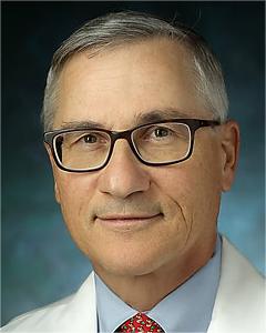 Gerald Louis Andriole合作论文数Brady Urologic Institute, Johns Hopkins University;Department of Surgery, Sibley Memorial Hospital1
 Chandru P. Sundaram合作论文数Department of Urology, Indiana University School of Medicine1
 John A Jane合作论文数Department of Neurosurgery, School of Medicine, University of Virginia;Virginia Tech Carilion School of Medicine1
 Pratim Biswas合作论文数Department of Chemical, Environmental and Materials Engineering, College of Engineering, University of Miami;Rosenstiel School of Marine, Atmospheric, and Earth Science, University of Miami1
 Steven M. Schwartz合作论文数The Hospital for Sick Children, University of Toronto School of Medicine1Michael Talcott合作论文数1Genoa G. Ferguson合作论文数1Kevin Morissey合作论文数1Salvatore P. Sutera合作论文数1
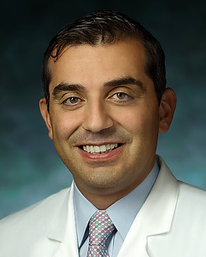 Mohamad E. Allaf合作论文数Department of Urology, School of Medicine, Johns Hopkins University1
 Kuk Cho合作论文数School of Engineering and Applied Sciences, Washington University in St. Louis1Josephine Hidalgo合作论文数1Mikel Gray合作论文数1
 Chris Hogan合作论文数Department of Chemistry, College of Science and Engineering, University of Minnesota;Department of Mechanical Engineering, College of Science and Engineering, University of Minnesota1J Skenazy合作论文数1James Sagar合作论文数1Keegan L. Maxwell合作论文数1Courtney Lee合作论文数1
 Myong-Hwa Lee合作论文数Aerosol and Air Quality Research Laboratory, Washington University in St. Louis1Fern G. Campbell合作论文数1Cathy Chen合作论文数1




