Latent human immunodeficiency virus type 1 (HIV-1) reservoirs in the central nervous system (CNS) may sustain viral persistence and neuroinflammation contributing to HIV-associated neurocognitive disorders (HAND) despite suppressive ART. AAV9-delivered CRISPR has successfully edited SIV proviral DNA in peripheral tissues with acceptable safety profiles, but the extent of in vivo genome editing in the brain remains unclear. Using SIV-infected rhesus macaques, we mapped intact proviral DNA across CNS regions and tested systemic AAV9-CRISPR/Cas9 targeting conserved sites within Ψ packaging signal and Gag region. Ten adult rhesus macaques were infected with genetically barcoded SIVmac239, suppressed with ART, then randomized to receive intravenous AAV9-SaCas9 with dual gRNAs (Ψ+Gag) or a Cas9-only control. At necropsy after viral rebound, SIV genomes were detected in multiple brain regions as well as lymphoid tissues, confirming the CNS as a persistent reservoir during ART. Barcode analysis revealed region-specific patterns consistent with compartmentalized CNS persistence. In CRISPR-treated animals, proviral editing was measurable across anatomically distinct CNS sites. These findings demonstrate that intact and potentially replication-competent virus persists in the primate brain under ART and that systemic AAV9-CRISPR can reach and edit proviral DNA in this sanctuary, supporting genome editing as a strategy toward durable remission of CNS reservoirs.
PurposeTo investigate the correlation between macular retinal and choroidal structural characteristics and refractive status in preschool-aged children with retinopathy of prematurity (ROP), based on optical coherence tomography angiography (OCTA), and to analyze the relationship between serum vascular endothelial growth factor (VEGF) levels and microcirculatory alterations.Methods310 preterm children with a history of ROP who were followed up at our hospital between January 2022 and June 2024 were included. According to the International Classification of Retinopathy of Prematurity (ICROP), participants were categorized into mild (n = 150, stage 1–2 ROP not meeting treatment criteria), moderate (n = 150, pre-threshold stage 3 ROP requiring treatment), and severe (n = 10, stage 3 threshold or above, or cases with retinal detachment) groups. Foveal avascular zone (FAZ) area, vessel densities of the superficial capillary plexus (SCP) and deep capillary plexus (DCP), choroidal thickness, number of vascular branches, vascular tortuosity, and foveal thickness were evaluated. Refractive parameters and VEGF levels were measured. The correlation between OCTA parameters, refractive parameters, and serum VEGF levels was analyzed.ResultsThe myopia rate in the severe group (80.0%) was higher than that in the moderate (52.0%) and mild (18.7%) groups (χ² = 68.45, P < 0.001). The spherical equivalent (SE) was (−2.35 ± 2.75) D, (−0.85 ± 2.15) D, and (+0.75 ± 1.25) D in the severe, moderate, and mild groups, respectively (F = 45.32, P < 0.001). The FAZ area was negatively correlated with the SE (r = −0.659, P < 0.001) and positively correlated with the axial length (r = 0.653, P < 0.001). The SCP vessel density was positively correlated with the SE (r = 0.618, P < 0.001). The choroidal thickness was positively correlated with the SE (r = 0.655, P < 0.001). Multivariable logistic regression analysis showed that a history of severe ROP (OR = 3.85, 95% CI: 2.52–5.86), SCP vessel density <40% (OR = 2.58, 95% CI: 1.78–3.76), FAZ area >0.35 mm² (OR = 2.24, 95% CI: 1.55–3.25), and choroidal thickness <250 μm (OR = 2.32, 95% CI: 1.62–3.36) were associated with the development of myopia. Serum VEGF level was positively correlated with FAZ area (r = 0.375, P < 0.001) and negatively correlated with SCP vessel density (r = −0.419, P < 0.001).ConclusionIncreased FAZ area, decreased SCP vessel density, and decreased choroidal thickness were associated with the development of myopia. Serum VEGF levels were correlated with macular microcirculatory parameters.
Without an effective antiviral, JC virus (JCV) has persisted throughout multiple epochs of immunosuppression, causing the opportunistic demyelinating disease, progressive multifocal leukoencephalopathy (PML). This study proposes a novel therapy using a dual-gRNA, SaCas9, CRISPR antiviral targeting JCV transcription factor, large tumor antigen (LT-Ag), and capsid protein, viral protein 1 (VP1). This treatment was validated using traditional two-dimensional cell culture. A recombinant cell line was produced from SVG astrocytes (SVGA) via lentiviral inoculation and puromycin selection. Following infection, sanger sequencing identified uniform excision of the circular dsDNA genome of JCV, significantly reducing viral load per genomic copy number on qPCR, viral proteins on western blot, and infectivity of viral progeny on adoptive transfer. Following this proof-of-concept using cell lines, translatability of results was advanced using three-dimensional, heterogeneous cerebral organoids (COs). COs were infected and treated with the lentivirus-packaged CRISPR antiviral. As observed in monolayer culture, a truncated genome was confirmed with sequencing, reducing viral load per genomic copy number on qPCR, protein levels on immunofluorescent imaging, and infectivity on adoptive transfer. The high efficacy of this JCV-targeting CRISPR antiviral in the context of cerebral organoids expounds on its value for the currently untreatable JCV and PML.
Elimination of HIV-1 requires clearance and removal of integrated proviral DNA from infected cells and tissues. Here, sequential long-acting slow-effective release antiviral therapy (LASER ART) and CRISPR-Cas9 demonstrate viral clearance in latent infectious reservoirs in HIV-1 infected humanized mice. HIV-1 subgenomic DNA fragments, spanning the long terminal repeats and the Gag gene, are excised in vivo, resulting in elimination of integrated proviral DNA; virus is not detected in blood, lymphoid tissue, bone marrow and brain by nested and digital-droplet PCR as well as RNAscope tests. No CRISPR-Cas9 mediated off-target effects are detected. Adoptive transfer of human immunocytes from dual treated, virus-free animals to uninfected humanized mice fails to produce infectious progeny virus. In contrast, HIV-1 is readily detected following sole LASER ART or CRISPR-Cas9 treatment. These data provide proof-of-concept that permanent viral elimination is possible.
We used NOD/SCID mice, also known as NRG, to assess the ability of lentivirus-mediated intravenous delivery of CRISPR in editing the HIV-1 genome from the circulating PBMC engrafts, some of which homed within several animal solid tissues. Lentivirus-mediated delivery of a multiplex of guide RNAs accompanied by Cas9 endonuclease led to the excision of the targeted region of the viral genome positioned within the HIV-1 LTR from the in-vitro-infected human peripheral blood mononuclear cells (PBMCs) embedded in the spleens of NRG mice. Similarly, the treatment of NRG mice harboring PBMC engrafts derived from HIV-1-positive patients with the therapeutic lentivirus eliminated the presence of the viral DNA fragment in the blood, as well as in the spleen, lung, and liver, of the engrafted animals. Sanger sequence analysis of the viral DNA after treatment with the lentiviral vectors expressing Cas9 and gRNAs verified the editing and removal of the proviral DNA fragment from the viral genome at the predicted sites. This proof-of-concept study, for the first time, demonstrates successful excision of the HIV-1 proviral DNA from patient immune cell engrafts in humanized mice upon treatment with lentivirus-expressing CRISPR and causes a decline in the level of replication-competent virus.
目的 观察脂多糖(LPS)、4(甲基亚硝氨基)-3-吡啶-1-丁酮(NNK)对小鼠巨噬细胞增殖及相关基因表达的影响.方法 体外培养小鼠巨噬细胞Raw264.7细胞,检测不同浓度的LPS及NNK刺激后Raw264.7细胞C-myc mRNA的表达情况,选择药物最佳浓度.用所选浓度的LPS、NNK及LPS+ NNK孵育Raw264.7细胞不同时间,利用细胞计数仪检测细胞增殖能力,采用实时定量PCR方法检测C-myc、核转录因子-κB(NF-κB)、精氨酸酶(Arginase)、一氧化氮合酶(iNOS)、肿瘤坏死因子α(TNF-α)、白细胞介素(IL)-1α及IL-6 mRNA表达,应用酶联免疫试剂盒检测炎性递质TNF-α及IL-6的含量.结果 选择浓度为10 μmol/L NNK与1 mg/L LPS进一步实验.NNK孵育Raw264.7细胞24 h,其细胞增殖和各基因表达及IL-6、TNF-α蛋白表达与对照组比较差异无显著性.LPS和LPS+ NNK孵育Raw264.7细胞24 h,其细胞增殖活性显著降低;C-myc、NF-κB、Arginase、iNOS、IL 1α、IL-6 mRNA及IL-6、TNF-α蛋白表达显著升高(F=19.4~5 895.0,t=4.04~97.53,P<0.05).NNK对LPS刺激的Raw264.7细胞C-myc mRNA、TNF-α蛋白的表达升高具有拮抗作用;对NF-κB、IL-1α mRNA的表达升高具有协同作用.结论 LPS可抑制Raw264.7细胞的增殖,并促进Raw264.7细胞活化;NNK单独孵育对巨噬细胞的增殖和相关基因表达无明显影响,但可以强化LPS诱导的NF-κB、IL-1α基因高表达,促使巨噬细胞向M1型活化.
目的 研究静脉移植的骨髓间充质干细胞(BMSCs)在急性肝损伤小鼠肝组织内的分布情况.方法 雌性昆明种小鼠42只,随机分为正常对照组、损伤模型组和干细胞移植组.损伤模型组和干细胞移植组小鼠均以橄榄油配制的含体积分数0.4的CCl4一次性灌胃制备急性肝损伤模型.分离同种小鼠BMSCs,体外扩增、BrdU标记后经尾静脉移植于灌胃后24 h的干细胞移植组小鼠体内.分别于CCl4灌胃后的第5、10、15天取损伤模型组和干细胞移植组小鼠的肝脏,每组每个时间点取材6只,正常对照组小鼠不做处理,直接取6只小鼠肝脏,各组均制备石蜡切片和连续冷冻切片,苏木精-伊红(HE)染色观察肝组织的结构变化,免疫组织化学染色追踪BrdU阳性标记干细胞在肝组织内的迁移和分布情况.结果 HE染色结果显示,干细胞移植组小鼠在CCl4灌胃后的第5、10、15天的肝组织修复状态明显好于相同时间点的损伤模型组;干细胞移植组小鼠CCl4灌胃后的第15天肝组织接近正常.免疫组化结果显示,干细胞移植组小鼠在CCl4灌胃后第5天肝组织内散在分布着大量的黄色或褐色的BrdU阳性细胞胞核;干细胞移植组小鼠CCl4灌胃后第10天时肝组织内的BrdU阳性细胞胞核数目较第5天时少,而第15天时较第10天时明显减少,第15天与第5、10天比较,差异有显著性(t=3.350、2.907,P<0.05).结论 静脉移植的BMSCs可以迁移至急性肝损伤小鼠肝脏,并可促进损伤肝脏的修复;随着移植时间的延长单位面积内BrdU阳性细胞数量减少.
目的 观察诱导型一氧化氮合酶(iNOS)在表没食子儿茶素没食子酸酯(EGCG)促A549细胞凋亡中的作用.方法 体外培养A549细胞,随机分为对照组、EGCG组,MTT方法检测筛选EGCG最佳作用浓度,流式细胞仪检测EGCG促A549细胞凋亡作用,免疫组化染色法、蛋白质印迹定量分析法与Realtime-PCR方法检测各组中iNOS蛋白和mRNA的表达情况.结果 200 mg/L的EGCG组细胞增殖活性与其他组比较差异有显著意义(F=1 000.35,q=3.43~60.00,P<0.05).EGCG组晚期A549细胞凋亡率与对照组比较,差异有显著性(x 2=65.23,P<0.05).与对照组比较,EGCG同样可以降低A549细胞中iNOS蛋白与mRNA的表达量,差异有显著性(t=9.4、5.7,P<0.05).结论 EGCG促A549细胞凋亡可能与下调iNOS蛋白与mRNA的表达量有关.
目的:研究表没食子儿茶素-3-没食子酸酯(epigallocatechin-3-gallate,EGCG)对炎性刺激的人肺腺癌A549细胞增殖和凋亡的影响及与CUGBP1表达的关系.方法:MTT法检测EGCG和LPS刺激A549细胞增殖活性的影响;流式细胞仪检测细胞凋亡;免疫细胞化学检测EGCG对LPS刺激人肺腺癌A549细胞内CUGBP1蛋白的表达.结果:与对照组相比,LPS体外显著促进A549细胞增殖,其胞核胞质内CUGBP1表达明显增强(P<0.01).加入EGCG可拮抗LPS促A549细胞增殖的作用,促进其凋亡,明显抑制LPS刺激的A549细胞内CUGBP1的表达(P<0.01).CUGBP1蛋白定量分析可知EGCG和LPS共同孵育A549细胞4h、24h时,细胞中的CUGBP1蛋白表达量较单纯LPS作用时降低.但EGCG和LPS共同孵育A549细胞24h,A549细胞中胞核CUGBP1蛋白表达量(1210.565± 3.46)较4h时胞核CUGBP1蛋白表达量(67.344± 3.68)高,差异有统计学意义(t=927.164,P<0.001).结论:EGCG可能通过干扰CUGBP1基因的表达抑制炎症刺激人肺腺癌细胞A549的增殖,促进其凋亡.
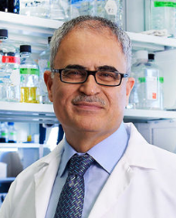 Kamel Khalili合作论文数Department of Neuroscience, Lewis Katz School of Medicine, Temple University7
 Hong Liu合作论文数Dept Med Oncol, Zhejiang Univ4
 Won-Bin Young合作论文数4
 JingJing Bian (卞晶晶)合作论文数4XiaoPing Liu (刘晓萍)合作论文数4
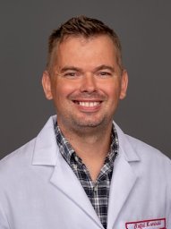 Rafal Kaminski合作论文数Center for Neurovirology, Temple University School of Medicine4
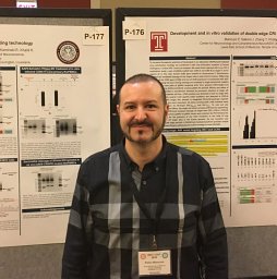 Pietro Mancuso合作论文数Temple Univ, Dept Neurosci, Lewis Katz Sch Med, Philadelphia, PA 19122 USA3Rahsan Sariyer合作论文数Temple Univ, Lewis Katz Sch Med, Ctr Neurovirol, Dept Neurosci, Philadelphia, PA 19122 USA3
 Ramona Bella合作论文数Temple Univ, Dept Neurosci, Lewis Katz Sch Med, Philadelphia, PA 19122 USA3Jing ZHANG (张静)合作论文数3
 Ilker K Sariyer合作论文数3Tongxi YANG (杨童茜)合作论文数2Liuxia Cao合作论文数2Zhongjie YU (于忠杰)合作论文数2Shohreh Amini合作论文数College of Science and Technology, Temple University2
 Pasquale Ferrante合作论文数Department of Biomedical Sciences and Technologies, University of Milan2
 Jennifer Gordon合作论文数Dept Neurosci, Temple Univ2Shuren Liao合作论文数2Conrad Cruz合作论文数1Senem Cakir合作论文数1Alison Kearns合作论文数1Angela Rocchi合作论文数1Ranieri B合作论文数1
 Zhiyi Lin合作论文数1Qi WU (吴琪)合作论文数1Dominika Nawrat合作论文数1
 Saumi Mathews合作论文数Univ Nebraska Med Ctr, Dept Pharmacol & Expt Neurosci, Omaha, NE USA1Mary Banoub合作论文数Department of Pharmacology and Experimental Neuroscience, University of Nebraska Medical Center1
 James R Hilaire合作论文数Univ Nebraska Med Ctr, Dept Pharmacol & Expt Neurosci, Omaha, NE 68198 USA1Martina Donadoni合作论文数Temple Univ, Lewis Katz Sch Med, Philadelphia, PA 19122 USA1Jake A Robinson合作论文数1
 Hang su合作论文数Univ Nebraska Med Ctr, Dept Pharmacol & Expt Neurosci, Omaha, NE USA1
 Brady Sillman合作论文数Univ Nebraska Med Ctr, Dept Pharmacol & Expt Neurosci, Omaha, NE 68198 USA1Yan REN (任妍)合作论文数1
 Larisa Poluektova合作论文数Univ Nebraska Med Ctr1KeLing Zhang (张克凌)合作论文数1
 Burdo Tricia H合作论文数Dept Biol, Boston Coll1
 Nagsen Gautam合作论文数Dept Pharmaceut Sci, Univ Nebraska Med Ctr1
 Benson Edagwa合作论文数Dept Pharmacol & Expt Neurosci, Univ Nebraska Med Ctr1Wollebo Hassen S合作论文数1
 Prasanta Dash合作论文数1Fischer Tracy合作论文数Center for Neurovirology, Temple University School of Medicine1
 Taha Mohseni Ahooyi合作论文数1
 Aditya N. Bade合作论文数University of Nebraska Medical Center, Univ. of Nebraska Medical Center1
 Jeffrey N. Martin合作论文数San Francisco General Hospital;Department of Epidemiology and Biostatistics, University of California, San Francisco1
 Howard E. Gendelman合作论文数Department of Pharmacology and Experimental Neuroscience, College of Medicine, University of Nebraska Medical Center1
 R. Lee Mosley合作论文数Center for Neurovirology and Neurodegenerative Disorders, University of Nebraska Medical Center1Catherine M. Bollard合作论文数1Santhi Gorantla合作论文数Department of Pharmacology and Experimental Neuroscience, University of Nebraska Medical Center1




