Cellular senescence was initially defined in vitro as a stable cell-cycle arrest that occurs after repeated replication, but it is now recognized as a heterogeneous state shaped by cell type, species, senescence-inducing stress, tissue microenvironment and time. To organize this complexity, we propose the term 'senotype' to classify senescent cells by their inputs, molecular features and functional effects. We outline a practical framework incorporating: (1) cell identity and context; (2) inducing mechanism; (3) temporal stage; (4) multimodal molecular and structural features; and (5) physiological or pathological functions. Experimentally defined senotypes can serve as references for interpreting tissue-derived senotypes, where parameters may be incomplete. Senotypes should be anchored in combinations of core hallmarks (that is, durable cell-cycle arrest, altered secretory profiles, macromolecular or organelle damage, disrupted homeostasis) rather than single markers. Advances in single-cell, spatial, proteomic and computational methods enable rigorous senotype characterization, improving consistency and accelerating development of targeted senotherapeutics.
Abstract The foreign body response (FBR) is an immune mediated event that occurs with every material implant. The extent of the fibrosis is dependent on many factors including the biomaterial design, tissue location, and host factors such as age, sex, ancestry, diet. There are known clinical outcomes of implants dependent on age and sex, including increased fibrosis and implant failure in aged and female patients. As the population ages, there is a growing need to understand how aging affects the FBR, and how preclinical models can capture this to guide biomaterial design. Here, we investigated how chronic fibrosis in a murine model of the FBR is altered by two biological factors: age and sex. We investigated changes in fibrosis using a volumetric muscle loss (VML) injury model coupled with polycaprolactone (PCL) or polyethylene (PE) microparticle implants. Fibrosis was quantified through gene expression, microscopic analysis of histologic sections, and the corresponding immune response measured via gene expression and flow cytometry data. We found gene expression differences with immune pathways enriched in female mice, and microscopy revealed collagen birefringence area increased in young male mice. Both the innate and adaptive immune response were altered by age and sex via T cell and macrophage phenotype, and the effects of aging differed between sexes. These results reveal both variables contribute to discrepant outcomes in both fibrosis and the local immune response to synthetic material implants. This demonstrates a clear need to understand and account for the influence of biological factors in biomaterial design.
Uterine leiomyomas (ULs) are prevalent benign tumors in women of reproductive age characterized by cellular senescence. Cellular senescence is a state of stable, irreversible cell cycle arrest characterized by discrete changes in cellular morphology and gene expression. This systematic review, following PRIMSA guidelines, evaluated the molecular pathways contributing to senescence in ULs and the use of novel therapeutic agents to target senescence. Two investigators independently screened and identified relevant articles written in English involving human subjects. Sixty-nine articles were identified; 11 studies met criteria. Multiple studies recognized a range of biomarkers of senescence in ULs including senescence associated beta galactosidase (SA-β-gal), senescent associated proteins (p16, p21, p14ARF), and telomere shortening. Key pathways such as AKT and p14ARF-TP53-p21, and genes such as HMGA2 and MED12 have been implicated in regulating the balance between tumor proliferation and growth arrest and senescence. However, the specific genetic and epigenetic mechanisms that induce and maintain senescence in ULs are not fully understood. There is growing interest in investigating whether senescent cells can be therapeutically targeted in ULs by senolytic agents that induce apoptosis, and senomorphic agents that modulate the senescence-associated secretory phenotype (SASP) to reduce its pro-tumorigenic effects. While limited, non-clinical data suggests this approach may be promising, further investigation is needed to establish their clinical efficacy in patients with ULs.
Aging of the hematopoietic system impairs hematopoietic stem cell (HSC) function and alters bone marrow niche behavior, increasing susceptibility to anemia, infections, and hematologic malignancies. Here, pharmacologic clearance of senescent cells with the PROTAC compound 753b simultaneously targeting BCL-xL and BCL-2 reverses key secretory, transcriptional, and functional hallmarks of hematopoietic aging with low toxicity, restoring balanced lineage output. Single-cell RNA sequencing further demonstrates that 753b treatment attenuates aging-associated transcriptional signatures in HSCs, while selectively eliminating senescent, pro-survival niche cells without grossly perturbing niche composition. Functionally, 753b suppresses pro-inflammatory cues from both niche and hematopoietic cells including those emanating from neutrophil progenitors, rebalancing global bone marrow secretory ecosystem across stromal and hematopoietic compartments. Collectively, we identify 753b-induced senescent cell clearance as a powerful strategy to rejuvenate aged hematopoiesis and re-establish homeostatic communication between HSCs and their microenvironment, with implications for mitigating age-related hematologic dysfunction and improving hematologic health in older individuals.
Uterine fibroids (leiomyomas) are highly prevalent benign tumors defined by excessive extracellular matrix (ECM) deposition, altered vascular structure, and progressive tissue stiffening, yet the cellular programs that coordinate these features remain poorly understood. Cellular senescence has been implicated in fibroid biology, but whether senescence represents a uniform state or distinct, functionally specialized cell identities within fibroids is unknown. Here, we identify the distinct heterogeneous populations of senescent cells ("senotypes") present in human fibroids and characterize their role in shaping the fibroid microenvironment. Using single-cell RNA sequencing (scRNA-seq) integrated with a senescence gene signature and protein-level validation, we identify senescent cells (SnC) distributed across fibroblast, mural, and endothelial compartments, each exhibiting distinct transcriptional programs. SnC endothelial cells (ECs) are enriched in fibroids relative to matched myometrium and activate TEAD4 -associated mechanosensing, angiogenic, and immune signaling pathways, despite being associated with impaired vessel maturation in situ . In parallel, SnC fibroblast and mural populations in fibroids upregulated SRF-associated cytoskeletal and ECM programs, accompanied by increased COL6A3 expression and collagen VI deposition, consistent with tissue stiffening. Ligand-receptor and spatial analyses reveal that these SnC populations function as interconnected signaling hubs, coordinating immune cell recruitment and stromal remodeling. Importantly, analysis of human fibroids treated with collagenase demonstrated a reduction in both ECM density and SnC burden, supporting a reinforcing relationship between matrix mechanics and senescence. Together, these findings establish senescence in fibroids as a heterogeneous, mechanically reinforced, and network-driven process that links vascular dysfunction, immune signaling, and fibrosis, highlighting distinct SnC states as potential translational targets for non-surgical therapies.
Fibrotic conditions contribute to significant global morbidity and mortality. Yet the underlying processes that orchestrate fibrosis remain poorly understood due to the cellular and spatial complexity of the stromal, immune, and vascular compartments that regulate fibrotic disease progression. Senescent cells (SnCs) have been implicated in fibrosis, but their roles are unclear, as evidence indicates that they serve both pathogenic and reparative functions. Here, we show that fibrosis-associated SnCs contain functionally divergent senotypes that are organized into distinct spatial niches. Using integrated single-cell and spatial transcriptomics analyses and hierarchical factorization in a murine fibrosis model, we identify fibroblast and perivascular SnC subpopulations that upregulate diverse programs related to extracellular matrix (ECM) production, immune signaling, and vascular remodeling. Fibroblast senotypes localize to discrete microenvironments with distinct tissue architectures, including niches associated with fibrotic signaling, immune activity, and cartilage development. Perivascular SnCs occupy interfaces between fibrotic signaling and immune-active niches and upregulate vascular and fibrotic remodeling pathways. Depletion of pericyte-lineage SnCs increases vascular maturation and fibrotic ECM deposition, providing mechanistic validation of the beneficial role these SnCs play in vascular remodeling and fibrosis modulation. In addition, using a new web-based infrastructure to query our senotype gene signatures in public datasets, we demonstrate that these senotypes are conserved across different murine and human fibrotic conditions. These findings establish senescence as a spatially organized regulator of fibrosis and identify perivascular senescence as a link between vascular remodeling and fibrotic outcomes.
Identifying the drivers of cellular senescence that contribute to the decline in tissue function related to aging- and disease is critical for developing restorative interventions. Here, we investigated how increased mechanical stress from extracellular matrix (ECM) stiffening shapes endothelial cell (EC) senescence. We developed a 3D human in vitro model that decouples mechanical stress from inflammatory or biochemical inputs, enabling the study of senescence responses to tissue stiffening alone. We found that matrix stiffening induces an EC senescence phenotype with elevated p16/p21 and an immunomodulatory senescence-associated secretory phenotype (SASP), in the absence of inflammatory signals. This mechano-induced senescence state engaged a Notch-JNK-FOS signaling axis, and pharmacologic inhibition of Notch attenuated stiffness-induced senescence. Supporting the translational relevance of this mechanism, analysis of fibrotic capsule tissue from patients with synthetic breast implants, a model of localized, mechanically driven fibrosis, revealed increased p16+Notch1+ endothelial populations. Complementary single-cell RNA sequencing data confirmed their enrichment in Notch/JNK- and SASP-related gene programs. Together, these findings define vascular senescence as a mechanosensitive process and identify tissue stiffening as an upstream aging signal. Our work offers a human-relevant platform for studying targetable stages of endothelial mechanoaging.
MOTIVATION:Algorithms for ligand-receptor network inference have emerged as commonly used tools to estimate cell-cell communication from reference single-cell data. Many studies employ these algorithms to compare signaling between conditions and lack methods to statistically identify signals that are significantly different. We previously developed the cell communication inference algorithm Domino, which considers ligand and receptor gene expression in association with downstream transcription factor activity scoring. We developed the dominoSignal software to innovate upon Domino and extend its functionality to test statistically differential cellular signaling. RESULTS:This new functionality includes the compilation of active signals as linkages from multiple subjects in a single-cell data set and testing condition-dependent signaling linkage. The software is applicable for analysis of single-cell data sets with multiple subjects as biological replicates as well as with bootstrapped replicates from data sets with few or pooled subjects. We use simulation studies to benchmark the number of subjects in compared groups and cells within an annotated cell type sufficient to accurately identify differential linkages. We demonstrate the application of the Differential Cell Signaling Test (DCST) in the dominoSignal software to investigate consequences of cancer cell phenotypes and immunotherapy on cell-cell communication in tumor microenvironments. These applications in cancer studies demonstrate the ability of differential cell signaling analysis to infer changes to cell communication networks from therapeutic or experimental perturbations, which is broadly applicable across biological systems. AVAILABILITY:dominoSignal is available through Bioconductor at https://www.bioconductor.org/packages/release/bioc/html/dominoSignal.html.
Acellular Adipose Tissue (AAT) is an off-the-shelf, cadaveric adipose-derived ECM-based biomaterial for soft tissue reconstruction. AAT has been validated preclinically to promote angiogenesis and adipogenesis and demonstrated safety, biocompatibility, and tolerability in a Phase I study. In this study we report the findings for the first ten patients in the Phase II study for permanent reconstruction of modest soft tissue defects. AAT promoted macrophages, CD3 + T cells, and CD34 + progenitor activity. Multiplex immunofluorescence staining using the PhenoCycler (formerly CODEX) imaging platform found that AAT can induce tertiary lymphoid structures (TLS). Nanostring GEOMx spatial transcriptional data analysis found significant differential gene expression between neighboring tissues with EGR1 , MCL1 , and NR4A1 upregulated in AAT. These genes have roles in angiogenesis, anti-apoptotic processes, and promotion of anti-inflammatory genes, respectively. AAT promoted anti-fibrotic CD74 + adipose-derived stromal cells, confirmed by immunofluorescence staining. Our findings demonstrate that AAT promotes angiogenesis, adipogenesis, and anti-fibrotic remodeling.
The role of glioma-associated myeloid cells in tumor growth and immune evasion remains poorly understood. We performed single-cell RNA sequencing of immune and tumor cells from 33 gliomas, identifying two distinct myeloid-derived suppressor cell (MDSC) populations in isocitrate dehydrogenase-wild-type (IDT-WT) glioblastoma: an early progenitor MDSC (E-MDSC) population with up-regulation of metabolic and hypoxia pathways and a monocytic MDSC (M-MDSC) population. Spatial transcriptomics demonstrated that E-MDSCs geographically colocalize with metabolic stem-like tumor cells in the pseudopalisading region. Ligand-receptor analysis revealed cross-talk between these cells, where glioma stem-like cells produce chemokines attracting E-MDSCs, which in turn produce growth factors for the tumor cells. This interaction is absent in IDH-mutant gliomas, associated with hypermethylation and repressed gene expression of MDSC-attracting chemokines. Our study elucidates specific MDSCs that may facilitate glioblastoma progression and mediate tumor immunosuppression.
Therapeutic biological scaffolds promote tissue repair primarily through the induction of type 2 immunity. However, systemic immunological factors, including aging, sex, and previous infections, can modulate this response. The gut microbiota is a well-established modulator of immune function across organ systems, yet its influence on type 2-mediated repair remains underexplored. Here, we establish a bidirectional relationship between the gut microbiota and biological scaffold-mediated tissue repair. Utilizing a conventionalized germ-free mouse, we demonstrate that scaffold implantation induces compositional and functional changes in the gut microbiome, particularly affecting amino acid biosynthesis. Additionally, in a model of antibiotic-induced microbiota depletion, we show that dysbiosis disrupts key immune regulators of type 2 immunity, including reductions in eosinophils, proregenerative macrophages, and interleukin-4 (IL-4)-producing CD4+T cells. At 6 wk post-scaffold implantation, we observed a significant decrease in myocytes with centrally located nuclei alongside an upregulation in profibrotic gene expression with antibiotic treatment. These findings provide insights into the influence of the gut microbiota on type 2-mediated tissue repair.
BACKGROUND:Crosslinked hyaluronic acid (HA)-based hydrogels are commonly used as dermal fillers where they interact with surrounding tissues including host stromal and immune cells. HA fillers are widely used for aesthetic applications, with products designed with varying properties depending on their indication. Although HA fillers have been demonstrated to have a strong biocompatibility profile, a small subset of patients' experiences delayed-onset adverse events hypothesized to be inflammatory and allergy-related outcomes such as delayed-onset hypersensitivity. AIMS:The overall goal of this study was to evaluate the innate and adaptive immune response to two clinically available HA filler formulations. METHODS:Using multiparametric flow cytometry, we characterized the immune response to Juvèderm Volbella (VYC-15 L) and Juvèderm Ultra 3 (SGD-30XP) in a murine quadricep muscle resection that enables implantation of larger volumes and exposure to muscle and adipose. RESULTS:Presence of the implanted HA filler increased recruitment of immune cells, specifically antigen presenting macrophages, eosinophils, and gamma-delta (γδ) T cells to the injury site compared to no implant (saline) controls. Comparing the two materials, VYC-15 L increased interleukin 17a (IL17a) production by lymphocyte subsets at the injury site and induced higher levels of circulating IgE relative to SGD-30XP and saline controls. CONCLUSION:Overall, these results provide insights into the immune response to HA fillers and how different formulations may alter the immune outcomes.
Cellular senescence is a complex biological process that plays a pathophysiological role in aging and age-related diseases. The biological understanding of senescence at the cellular and tissue levels remains incomplete due to the lack of specific biomarkers as well as the relative rarity of senescent cells, their phenotypic heterogeneity and dynamic features. This Review provides a comprehensive overview of multiomic approaches for the characterization and biological understanding of cellular senescence. The technical capability and challenges of each approach are discussed, and practical guidelines are provided for selecting tools for identifying, characterizing and spatially mapping senescent cells. The importance of computational analyses in multiomics research, including senescent cell identification, signature detection and interactions of senescent cells with microenvironments, is highlighted. Moreover, tissue-specific case studies and experimental design considerations for individual organs are presented. Finally, future directions and the potential impact of multiomic approaches on the biological understanding of cellular senescence are discussed.
 Qiongyu Guo合作论文数Dept. of Biomed. Eng., Johns Hopkins Univ.;c;Dept. of Biomed. Eng., Johns Hopkins Univ.22
 Morgana M. Trexler合作论文数21Oliver Schein合作论文数Epidemiology, Bloomberg School of Public Health, Johns Hopkins University20
 Anirudha Singh合作论文数Alcon19QiaoZhi Lu合作论文数19Xiaokun Wang (王晓鲲)合作论文数School of Information Science and Engineering, Shandong University19Majumdar Shoumyo合作论文数Wilmer Eye Institute and Department of Biomedical Engineering, Johns Hopkins School of Medicine18Cherry Christopher M合作论文数Department of Cell Biology, Johns Hopkins University School of Medicine18
 Drew Mark Pardoll合作论文数Department of Oncology, School of Medicine, Johns Hopkins University;Bloomberg~Kimmel Institute for Cancer Immunotherapy, School of Medicine, Johns Hopkins University;Sidney Kimmel Comprehensive Cancer Center, School of Medicine, Johns Hopkins University18
 Matthew T. Wolf合作论文数Department of Bioengineering, University of Pittsburgh18
 Elana J. Fertig合作论文数Institute for Genome Sciences, University of Maryland School of Medicine16
 Alexander Hillel合作论文数Department of Otolaryngology, Johns Hopkins University;Johns Hopkins School of Medicine14
 David R. Maestas Jr合作论文数Translational Tissue Engineering Center, Johns Hopkins University13Sven D. Sommerfeld合作论文数Translational Tissue Engineering Center, Johns Hopkins University12Jin Han合作论文数11Anna Ruta合作论文数11
 Liam Chung合作论文数11
 Kaitlyn Sadtler合作论文数National Institute of Biomedical Imaging and Bioengineering, National Institutes of Health11
 Franck Housseau合作论文数Department of Otolaryngology-Head and Neck Surgery, School of Medicine, Johns Hopkins University11Calderón-Colón Xiomara合作论文数11
 Shyni Varghese合作论文数Department of Biomedical Engineering,Johns Hopkins University11Tam Ada合作论文数10
 Nathaniel S. Hwang合作论文数Department of Biomedical Engineering;Johns Hopkins University;Department of Biomedical Engineering, Johns Hopkins University10Iossif Strehin合作论文数10Jeffrey P Maranchi合作论文数9
 Robert S. Langer合作论文数Department of Chemical Engineering, Massachusetts Institute of Technology9
 Blanka Sharma合作论文数Univ Florida9
 Katlin B Stivers合作论文数8Wu Iwen合作论文数8
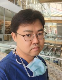 Jemin Jeremy Chae合作论文数8
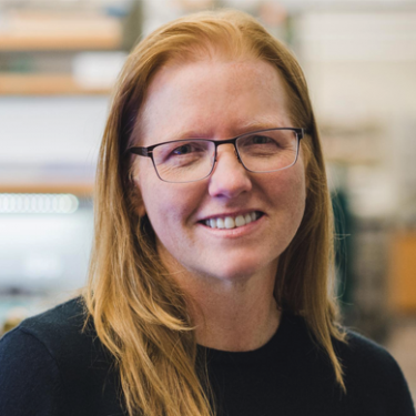 Kristi Anseth (克里斯蒂·安塞斯)合作论文数Department of Chemical and Biological Engineering, College of Engineering and Applied Science, University of Colorado, Boulder;The BioFrontiers Institute, University of Colorado, Boulder8Winnette Mcintosh Ambrose合作论文数8Elise Gray-Gaillard合作论文数7
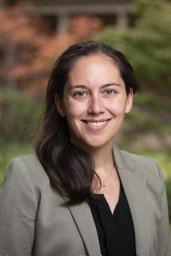 Joscelyn C Mejías合作论文数Johns Hopkins University7
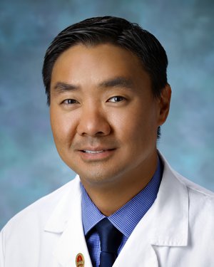 Albert S. Jun合作论文数Department of Otolaryngology-Head and Neck Surgery, School of Medicine, Johns Hopkins University7
 Sudipto Ganguly合作论文数Johns Hopkins Sidney Kimmel Comprehensive Cancer Research Center7Nahas Zayna合作论文数7
 Ludmila Danilova合作论文数Sidney Kimmel Comprehensive Cancer Center, Johns Hopkins University;School of Medicine, Johns Hopkins University7
 Jeannine Coburn合作论文数Department of Biomedical Engineering, Tufts University7
 Elizabeth M. Jaffee合作论文数Department of Pathology, School of Medicine, Johns Hopkins University;Institute for Clinical & Translational Research, Johns Hopkins University7Kagohara Luciane T合作论文数Sidney Kimmel Comprehensive Cancer Center, Johns Hopkins University School of Medicine7
 Walter Stark合作论文数Department of Ophthalmology, Wilmer Eye Institute, Johns Hopkins University;Johns Hopkins Hospital7
 Christopher G. Williams合作论文数7
 Alexandra Bolm Pucsek合作论文数6Samantha Guinn合作论文数6Melissa R. Lyman合作论文数6Haley Zlomke合作论文数6Reecha Suri合作论文数6Helen Hieu Nguyen合作论文数6




