Previous in vivo studies demonstrated that JHU083/6-Diazo-5-oxo-L-norleucine (DON), a glutamine analog drug, potently reprograms M1/M2 macrophages. To determine whether these effects are direct or indirect, we utilized an in vitro murine bone marrow-derived macrophage model, which recapitulates macrophage differentiation and polarization processes, to examine the impact of DON on the M1 macrophages. DON was applied during M1 differentiation or to fully polarized M1 macrophages, revealing that glutamine inhibition initially suppressed M1 activity but later enhanced it, resulting in sustained pro-inflammatory activation. Multi-omics analyses (bulk RNA-sequencing and LC-MS), time-course assays, and glutamine depletion experiments consistently suggested that prolonged glutamine inhibition elevated glutamine levels, which sustained pro-inflammatory gene transcription. In contrast, M2 and tumor-associated macrophages, which are immunosuppressive, were more susceptible to DON, leading to functional suppression. Collectively, our findings uncover stage-specific mechanisms by which glutamine inhibition modulates M1 polarization, offering a mechanistic rationale for therapeutic strategies that sustain pro-inflammatory, anti-tumor macrophage activity while concurrently suppressing immunosuppressive myeloid subsets in cancer.
A critical challenge in therapies for glioblastoma is systemic immunosuppression which is associated with poor response to therapies and thus correlated with worse outcomes. In this study we assess whether immune cytokines at baseline after diagnosis and during therapy are associated with outcome and may serve as an early biomarker predictive of treatment outcome. Patients were enrolled in a prospective, single institution, immune surveillance study. Peripheral blood was collected prior to initiating treatments and weekly during concurrent radiation and chemotherapy. Cytokine levels were measured from plasma samples isolated from peripheral blood. The cytokines were categorized as proinflammatory or anti-inflammatory. Baseline levels and dynamic changes in the levels of cytokines were analyzed for association with survival. 15 patients and 8 healthy controls were enrolled. At baseline, a majority immunosuppressive cytokines (IL-10, M-CSF, BTLA, PD-L1, LAG-3, PD-1, TIM-3, CTLA-4, TGFβ2 and TGFβ3) were elevated. The only proinflammatory cytokines associated with survival were IP-10, MCP-1, and IL-12p70 (according to the ANOVA analysis (p < 0.05)). A dynamic increase in the levels of a select proinflammatory cytokines at end of radiation (IL-34, and IL-12 p70) was associated with poor survival as was increased MCP-1 in those with unmethylated glioblastoma. No association between outcomes and dynamic changes in remaining proinflammatory cytokines or any of the immunosuppressive cytokines was noted. In this exploratory study, our data suggests that in patients with glioblastoma, measurements of plasma cytokines at diagnosis may predict outcomes. In addition, the dynamic changes in the cytokine levels could similarly serve as a biomarker guiding treatments. Future studies integrating clinical and patient specific immunological variables are required.
Background A critical challenge in therapies for glioblastoma is systemic immunosuppression. The immunosuppressed state is associated with poor response to therapies and thus correlated with worse outcomes. Immunosuppression is mediated by multiple factors including glioblastoma tumor and secreted cytokines, as well as radiation and chemotherapy. Methods Patients were enrolled in a single-institution, prospective, immune surveillance study. Peripheral blood was collected prior to initiating treatments and weekly during concurrent radiation and chemotherapy. Cytokine levels were measured from plasma samples isolated from peripheral blood. The cytokines were categorized as proinflammatory or anti-inflammatory. Baseline levels and dynamic changes in the levels of cytokines were analyzed for association with survival. Results 16 patients and 8 healthy controls were enrolled. A higher level of immunosuppressive cytokines at baseline (IL-10, IP-10, MCP-1, IL-34, M-CSF, PD-L1, LAG-3, PD-1, TIM-3, and CTLA-4) was inversely related with shorter survival (according to the ANOVA analysis (p < 0.05)). No association of baseline levels of proinflammatory cytokines with survival was observed. However, an increase in the levels of select proinflammatory cytokines at end of radiation (IL-34, IL-12 p70) was associated with poor survival as was increased MCP-1 in those with unmethylated tumor No association between outcomes and dynamic changes in remaining proinflammatory cytokines or any of the immunosuppressive cytokines was noted. Conclusions Our data suggests that in patients with glioblastoma, measurements of plasma cytokines at diagnosis may predict response to treatments and overall survival. In addition, the dynamic changes in the cytokine levels may similarly serve as a biomarker.
Previous in vivo studies demonstrated that JHU083/DON, a glutamine analog drug, potently reprograms M1/M2 macrophages. To determine whether these effects are direct or indirect, we utilized an in vitro murine bone marrow-derived macrophage (BMDM) model, which recapitulates macrophage differentiation and polarization processes, to examine the impact of DON on the M1 macrophages. DON was applied during M1 differentiation or to fully polarized M1 macrophages, revealing that glutamine inhibition initially suppressed M1 activity but later enhanced it, resulting in sustained pro-inflammatory activation. Multi-omics analyses (bulk RNA-seq and LC-MS), time-course assays, and glutamine depletion experiments consistently suggested that prolonged glutamine inhibition elevates glutamine levels, which sustain pro-inflammatory gene transcription. In contrast, M2 and tumor-associated macrophages (TAM), which are immunosuppressive, were more susceptible to DON, leading to functional suppression. Collectively, our findings uncover stage-specific mechanisms by which glutamine inhibition modulates M1 polarization, offering a mechanistic rationale for therapeutic strategies that sustain pro-inflammatory, anti-tumor macrophage activity while concurrently suppressing immunosuppressive myeloid subsets in cancer.
OBJECTIVE:PARP inhibitors may work synergistically to improve the efficacy of immunotherapy in patients with epithelial ovarian cancer (EOC). We performed a parallel-arm study of tremelimumab, alone or with olaparib, in patients with recurrent EOC. METHODS:Eligibility criteria included measurable disease and progression <12 months from last platinum. Participants were randomized to Arm A (tremelimumab monotherapy, 10 mg/kg/dose intravenously [IV]) or Arm B (dose level 1 [DL1] olaparib orally 150 mg twice daily with tremelimumab IV 3 mg/kg/dose and DL2 olaparib orally 150 mg twice daily with tremelimumab IV 10 mg/kg/dose). Primary objectives were safety, change in peripheral ICOS+ T cells, and identification of optimal dose combination. RESULTS:Among 24 total patients (12 on Arm A, 6 on Arm B-DL1, 6 on Arm B-DL2), the most common grade 3 toxicities were rash (13 %), immune-mediated hepatitis (8 %), and colitis (8 %). No grade ≥ 4 toxicities were identified. No dose-limiting toxicities were identified. One patient (Arm B-DL2) experienced a partial response; no complete responses were observed. Ten patients (7 on Arm A, 2 on Arm B-DL2, and 1 on Arm B-DL1) had a best response of stable disease. There was a significant increase in CD4+ICOS+ and CD8+ICOS+ T cells at both C1D15 and C1D22 in groups treated with tremelimumab IV 10 mg/kg/dose, but not in those treated with tremelimumab 3 mg/kg/dose. CONCLUSIONS:Tremelimumab IV 10 mg/kg/dose with olaparib 150 mg orally twice daily was safe and feasible. Tremelimumab 10 mg/kg/dose (as opposed to 3 mg/kg/dose) was required for immune activation, although this did not translate into clinical responses.
Aberrant microRNA expression is common in cancer, yet cell-type-specific microRNA activity in the tumor microenvironment (TME) remains poorly understood. Here, we show that germline deletion of miR-21 significantly attenuated the progression of MYC-driven prostate cancer (PCa), reducing prostate weight, tumor burden, and proliferation index in Hi-Myc mice. In situ hybridization revealed elevated miR-21 expression in multiple cell types during disease progression. Inflammatory and premalignant lesions in mouse and human prostate showed increased miR-21 in both stroma and epithelium, with further enrichment in the stroma of invasive adenocarcinoma. In Hi-Myc mice, single cell RNA-sequencing revealed miR-21 gene regulation in neoplastic, stromal, and immune cells in a cell-type-specific manner, impacting both direct and indirect targets. Notably, miR-21 deletion reduced immune infiltration into the prostate TME, particularly Trem2 -expressing macrophages and regulatory T cells. The Timp1-fibroblast gene signature in MYC-driven PCa was suppressed in miR-21 knockout prostates. Cell-cell communication analysis showed that miR-21 suppressed TGF-beta signaling in the TME, partially through Ski and Smad7 suppression in cancer-associated fibroblasts. These findings underscore the crucial role of miR-21 in PCa and provide some of the first in situ insights into cell-type-specific miRNA activity in solid tumors.
Radiation therapy affects a wide range of circulating lymphocyte subpopulations that are integral to mounting a successful lymphocyte-mediated immune response. Lymphopenia is associated with inferior tumor control and could be addressed to improve outcomes in glioblastoma. RT is a significant and potentially actionable iatrogenic suppressor of immune response that may limit the success of therapy. We hypothesized that a comprehensive evaluation of serial cytokine measurements during concurrent radiation and chemotherapy in glioblastoma will provide information about systemic immune status during treatment. 16 patients were enrolled in a single-institution, observational, immune surveillance study between 2018 and 2019. Blood was collected prior to starting radiation, and then weekly for a total of 7 timepoints. Plasma was isolated from blood, and cytokine levels were measured with a Luminex 26-cytokine panel. Survival was defined as a binary outcome at two years post-diagnosis: patients living beyond two years were classified as survivors, while those who died within two years were non-survivors. Cytokine levels were compared with a cohort of 8 healthy controls. Data are log2 transformed to reduce skewness. ANOVA was used to find cytokines that differ among 3 groups. Only for those cytokines with significant ANOVA results, pair-wise t-test was performed. Increase in IL-34 levels, a proinflammatory cytokine corresponded to a shorter OS. The last measure of pro-inflammatory cytokine IL-12 p70 greater than 1, one month after conclusion of chemotherapy and radiotherapy, corresponded to an increased mortality rate (HR=6.693, p=0.0216). In patients with unmethylated MGMT promoter (<0.79) and a rapid increase in MCP-1, had worse OS (HR=23.69, p=0.0071). Cytokine levels during chemotherapy and radiation is in glioblastoma are dynamic. We showed previously the immunosuppressive cytokines at baseline predict survival, but increasing proinflammatory cytokine levels were associated with a poor survival. Additional studies may help decode the profoundly immunosuppressive glioblastoma microenvironment.
IntroductionThis is a prospective, rigorous inquiry into the systemic immune effects of standard adjuvant chemoradiotherapy, for WHO grade 4, glioblastoma. The purpose is to identify peripheral immunologic effects never yet reported in key immune populations, including myeloid-derived suppressor cells, which are critical to the immune suppressive environment of glioblastoma. We hypothesize that harmful immune-supportive white blood cells, myeloid derived suppressor cells, expand in response to conventionally fractionated radiotherapy with concurrent temozolomide, essentially promoting systemic immunity similar what is seen in chronic diseases like diabetes and heart disease.Methods16 patients were enrolled in a single-institution, observational, immune surveillance study where peripheral blood was collected and interrogated by flow cytometry and RNAseq. Tumor tissue from baseline assessment was analyzed with spatial proteomics to link peripheral blood findings to baseline tissue characteristics.ResultsWe identified an increase in myeloid-derived suppressor cells during the final week of a six-week treatment of chemoradiotherapy in peripheral blood of patients that were not alive at two years after diagnosis compared to those who were living. This was also associated with a decrease in CD8+ T lymphocytes that produced IFNγ, the potent anti-tumor cytokine.DiscussionThese data suggest that, as in chronic inflammatory disease, systemic immunity is impaired following delivery of adjuvant chemoradiotherapy. Finally, baseline investigation of myeloid cells within tumor tissue did not differ between survival groups, indicating immune surveillance of peripheral blood during adjuvant therapy may be a critical missing link to educate our understanding of the immune effects of standard of care therapy for glioblastoma.
Abstract Radiation Therapy affects a wide range of circulating lymphocyte subpopulations that are integral to mounting a successful lymphocyte-mediated immune response. Lymphopenia is associated with inferior tumor control and could be addressed to improve outcomes in glioblastoma. RT is one significant and potentially actionable iatrogenic suppressor of immune response that may limit the success of therapy. We hypothesized that a comprehensive evaluation of serial cytokine measurements during concurrent radiation and chemotherapy in glioblastoma will provide information about systemic immune status during treatment. 16 patients were enrolled in a single-institution, observational, immune surveillance study between 2018 and 2019. Peripheral blood was collected prior to starting radiation, and then weekly for a total of 7 timepoints. Plasma was isolated from blood, and cytokine levels were measured with a Luminex 23-cytokine panel. Survival was defined at 2 years since diagnosis. Patients >2 years were called survivors and <2 years were called non-survivors. Cytokine levels were compared with a cohort of 4 healthy controls. All data are log2 transformed using formula log2(x+1). ANOVA was used to find cytokines that differ among 3 groups. Only for those cytokines with significant ANOVA results, pair-wise t-test was performed. We found a total of 12 cytokines that were increased in plasma in glioblastoma patients undergoing radiation compared to control. Top cytokines to show differences were IL-10, M-CSF, TGF-b3 and TIM-3 (p<0.05). The others to show a difference were IP-10, MCP-1, TGF-b2, IL-34, PD-L1, LAG-3, PD-1, and CTLA-4. These cytokines are known to be immunosuppressive, and our results demonstrate a soluble mechanism of immunosuppression in this patient population. This cytokine signature can predict early during treatment the overall survival in patients with glioblastoma.
Abstract Advanced or recurrent high-grade serous ovarian cancer (HGSOC) are associated with poor prognosis. While HGSOC is moderately immunogenic, anti-PD-1/L1 therapy has fared poorly in trials. PVRL2 (Nectin2) is both a cell-adhesion molecule as well as a ligand for PVRIG, a novel co-inhibitory receptor. While PVRL2 is constitutively expressed by epithelial/tumor cells, it can also be upregulated by macrophages. Whether macrophage-expressed PVRL2 functions exclusively as an immunoregulatory ligand in HGSOC is not known. CD45+ tumor infiltrate from 6 treatment-naïve and 3 neoadjuvant chemotherapy-treated patients with HGSOC was evaluated by CITE-seq. Multiplex immunofluorescence (mIF) of PanCK, CD68 and PVRL2 was performed on 28 FFPE sections. In vivo, we monitored survival of wild-type and PVRL2−/− mice as well as corresponding bone marrow chimeras orthotopically implanted with ID8-Vegf-Defb29 ovarian tumors. CITE-seq was run on CD45+ cells sorted from wild-type vs PVRL2−/− tumors and ascites. In tumor-bearing mice, macrophages, CD8 or NK cells were depleted and differences in survival assessed. In vitro, M1 and M2 macrophages were differentiated from steady-state bone marrow progenitors and myeloid gene profiles were analyzed. CITE-seq of patient-derived immune cells revealed that PVRL2 expression was restricted to macrophage clusters. Macrophages with low PVRL2 expression showed M1 polarity and abundant expression of inflammatory cytokines and chemokines. Overall macrophage counts and PVRL2 expression were lower in post-chemotherapy cases. In mIF, PVRL2 staining was observed on CD68-positive cells as well as tumor cells. In addition, PVRL2-expressing macrophages accumulated in tumor areas, especially in residual tumor areas after chemotherapy. PVRL2−/− mice and PVRL2 bone marrow chimeras had significantly improved survival compared to their respective control cohorts. Macrophage depletion significantly shortened the prognosis of PVRL2−/− mice compared to wild-type mice. In contrast, depletion of CD8 T cells or NK cells had no effect on survival of wild-type and PVRL2−/− mice. M1 macrophages from PVRL2−/− mice expressed higher levels of inflammatory cytokines and chemokines compared to their wild-type counterparts. Furthermore, CITE-seq showed that tumor-infiltrating macrophages in PVRL2−/− mice exhibited M1 polarization compared to those from wild-type tumors. However, the inflammatory phenotype appeared to be linked more with the tumor-infiltrating macrophages than with ascites macrophages. In conclusion, our research suggests that PVRL2 upregulation by tumor-infiltrating macrophages polarizes towards a pro-tumor phenotype and affects prognosis of HGSOC. PVRL2-deficient macrophages exert their antitumor effects via inflammatory tumor killing and not via antigen-presentation to effector T cells. Citation Format: Kosuke Murakami, Hirofumi Ando, Michele Doucet, Dacheng Ding, William Huang, Sudipto Ganguly. PVRL2 upregulation by tumor-associated macrophages confers a regulatory phenotype and is associated with poor prognosis in high-grade serous ovarian cancer [abstract]. In: Proceedings of the American Association for Cancer Research Annual Meeting 2024; Part 1 (Regular Abstracts); 2024 Apr 5-10; San Diego, CA. Philadelphia (PA): AACR; Cancer Res 2024;84(6_Suppl):Abstract nr 165.
In recent years, immunotherapy has emerged as a crucial component of cancer treatment. However, its efficacy remains limited across various cancer types, highlighting unmet needs. Poliovirus receptor-related 2 (PVRL2) and Poliovirus receptor (PVR) are members of the Nectin and Nectin-like Molecules family, known for their role as cell-cell adhesion molecules. With the development of immunotherapy, their involvement in tumor immune mechanisms as immune checkpoint factors has garnered significant attention. PVRL2 and PVR are predominantly expressed on tumor cells and antigen-presenting cells, binding to PVRIG and TIGIT, respectively, which are primarily found on T and NK cells, thereby suppressing antitumor immunity. Notably, gynecological cancers such as ovarian and endometrial cancers exhibit high expression levels of PVRL2 and PVR, with similar trends observed in various other solid and hematologic tumors. Targeting these immune checkpoint pathways offers a promising therapeutic avenue, potentially in combination with existing treatments. However, the immunomodulatory mechanism involving these bindings, known as the DNAM-1 axis, is complex, underscoring the importance of understanding it for developing novel therapies. This article comprehensively reviews the immunomodulatory mechanisms centered on PVRL2 and PVR, elucidating their implications for various cancer types.
List of 145 differentially-expressed genes in PVRIG-knockout vs wild-type CD8 TILs (day 18 post-implantation).
Supplementary Table S3. Demographic and pathologic characteristics of the Malaysian CRC patients.
Supplementary Table S4. Histology scores for CD4Stat3-/- and WT (CD4Stat3+/+) C57BL/6 mice colonized 7 days with ETBF.
Radiation therapy (RT) has been a primary treatment modality in cancer for decades. Increasing evidence suggests that RT can induce an immunosuppressive shift via upregulation of cells such as tumor-associated macrophages (TAMs) and myeloid-derived suppressor cells (MDSCs). MDSCs inhibit antitumor immunity through potent immunosuppressive mechanisms and have the potential to be crucial tools for cancer prognosis and treatment. MDSCs interact with many different pathways, desensitizing tumor tissue and interacting with tumor cells to promote therapeutic resistance. Vascular damage induced by RT triggers an inflammatory signaling cascade and potentiates hypoxia in the tumor microenvironment (TME). RT can also drastically modify cytokine and chemokine signaling in the TME to promote the accumulation of MDSCs. RT activation of the cGAS-STING cytosolic DNA sensing pathway recruits MDSCs through a CCR2-mediated mechanism, inhibiting the production of type 1 interferons and hampering antitumor activity and immune surveillance in the TME. The upregulation of hypoxia-inducible factor-1 and vascular endothelial growth factor mobilizes MDSCs to the TME. After recruitment, MDSCs promote immunosuppression by releasing reactive oxygen species and upregulating nitric oxide production through inducible nitric oxide synthase expression to inhibit cytotoxic activity. Overexpression of arginase-1 on subsets of MDSCs degrades L-arginine and downregulates CD3ζ, inhibiting T-cell receptor reactivity. This review explains how radiation promotes tumor resistance through activation of immunosuppressive MDSCs in the TME and discusses current research targeting MDSCs, which could serve as a promising clinical treatment strategy in the future.
Background Immunotherapy holds great potential to treat cancers such as sarcomas. A major impediment for immune mediated tumor killing in sarcomas is a strong presence of suppressive cell types such as Myeloid Derived Suppressor Cells (MDSC) dominating the Tumor Immune Micro-Environment (TIME) and a dearth of effector cell types such as T Cells.1 Cellular metabolism has emerged as a novel checkpoint to modulate immune responses by targeting various metabolic pathways. Glutamine is a key metabolite participating in the TCA cycle and is implicated in sarcoma-genesis2 and its blockade has shown to skew immune cell function and phenotype.3 We used JHU083, a novel prodrug of a glutamine antagonist (6-Diazo-5-oxo-L-norleucine) to rid the TME of glutamine and interrogate its downstream effects on the TIME. Methods Cells derived from primary tumors from LSL-KrasG12D/+ p53flox/flox mice (KP Cells)4 were subcutaneously injected in C57BL/6J mice. The mice were treated with JHU083 (1mg/kg on days 7–11 and 0.3mg/kg daily till end) and anti-PD1 monoclonal antibody (100ug on days 7, 9, 11 and 13) or saline and euthanized at Day 20. We studied the 3 major tumor infiltrating myeloid cells, namely, Monocytic MDSC, Granulocytic MDSC (GMDSC) and Macrophages by flow cytometry and FACS sorted them to profile their transcriptome using NanoString Mouse Myeloid Innate Immunity Panel. Results The combination treated group had significantly less tumor burden at the end of Day 20 than the control group. We saw a significant reduction in the percentage of GMDSC in the combination treated group. We observed a consistent reduction in expression of Csf3 and Csf3r and an upregulation of complement related genes specifically C1qa, C1qb and C1qc across the three subsets in the treated group. Transcriptionally, the most affected cell type was the GMDSC subset which saw an upregulation of Apoe and other genes such as Acly, Calr, Pf4, Grn, Trem2 upon glutamine blockade. These genes are known to be expressed in a subset of myeloid cells in multiple human cancers.5 Conclusions Combination of glutamine blockade and anti-PD1 therapy can be an effective strategy to modulate myeloid cell frequency and phenotype in this model of soft tissue sarcomas. Transcriptional changes upon glutamine blockade, especially upregulation of Apoe, a key apolipoprotein implicated in cholesterol transport points towards rewiring of metabolism of the myeloid cells. This hints that the heterogenous metabolome/lipidome might hold clues to the differential responses to glutamine blockade in the myeloid subsets. References L Chen. The Immunosuppressive Niche of Soft-Tissue Sarcomas is Sustained by Tumor-Associated Macrophages and Characterized by Intratumoral Tertiary Lymphoid Structures, Clinical Cancer Research, 26:4018–4030 Lee P, Malik D, Perkons N, et al. Targeting glutamine metabolism slows soft tissue sarcoma growth. Nat Commun 2020;11:498. Min-Hee Oh. Targeting glutamine metabolism enhances tumor-specific immunity by modulating suppressive myeloid cells. J Clin Invest. 2020;130(7):3865–3884 D Kirsch. A spatially and temporally restricted mouse model of soft tissue sarcoma. Nat Med. 2007 Aug;13(8):992–7 RY Ma. Macrophage diversity in cancer revisited in the era of single-cell omics. Trends in Immunology. July 2022;43(7):546–563. Ethics Approval All animal procedures performed were approved by the Johns Hopkins University Animal Care Committee.
The immune system is increasingly recognized as an important regulator of tissue repair. We developed a regenerative immunotherapy from the helminth Schistosoma mansoni soluble egg antigen (SEA) to stimulate production of interleukin (IL)-4 and other type 2-associated cytokines without negative infection-related sequelae. The regenerative SEA (rSEA) applied to a murine muscle injury induced accumulation of IL-4-expressing T helper cells, eosinophils, and regulatory T cells and decreased expression of IL-17A in gamma delta (γδ) T cells, resulting in improved repair and decreased fibrosis. Encapsulation and controlled release of rSEA in a hydrogel further enhanced type 2 immunity and larger volumes of tissue repair. The broad regenerative capacity of rSEA was validated in articular joint and corneal injury models. These results introduce a regenerative immunotherapy approach using natural helminth derivatives.
 Drew Mark Pardoll合作论文数Department of Oncology, School of Medicine, Johns Hopkins University;Bloomberg~Kimmel Institute for Cancer Immunotherapy, School of Medicine, Johns Hopkins University;Sidney Kimmel Comprehensive Cancer Center, School of Medicine, Johns Hopkins University31
 Mitali Chatterjee合作论文数Department of Pharmacology, Institute of Postgraduate Medical Education & Research, Infectious Diseases Data Observatory18
 Rupashree Sen合作论文数Institute of Post Graduate Medical Education & Research Department of Pharmacology Kolkata 700020 India18
 Tam Ada合作论文数17
 Franck Housseau合作论文数Department of Otolaryngology-Head and Neck Surgery, School of Medicine, Johns Hopkins University14Michele Doucet合作论文数12
 Leo Luznik合作论文数Department of Medicine, University of California San Diego12
 Cynthia Louise Sears合作论文数The Johns Hopkins Hospital;School of Medicine, Johns Hopkins University;Bloomberg School of Public Health, Johns Hopkins University9L. Sloan合作论文数8Benjamin Murter合作论文数8Fan Hong-Ni合作论文数8
 Nicolas Llosa合作论文数Sidney Kimmel Comprehensive Cancer Center, School of Medicine, Johns Hopkins University8Elizabeth C. Wick合作论文数8David L. Huso合作论文数8Eran Ophir合作论文数8
 Shaoguang Wu合作论文数8Xinqun Wu合作论文数8Sara Van Meerbeke合作论文数7ausama a malik合作论文数7
 Zoya Alteber合作论文数7
 Jane Wanyiri合作论文数7
 Thevambiga Iyadorai合作论文数Department of Microbiology, University of Malaya7
 April Roslani合作论文数7
 Ofer Levy合作论文数Division of Infectious Diseases, Boston Children's Hospital7
 Kellie N Smith合作论文数Department of Oncology, The Johns Hopkins University School of Medicine7Liat Dassa合作论文数7
 Jennifer H. Elisseeff合作论文数Department of Chemical and Biomolecular Engineering, Whiting School of Engineering, Johns Hopkins University;Institute for Basic Biomedical Science, Johns Hopkins University;Translational Tissue Engineering Center, Johns Hopkins University7Lawrence Kleinberg合作论文数Department of Radiation Oncology, Johns Hopkins Hospital7
 Piu Saha合作论文数Department of Pharmacology, Institute of Postgraduate Medical Education and Research, 244 B, Acharya JC Bose Road, Kolkata, West Bengal 700 020, India7Ilan Vaknin合作论文数6Jeffrey M. Dodd-O合作论文数6Lee Blosser合作论文数5Jamunarani S. Vadivelu合作论文数5
 Gautam Mandal合作论文数5Ran Salomon合作论文数5Sushant Puri合作论文数Johns Hopkins Univ Hosp5
 David R. Maestas Jr合作论文数Translational Tissue Engineering Center, Johns Hopkins University5Thomas R Nirschl合作论文数5Meir Azulay合作论文数5Kanakry Christopher G合作论文数Expt Transplantat & Immunol Branch, NIH5
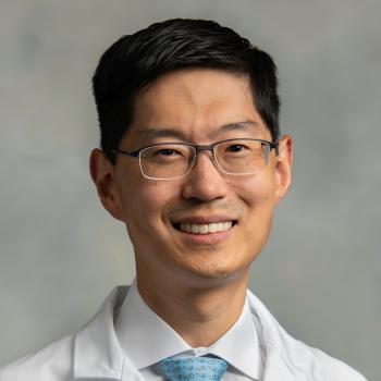 Michael Lim合作论文数School of Medicine, Johns Hopkins University;School of Medicine, Stanford University5
 Stuart Grossman合作论文数Johns Hopkins Sidney Kimmel Comprehensive Cancer Center;The Johns Hopkins Hospita5Spencer C Liang合作论文数4
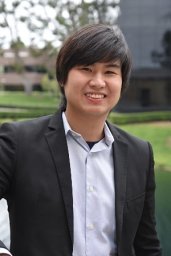 Liam Chung合作论文数4
 Madeline Fryer合作论文数4Ling Leung合作论文数4
 Byoung Chol Oh合作论文数4
 Joscelyn C Mejías合作论文数Johns Hopkins University4
 Giorgio Raimondi合作论文数Department of Otolaryngology-Head and Neck Surgery, School of Medicine, Johns Hopkins University4




