PURPOSE:Neurofibromatosis type 2 (NF2) is a tumor predisposition syndrome characterized by bilateral vestibular schwannomas (VSs) resulting in deafness and brainstem compression. This study evaluated efficacy and biomarkers of bevacizumab activity for NF2-associated progressive and symptomatic VSs.PATIENTS AND METHODS:Bevacizumab 7.5 mg/kg was administered every 3 weeks for 46 weeks, followed by 24 weeks of surveillance after treatment with the drug. The primary end point was hearing response defined by word recognition score (WRS). Secondary end points included toxicity, tolerability, imaging response using volumetric magnetic resonance imaging analysis, durability of response, and imaging and blood biomarkers.RESULTS:Fourteen patients (estimated to yield > 90% power to detect an alternative response rate of 50% at alpha level of 0.05) with NF2, with a median age of 30 years (range, 14 to 79 years) and progressive hearing loss in the target ear (median baseline WRS, 60%; range 13% to 82%), were enrolled. The primary end point, confirmed hearing response (improvement maintained ≥ 3 months), occurred in five (36%) of 14 patients (95% CI, 13% to 65%; P < .001). Eight (57%) of 14 patients had transient hearing improvement above the 95% CI for WRS. No patients experienced hearing decline. Radiographic response was seen in six (43%) of 14 target VSs. Three grade 3 adverse events, hypertension (n = 2) and immune-mediated thrombocytopenic purpura (n = 1), were possibly related to bevacizumab. Bevacizumab treatment was associated with decreased free vascular endothelial growth factor (not bound to bevacizumab) and increased placental growth factor in plasma. Hearing responses were inversely associated with baseline plasma hepatocyte growth factor (P = .019). Imaging responses were associated with high baseline tumor vessel permeability and elevated blood levels of vascular endothelial growth factor D and stromal cell-derived factor 1α (P = .037 and .025, respectively).CONCLUSION:Bevacizumab treatment resulted in durable hearing response in 36% of patients with NF2 and confirmed progressive VS-associated hearing loss. Imaging and plasma biomarkers showed promising associations with response that should be validated in larger studies.
Background:Salvage therapies for adults with recurrent ependymoma are limited. A prior retrospective review of patients with recurrent ependymomas treated with bevacizumab and carboplatin reported a 75% radiographic response rate. This prospective single-arm, open-label Phase 2 study was designed to assess clinical efficacy of this regimen. Methods:Twenty-two patients were evaluated in this CERN Adult Clinical Trials network study. Adult patients (age ≥18) with recurrent ependymomas received carboplatin (AUC = 5-6) every 4 weeks and bevacizumab 10mg/kg every 2 weeks for 6 cycles, after which carboplatin was discontinued, while bevacizumab could be continued at physician's discretion. Imaging of areas involved and patient-reported outcomes (PRO) with the MD Anderson Symptom Inventory (brain and/or spine modules) were assessed at baseline and every 2 cycles. Results:With a median follow-up time of 25.9 months (mo), the primary endpoint of 12-mo progression-free survival rate (PFS-12) greater than 50% was reached, with a rate of 76.4% (95% CI, 52.2, 89.4). The median PFS of this cohort was 18.0 mo. Two patients achieved objective partial responses (9.1%). There were no treatment-related grade ≥4 toxicities. Brain tumor responders (radiographic objective response or stable disease) experienced improved cognitive and neurological symptoms, while spine tumor patients reported worsening symptom outcomes regardless of response. Conclusions:Treatment with carboplatin and bevacizumab in adult recurrent ependymomas met the PFS-12 clinical efficacy endpoint. However, symptomatic worsening in spinal tumors suggests imaging stability and symptom improvement in brain disease may be related to bevacizumab pseudo-response.
Precision oncology lacks scalable tools to assess, at the patient level, systems-level tumor microenvironment (TME) programs driving therapeutic resistance. To address this gap, we trained a weakly-supervised deep learning model, using routine H&E slides as input, to derive quantitative activity for therapeutically-relevant TME phenotypes, spanning immune, metabolic, and tumor cell-intrinsic programs. Using 3111 breast cancer H&E WSIs with matched bulk transcriptomics, our model accurately infers these biological states (AUROC>0.80; PCC>0.64). Validation spanned three levels: (i) tissue-matched multiplexed immunofluorescence, showing concordance between inferred functional states and immune cell fractions (p=0.006-0.106), (ii) blinded reader assessments, confirming localization of phenotype-specific morphology (p<3×10⁻⁵), and (iii) multi-institutional patient cohorts, where model-derived phenotypes stratified for clinical response (p<0.045). Despite relying on slide-level labels for training, our model's attention mechanism identifies focal regions of tumor tissue that drive the overarching clinical phenotype or treatment response. By extracting spatially resolved TME biology from routine histology, this strategy can be applied to massive legacy biobanks to enable discovery of new morpho-molecular mediators of resistance across real-world patient populations.
Glioblastoma (GBM) is a devastating primary brain tumor of adults with few treatment options. While there has been long-standing interest in engaging the immune system to combat this disease, monoclonal antibody therapies – including checkpoint modulating agents – have yet to result in widespread clinically meaningful outcomes. By contrast, adoptive cell therapies including chimeric antigen receptor (CAR)-T cell therapies have demonstrated promising early indications of efficacy in select GBM patients. However, heterogeneity of target molecule expression remains a significant barrier to long-term disease control. In a recent report, a first-in-human trial of CARv3-TEAM-E T cells engineered to target the epidermal growth factor receptor variant III tumor-specific antigen (EGFRvIII) as well as wild-type EGFR through secretion of a T-cell-engaging antibody molecule (TEAM) showed preliminary evidence of CAR activity. We now report the results of single cell RNA-sequencing (scRNA seq) of lymphocytes isolated from the cerebrospinal fluid (CSF) of CARv3-TEAM-E treated GBM patients. Longitudinal CSF sampling in these patients was performed via an Ommaya reservoir. Sampled cells were interrogated by scRNA Seq. We demonstrate the ability to detect transduced CARv3-TEAM-E expressing T cells in the CSF from these patients. Unsupervised analysis revealed evidence of the expression of cytotoxic effector gene expression programs, which we explore over the course of longitudinal sampling in this initial cohort. Additionally, we compare expression signatures in the CSF-sampled T cells to those present in the CARv3-TEAM-E infusion products. Taken together, our findings offer insight into the longitudinal dynamics of T cell gene expression programs in the setting of this first-in-human trial of CARv3-TEAM-E T cells in GBM patients. Christopher Mount, Demi Gerovasilis, Sophia Kovatsis, Jun Zhong, Md Raihan Chowdhury, Maxx King, William T. Curry, Elizabeth R. Gerstner, Kathleen M. Gallagher, Bryan D. Choi, Mario Suva, Marcela V. Maus. Single cell RNA sequencing of cerebrospinal fluid lymphocytes in CARv3 TEAM E treated glioblastoma patients [abstract]. In: Proceedings of the American Association for Cancer Research Annual Meeting 2025; Part 2 (Late-Breaking, Clinical Trial, and Invited Abstracts); 2025 Apr 25-30; Chicago, IL. Philadelphia (PA): AACR; Cancer Res 2025;85(8_Suppl_2):Abstract nr LB358.
2008 Background: Chimeric Antigen Receptor (CAR) T cells for glioblastoma (GBM) have been limited by the challenge of targeting a single tumor antigen in a heterogeneous disease. To address this barrier, we generated a novel engineered T-cell product (CARv3-TEAM-E) that targets the EGFRvIII antigen while also secreting T-cell-Engaging Antibody Molecules (TEAMs) against wild-type EGFR. Methods: The INCIPIENT clinical trial is a first-in-human study of CARv3-TEAM-E in patients with recurrent GBM (NCT05660369). Patients were treated with intraventricular CARv3-TEAM-E T cells (10E6 cells per infusion). A subset of patients were conditioned with lymphodepleting chemotherapy (LDC) consisting of cyclophosphamide and fludarabine. Immune cells were profiled in the cerebrospinal fluid (CSF) and peripheral blood of patients by flow cytometry. Results: CAR T cells were detected in the CSF of all patients for an average of 33.6 days ( SD = 10.33). Granulocytes, NK cells, B cells, and monocytes appeared in the CSF immediately after infusion, decreasing to low levels over the course of several weeks. TEAM-positive T cells persisted in CSF until (median) day 33.6 ( SD = 10.8) with a range of 21-56 days. CAR T cells were transiently detected in the peripheral blood of 9/10 patients at an average of 14 days ( SD = 3.5) after infusion. Prior to infusion, CAR T cells were predominantly CD4-positive and remained as such in the CSF over time. Those in the periphery exhibited CD4-to-CD8 polarization. Of patients who received multiple infusions, 3 out of 6 had CAR-positive T cells in the CSF after a second infusion, although their persistence was short-lived and was not detected in the periphery following repeat infusions. LDC increased engraftment of CAR T cells in CSF but not in peripheral blood. Patients with poor CAR persistence demonstrated the development of anti-CARv3-TEAM-E antibodies in the CSF and serum, which increased with reinfusion. Conclusions: Following initial infusion, intraventricularly delivered CARv3-TEAM-E T cells were detected in the CSF and peripheral blood in patients with recurrent GBM. Reduced persistence was observed with subsequent infusions. This corresponded with the emergence of anti-CARv3-TEAM-E antibodies in treated patients. Clinical trial information: NCT05660369 .
Brain metastases (BM) are an emerging challenge in modern oncology due to increasing incidence and limited treatments. Recent work by our group and others has illustrated that immune checkpoint inhibitors (ICI) are a promising therapy for treatment-refractory BM of diverse histologies. To build upon our findings and integrate ICI into precision medicine strategies for BM, scalable tools that quantify likelihood of response are needed. We curated a multi-institutional dataset of longitudinal mpMRI for 860 BM patients treated with ICI, assessing 2,542 BM responses based on pre- and 6-month post-treatment MRIs using standardized RANO criteria. A convolutional neural network (CNN) trained on pre-treatment mpMRI sequences predicted 6-month ICI efficacy. To ensure class balance and clinical relevance, four RANO classes were converted into a binary model: intracranial benefit including complete response (CR), partial response (PR) and stable disease (SD) vs. progressive disease (PD). Our custom CNN consists of three convolutional and two fully connected layers, with an 80/20 train-validation split. We also developed a "foundation model" for brain metastases using self-supervised contrastive learning on 21,179 BM private and public mpMRIs from more than 15,000 patients with a 3D ResNet50 architecture. The model was fine-tuned for ICI efficacy prediction using the same dataset that trained the CNN, held out during pretraining. A logistic regression model was trained on features extracted from the foundation model. Our CNN achieved an AUROC score of 0.650 on the validation set. Fine-tuning the foundation model with a linear classifier improved AUROC to 0.7904. Ongoing work includes optimizing models, developing multi-class response models (e.g., CR/PR vs. SD vs. PD), and conducting histology-specific analyses. Our study presents one of the first Deep Learning-based efforts to predict ICI efficacy for BM. There is emerging promise in using Deep Learning to identify imaging patterns of biological significance within clinically acquired imaging.
2017 Background: CAR T therapy is a novel, promising approach in glioblastoma (GBM) but tumor heterogeneity can limit efficacy when a single antigen is targeted. We designed a second-generation CAR T molecule that targets epidermal growth factor receptor vIII (EGFRvIII) and also secretes a T-cell–engaging antibody molecule (TEAM) against wild-type EGFR. Methods: In a phase 1, first-in-human study (INCIPIENT, NCT05660369), patients with recurrent GBM with EGFRvIII mutation and/or EGFR amplification were eligible to receive up to 6 intraventricular doses of 10x106 CAR T cells via Ommaya catheter after lymphodepleting chemotherapy (LDC) with fludarabine and cyclophosphamide. Primary objective was safety and tolerability and secondary objective was preliminary tumor response determined by iRANO criteria. Results: CAR T manufacturing was successful for all patients. Seven patients (5 male) received at least 1 intraventricular infusion. Two patients received 2 infusions (1 for progressive disease (PD) and 1 without PD). One patient received 3 infusions after experiencing initial PD. No DLTs occurred. All patients experienced cytokine release syndrome (CRS) grade 1 lasting 0-9 days with only 1 patient experiencing CRS grade 2 for 1 day. One patient experienced ICANS grade 1 that lasted 2 days. All patients experienced tumor inflammation-associated neurotoxicity grade 1 with a duration of 2-9 days. Adverse events (grade 3-4) at least possibly related to CAR T were febrile neutropenia (N = 1) and neutrophil count decrease (N = 1). Toxicity was managed with supportive care without need for ICU monitoring and 3 patients received at least 1 dose of anakinra (max duration = 4 days, median = 1 day). Best response was stable disease (SD) in 5 patients with 1 patient achieving SD for 6 months after a single infusion and another experiencing a 33% decrease in tumor diameter after 2 infusions. All patients are alive 3-8 months after first infusion. From the preceding safety run-in arm of the study (without LDC), one patient survived 12 months and another is still alive > 20 months after infusion. Conclusions: Intraventricular CARv3-TEAM-E infusions were well tolerated, even with multiple doses, and no DLTs were noted. Toxicity was manageable in all patients with supportive care and anakinra was administered to 3 patients. Steroids were not required to manage toxicity. A subset of patients experienced SD for several months. Clinical trial information: NCT05660369 .
Background Fibroblast growth factor receptor (FGFR) alterations are potential oncogenic drivers that occur in a subset of patients with gliomas, but the natural history of these tumors is not clearly defined. An understanding of outcomes of FGFR-driven glioma has implications for targeted drug development for FGFR inhibitors, which are approved for other cancers.Methods We performed a retrospective cohort study of patients with gliomas who harbored FGFR alterations, including fusions, single nucleotide variant (SNV), and copy number variant (CNV) alterations seen at Massachusetts General Hospital between 2003 and 2023. The electronic medical record was searched to identify additional molecular data, treatment, and MRI scans. Kaplan-Meier analysis was used to assess progression-free and overall survival (OS).Results Thirty-one patients with glioblastoma (GBM), diffuse astrocytoma, and glioneuronal tumors with FGFR alterations were identified: 17 with FGFR fusions, 10 with SNV, and 4 with CNV. FGFR3-TACC3 was the most common fusion in patients with GBM or diffuse astrocytoma. Median OS in patients with GBM was 2.75 years, despite 55% of tumors having an unmethylated MGMT promoter. There were no clearly co-occurring mutations with an FGFR alteration. Of 7 patients who underwent subsequent surgery, 6 lost the original FGFR alteration. No patient received an FGFR inhibitor.Conclusions While FGFR alterations are rare in glioma, patients with FGFR-altered GBM may have prolonged survival, which has implications for clinical trial design. We found loss of FGFR alteration at the time of subsequent surgery, raising concern for the therapeutic potential of FGFR-targeting agents in recurrent gliomas.
Salvage therapies for adults with recurrent ependymoma are limited. A prior retrospective review of patients with recurrent ependymoma treated with bevacizumab and carboplatin reported a 75% radiographic response rate. This prospective single arm, open label Phase 2 study was designed to confirm these results. 22 patients were evaluated in this CERN Adult Clinical Trials network study. Adult patients (age≥18) with recurrent ependymoma were treated with both carboplatin (AUC = 5 -6) every 4 weeks and bevacizumab 10mg/kg every 2 weeks for 6 cycles, after which carboplatin was discontinued, and bevacizumab could be continued at physician’s discretion. Imaging of areas involved by tumor, and patient reported outcomes with the MD Anderson Symptom Inventory questionnaires (brain and/or spine modules) were assessed at baseline and every 2 cycles to evaluate response and symptom burden, respectively. With a median follow-up time of 25.9 months, the primary end point of 12-month progression free survival rate (PFS-12) greater than 50% was reached with a rate of 76.4% (95% CI, 52.2, 89.4). The median PFS of this cohort was 18.0 months. Two patients achieved a partial response (9.1%). There were no treatment-related grade 4 toxicities. Improved cognitive and neurological symptoms were observed in brain tumor responders, while spine tumor patients had worsening symptom outcomes regardless of response. Treatment with carboplatin and bevacizumab in adult recurrent ependymoma reached the PFS-12 clinical efficacy threshold, suggesting that this treatment has efficacy in this patient population. However, symptomatic worsening in patients with spinal cord disease does raise concerns that bevacizumab pseudoresponse may account for some imaging stability and symptom improvement with brain disease.
Craniopharyngiomas are a rare tumor along the pituitary-hypothalamic axis. Genetic analysis revealed that 95% of papillary craniopharyngiomas (PCP) have BRAF V600E mutations (Brastianos et al. Nature Genetics 2014). We previously reported that BRAF/MEK inhibition was associated with significant anti-tumor activity in newly-diagnosed PCP patients (Brastianos et al. NEJM 2023). We have now evaluated the efficacy of vemurafenib/cobimetinib in a separate cohort of patients with recurrent PCP whose tumors progressed after prior radiation. In this multicenter, NCI-sponsored cooperative group trial, patients with histologically confirmed BRAF-positive PCP who progressed after radiation were treated with vemurafenib/cobimetinib. The primary endpoint of response rate (RR) based on centrally determined imaging data was to be evaluated in 16 patients, where a partial response was defined as ≥20% decrease in volume. This single arm, Simon two-stage phase 2 trial had 89% power to detect a true RR ≥ 30% (vs. null 5%; α=0.04). In this design, 3 or more observed responses in 16 evaluable patients would be considered evidence of promising activity. Of the planned 16 patients, a total of 5 patients enrolled before the study was closed due to slow accrual. Median follow-up was 50 months (95% CI: 32 to not yet reached). Based on volumetric response criteria, all 5 patients had a response to treatment (100%; 95% CI: 47.8% to 100%). To date, 2 patients progressed, one of whom died 22 months after study entry. This cohort met the criteria for efficacy under the original design with >3 responders. No grade 4 or 5 toxicities were observed. Vemurafenib/cobimetinib resulted in an objective response in all patients with previously treated PCP. The study met primary endpoint, similar to that reported for previously untreated PCP. Thus, BRAF/MEK inhibitors could be an effective tool for previously treated PCP and warrants further evaluation. U10CA180821; U10CA180882; Genentech, https://acknowledgments.alliancefound.org;ClinicalTrials.gov: NCT03224767
BACKGROUND:Meningioma is the most common primary CNS tumor, with high-grade cases exhibiting aggressive behavior, frequent recurrence, and poor prognosis. Currently, no systemic therapies are approved for recurrent or malignant meningiomas. Chimeric antigen receptor (CAR) T-cell therapy has shown efficacy in hematologic malignancies and promise for solid tumors, but its use for meningiomas has been underexplored. Mesothelin, a glycoprotein overexpressed in several solid tumors of mesodermal origin, may serve as a viable immunotherapeutic target. This study aimed to evaluate mesothelin as a CAR T-cell target in meningiomas. METHODS:Mesothelin expression was analyzed in patient-derived meningioma samples using immunohistochemistry, flow cytometry, and droplet digital PCR. Mesothelin-specific CAR T-cells were generated and evaluated invitro, exvivo using patient-derived organotypic tumor spheroids (PDOTS), and invivo using orthotopic meningioma mouse models of human xenografts. Cytotoxicity, T-cell proliferation, cytokine secretion, and tumor clearance were assessed. RESULTS:Mesothelin was detected in a subset of tumors across all meningioma grades at the transcript and protein levels, with surface expression confirmed in patient-derived primary cells. Mesothelin-specific CAR T-cells exhibited potent and specific cytotoxicity, T-cell activation, and cytokine secretion in vitro and effectively eliminated PDOTS. In orthotopic human xenograft models, mesothelin CAR T-cell therapy led to significant tumor regression and prolonged survival. CONCLUSIONS:Mesothelin is a viable CAR T-cell target for meningiomas, and mesothelin-specific CAR T-cell therapy shows strong preclinical efficacy. These findings provide a rationale for early-phase clinical trials of mesothelin CAR T-cell therapy in patients with refractory meningiomas.
Background:Cancer morbidity disproportionately affects patients in low- and middle-income countries (LMICs), where timely and accurate tumor profiling is often nonexistent. Immunohistochemistry-based assessment of estrogen receptor (ER) status, a critical step to guide use of endocrine therapy (ET) in breast cancer, is often delayed or unavailable. As a result, ET is often prescribed empirically, leading to ineffective and toxic treatment for ER-negative patients. To address this unmet need, we developed ESPWA (Estrogen Receptor Status Prediction for Haitian patients using deep learning-enabled histopathology Whole Slide Imaging Analysis), a deep-learning (DL) model that predicts ER status directly from hematoxylin-and-eosin (H&E)-stained whole slide images (WSIs). Methods:We curated two cohorts of H&E WSIs with tissue-matched ER status: The Cancer Genome Atlas (TCGA, n = 1085) and Zanmi Lasante (ZL, n = 3448) from Haiti. We trained two models using weakly supervised attention-based multiple instance learning: a "TCGA" model, trained on TCGA data, and ESPWA, trained on the ZL dataset. Model performance was evaluated using 10-fold cross validation. Results:Performance of the "TCGA" model was sensitive to the domain shift between the TCGA and ZL datasets, with a performance of an area under receiver operating characteristic (AUROC) of 0.846 on the TCGA test sets and 0.671 on the ZL test sets. Compared to the "TCGA" model, ESPWA demonstrated improved performance on the ZL cohort (AUROC=0.790; p=0.005). Subgroup analyses revealed clinically relevant populations in which ESPWA demonstrated improved performance relative to the overall cohort. Finally, ESPWA outperformed an academic breast pathologist (accuracy: 0.726 vs 0.639 respectively; p <0.001) in determining ER status from H&E WSIs. Conclusion:ESPWA ("Hope" in Haitian Creole) offers an accessible framework to identify individualized therapeutic insights from H&E WSIs in LMICs. We have initiated clinical trials, using ESPWA, in ZL and sub-Saharan African countries to inform precision-based use of ET for prospective patients.
Background:Mutant isocitrate dehydrogenase (IDHm) inhibitors represent a novel targeted approach for treating IDHm glioma patients, yet their optimal use in clinical practice outside of clinical trials remains undefined. This study describes the real-world utilization of the mutant IDH1 inhibitor (IDHi), ivosidenib, in patients with IDHm glioma. Methods:We retrospectively reviewed clinical and radiographic data from patients with IDHm glioma treated with ivosidenib monotherapy from 2020 to 2024 at the Dana-Farber Cancer Institute and Massachusetts General Hospital. Results:This cohort included 74 patients with a median age of 39. There were 35 astrocytomas and 39 oligodendrogliomas, with 49, 23, and 2, grade 2, 3, and 4 tumors, respectively. Nineteen patients (26%) experienced an adverse event, although only 1 patient discontinued ivosidenib for adverse events. Median progression-free survival was 31 months and median overall survival was not reached. Seven patients (9%) had partial response, 3 (4%) had minor response, 47 (64%) had stable disease, and 17 (23%) had progressive disease. The presence of enhancing disease at ivosidenib initiation was associated with lower disease control rates (DCR) whereas DCR differences were not detected based on grade (grade 2 vs. 3), tumor histology, or age. Subsequent-line ivosidenib use had lower DCR although this may have been explained by enrichment of patients with enhancing disease. Conclusions:In this large cohort of IDHm glioma patients, ivosidenib was well tolerated. Our results support the use of IDHi therapy in patients with grade 2 or 3 astrocytoma or oligodendroglioma and highlight limited effectiveness in patients with enhancing disease.
Background Fibroblast growth factor receptor (FGFR) alterations are potential oncogenic drivers that occur in a subset of patients with gliomas, but the natural history of these tumors is not clearly defined. An understanding of outcomes of FGFR-driven glioma has implications for targeted drug development for FGFR inhibitors, which are approved for other cancers. Methods We performed a retrospective cohort study of patients with gliomas who harbored FGFR alterations including fusions, single nucleotide variant (SNV), and copy number variant (CNV) alterations seen at MGH between 2003 and 2023. The electronic medical record was searched to identify additional molecular data, treatment, and MRI scans. Kaplan-Meier analysis was used to assess progression free and overall survival (OS). Results Thirty-one patients with glioblastoma (GBM), diffuse astrocytoma, and glioneuronal tumors with FGFR alterations were identified: 17 with FGFR fusions, 10 with SNV, and 4 with CNV. FGFR3-TACC3 was the most common fusion in patients with GBM or diffuse astrocytoma. Median OS in patients with GBM was 2.75 years despite 55% of tumors having an unmethylated MGMT promoter. There were no clearly co-occurring mutations with an FGFR alteration. Six of 7 patients who underwent subsequent surgery lost the original FGFR alteration. No patient received an FGFR inhibitor. Conclusions While FGFR alterations are rare in glioma, patients with FGFR altered GBM may have prolonged survival which has implications for clinical trial design. We found loss of FGFR alteration at the time of subsequent surgery, raising concern for the therapeutic potential of FGFR targeting agents in recurrent gliomas.
Recent progress in deep learning (DL) is producing a new generation of tools across numerous clinical applications. Within the analysis of brain tumors in magnetic resonance imaging, DL finds applications in tumor segmentation, quantification, and classification. It facilitates objective and reproducible measurements crucial for diagnosis, treatment planning, and disease monitoring. Furthermore, it holds the potential to pave the way for personalized medicine through the prediction of tumor type, grade, genetic mutations, and patient survival outcomes. In this review, we explore the transformative potential of DL for brain tumor care and discuss existing applications, limitations, and future directions and opportunities.
INTRODUCTION: Chimeric antigen receptor (CAR) T cells represent a promising approach to cancer and have proven efficacy against hematological malignancies, for which they have become the standard of care. However, the use of CAR T cells in solid tumors has been limited. METHODS: This is a nonrandomized, open-label, single-site Phase I clinical trial. Three patients with EGFRvIII-positive recurrent glioblastoma were enrolled in a safety run-in cohort. Patients were treated with 10 million CARv3-TEAM-E T cells and monitored for toxic effects. Cerebrospinal fluid (CSF) and blood were sampled longitudinally and subjected to correlative analyses. RESULTS: No dose-limiting toxic effects were noted. Radiographic tumor regression occurred in all three patients within days after treatment, but this response was transient in two of the patients. Tumor regression correlated with decreased detection of antigen-specific RNA derived from extracellular vesicles (EVs) in both CSF and peripheral blood. CONCLUSIONS: Early data suggest safety and anti-tumor activity of CARv3-TEAM-E T cells in recurrent glioblastoma. EV-based liquid biopsy may assist in monitoring response to cell therapy. Ongoing enrollment has been modified to enhance durability using lymphodepletive chemotherapy. Additional arms will evaluate this approach in the setting of EGFRvIII-negative tumors and newly-diagnosed disease.
Excess D-2-hydroxyglutarate (2-HG) produced by mutant IDH enzymes hampers the ability of gliomas to repair DNA damage. We conducted a surgical window-of-opportunity trial of the selective PARP1/2 inhibitor niraparib in recurrent IDH-mutant glioma to assess tumor drug penetration and pharmacodynamics. Subjects with progressive, surgically accessible IDH-mutant glioma were randomized 1:1 to receive niraparib or no treatment for 4 weeks prior to tumor resection. Following surgery, all patients received niraparib (300 or 200 mg daily) until disease progression. Enhancing and non-enhancing tumor tissue were collected at surgery. Drug concentration and 2-HG levels were measured with MALDI-MSI. The primary objective was to measure intratumoral drug concentration. Secondary objectives included safety, target engagement and anti-tumor effect. Seven patients (F=0%, M=100%) with a median age of 37 years (range 23-47 years) were enrolled; three received pre-operative niraparib. The mean intratumoral niraparib concentration was 19.0 mM (n=5 samples, 3 patients), with 30.8 mM in enhancing (n=2) and 16.4 mM in non-enhancing (n=3) tissue. Mean 2-HG concentration was 2.3 mM (n=9 samples from 6 patients; range 0.3 – 7.9), 2-HG was below the level of detection in one patient who was subsequently found to not have active tumor at surgery. No correlation was found between 2HG level and receipt of pre-operative niraparib. Grade ≥ 3 hematologic toxicities occurred in 2/6 patients, including one grade 4 lymphopenia and one grade 3 anemia. Other treatment-related adverse events were grade 1–2. Median PFS and OS were 1.94 (95 % CI, 1.0–2.7) and 15.5 months (95% CI of 4 - NA), respectively. Niraparib was generally well tolerated but demonstrated limited anti-tumor activity in this patient population despite evidence of sufficient drug penetration in both enhancing and non-enhancing tumor. Results from ongoing spatial metabolomics and transcriptomics (Visium) to further evaluate pharmacodynamic effects will be presented. GSK provided funding and drug [NCT05406700].
 Tracy Batchelor合作论文数Harvard Medical School;Department of Neurology, Brigham and Women's Hospital;Cancer Center, Mass General Research Institute, Massachusetts General Hospital138
 Patrick Y. Wen合作论文数Department of Neurology, Harvard Medical School;Center For Neuro-Oncology, Dana-Farber Cancer Institute94
 Jayashree Kalpathy-Cramer合作论文数Harvard Medical School;Massachusetts General Hospital76
 Bruce Rosen合作论文数Harvard Medical School;Athinoula A. Martinos Center for Biomedical Imaging, Massachusetts General Hospital;Division of Health Sciences and Technology, Harvard-MIT60
 Daniel Cahill合作论文数Stephen E. and Catherine Pappas Center for Neuro-Oncology, Massachusetts General Hospital, Harvard Medical School, Harvard University;Department of Neurosurgery, Massachusetts General Hospital, Harvard Medical School, Harvard University60
 Jorg Dietrich合作论文数59
 Eudocia Q. Lee合作论文数Division of Cancer Neurology, Department of Neurology, Brigham and Women's Hospital, Harvard Medical School;Center for Neuro-Oncology, Dana-Farber Cancer Institute45
 Priscilla Brastianos合作论文数Division of Medical Sciences, Harvard Medical School42
 A. Gregory Sorensen合作论文数DeepHealth, Inc.;IMRIS - A Deerfield Imaging Brand37
 Rakesh K. Jain合作论文数Harvard Medical School, Harvard University;Edwin L. Steele Laboratories for Tumor Biology, Department of Radiation Oncology, Massachusetts General Hospital37
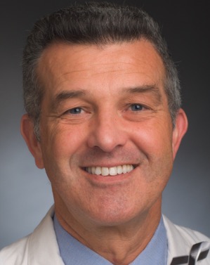 David Reardon合作论文数Center for Neuro-Oncology, Dana-Farber Cancer Institute;Harvard Medical School33
 William T Curry合作论文数32
 Scott Plotkin合作论文数The Stephen E. and Catherine Pappas Center for Neuro-Oncology, Massachusetts General Hospital, Harvard Medical School;Family Center for Neurofibromatosis, Massachusetts General Hospital, Harvard Medical School31
 Deborah Forst合作论文数29Arrillaga-Romany Isabel合作论文数28
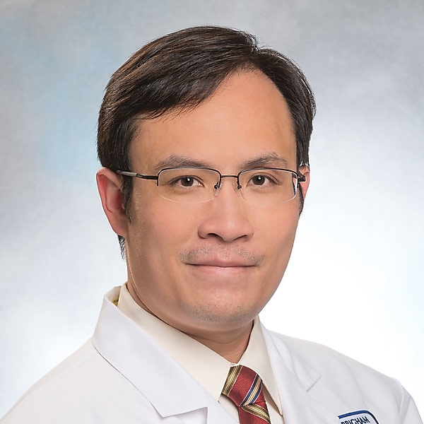 Raymond Y. Huang合作论文数Harvard Medical School;Department of Radiology, Brigham and Women's Hospital28Ugonma Chukwueke合作论文数28Andrew S. Chi合作论文数Laura and Isaac Perlmutter Cancer Center, NYU Langone Health/Department of Neurosurgery, NYU Langone Health27
 Kyrre Eeg Emblem合作论文数Department of Physics and Computational Radiology, Oslo University Hospital27Lakshmi Nayak合作论文数27
 Ken Chang合作论文数Stanford Health Care25
 Helen Shih合作论文数MGH Proton Therapy Centers, Massachusetts General Hospital;Harvard Medical School25
 April F Eichler合作论文数25
 Benjamin Ellingson合作论文数University of California Los Angeles24Kim Albert合作论文数22
 Evanthia Galanis合作论文数Division of Medical Oncology, Department of Oncology, Mayo Clinic;Mayo Clinic Comprehensive Cancer Center, Mayo Clinic21
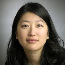 Nancy U. Lin合作论文数Department of Medical Oncology, Dana-Farber Cancer Institute;Harvard Medical School20
 Alona Muzikansky合作论文数Biostatistics Center, Massachusetts General Hospital20
 Justin Jordan合作论文数Department of Neurology, Massachusetts General Hospital, Boston, Massachusetts20
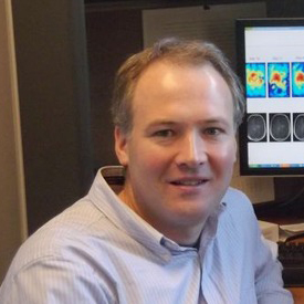 Ovidiu Andronesi合作论文数Harvard Medical School, Harvard University;Athinoula A. Martinos Center for Biomedical Imaging, Mass General Research Institute, Massachusetts General Hospital, Mass General Brigham20
 Dan G. Duda合作论文数Houston Methodist Research Institute19
 Timothy Cloughesy合作论文数Department of Molecular and Medical Pharmacology, David Geffen School of Medicine, University of California, Los Angeles;Brain Research Institute, University of California, Los Angeles19
 Wenya Linda Bi合作论文数Department of Neurosurgery, Brigham and Women's Hospital;Harvard Medical School18
 Kevin Oh合作论文数Massachusetts General Hospital;Harvard Medical School18A. Giobbie-Hurder合作论文数Cooperative Studies Program Coordinating Center, Hines, IL18
 Anthony Iafrate合作论文数Department of Pathology, Massachusetts General Hospital Cancer Center;Harvard Medical School17
 Ryan Sullivan合作论文数Massachusetts General Hospital17Maria Martinez-Lage合作论文数17
 Marco Pinho合作论文数Peter O'Donnell Jr. Brain Institute, University of Texas Southwestern Medical Center17
 Andrew Beers合作论文数Massachusetts Gen Hosp, Athinoula A Martinos Ctr Biomed Imaging, Charlestown, MA USA16
 Sandro Santagata合作论文数Laboratory of Systems Pharmacology, Harvard Medical School;Department of Pathology, Brigham and Women's Hospital;Dana-Farber Cancer Institute16Ina Ly合作论文数15
 Timothy J Kaufmann合作论文数15
 Bryan D. Choi合作论文数Department of Surgery, Duke University Medical Center15
 Jan Drappatz合作论文数Center for Neuro-Oncology, Department of Medical Oncology, Dana-Farber/Brigham and Women’s Cancer Center, 44 Binney St, Boston, MA 02115, USA15
 Gilbert Youssef合作论文数Dana-Farber/Brigham and Women's Cancer Center, Harvard Medical School14
 Christopher P. Bridge合作论文数Univ Oxford, Inst Biomed Engn, Oxford, England14
 Justine V. Cohen合作论文数Division of Hematology and Oncology, Department of Medicine, Perelman School of Medicine, University of Pennsylvania14Catana Ciprian合作论文数14




