Across cancer, one of the most frequent examples of histologic transformation is the evolution of follicular lymphoma (FL) to an aggressive large cell lymphoma. Despite recent progress, understanding of the molecular and cellular underpinnings of transformation remains incomplete. Here, we dissect the interplay of tumor and microenvironment cell populations across transformation through a multimodal investigation of 95 FL and transformed FL (tFL) samples, including single-cell and bulk RNA-sequencing alongside spatial transcriptomics and proteomics, and validate findings across independent FL-tFL pairs. Upon transformation, fibroblasts and GPNMB+ macrophages increase while lymph-node organizing follicular dendritic and CCL21+ fibroblastic reticular cells were lost, resulting in an altered spatial distribution of cytokines that impacts T cell infiltration and macrophage differentiation and function. Secreted stromal and macrophage signals were further evident by non-invasive plasma proteomics. Taken together, our data reveal expansion of macrophages and fibroblasts as key features of transformation with potential diagnostic and therapeutic implications.
ABSTRACT:Synchronous systemic and de novo secondary central nervous system (CNS) large B-cell lymphoma (LBCL) is an aggressive clinical entity with a historically poor prognosis. Given the rarity of this presentation, prospective studies are limited, and treatment paradigms and outcomes are extrapolated from small, heterogenous retrospective studies. We performed a retrospective study with extended follow-up for 63 consecutive patients with previously untreated synchronous systemic and de novo secondary CNS LBCLs presenting to 2 institutions over 21 years. Most patients had diffuse LBCL (73%) and were treated with an average of 6 cycles of R-CHOP (rituximab, cyclophosphamide, doxorubicin, Oncovin [vincristine], and prednisone) intercalated with high-dose IV methotrexate (R-CHOP-M). The overall response rate was 84% (75% complete). With a median follow-up of 8.1 years, the median progression-free survival (PFS) was 1.2 years, and the 6-year PFS rate was 37%. The median overall survival (OS) was 7.9 years, and the 6-year OS rate was 52%. Normal lactate dehydrogenase (LDH), low International Prognostic Index (IPI) score, and brain parenchymal-only CNS disease were associated with improved PFS, whereas normal LDH and parenchymal disease were associated with OS. The 25% of patients who underwent consolidation with high-dose chemotherapy (HDC) and autologous stem cell transplant (ASCT) had superior PFS and OS than patients who did not receive a transplant, with particular benefit in those with IPI score of ≥3. This study demonstrates that a proportion of patients presenting with secondary CNS involvement are cured with upfront chemoimmunotherapy with or without HDC/ASCT and helps identify prognostically favorable subgroups, which can guide counseling of patients with this rare, high-risk clinical presentation.
ABSTRACT:Langerhans cell histiocytosis (LCH) and non-Langerhans cell (non-LC) histiocytosis are rare neoplastic diseases with variable behavior. Although chemotherapy is often effective, some patients respond poorly to treatment, experience disease recurrence and/or develop long-term complications. Clofarabine (Clolar), a deoxyadenosine analog, has shown encouraging activity in histiocytic disorders. Here, we report a prospective, multicenter, phase 2 study evaluating the efficacy and toxicity of clofarabine in recurrent/refractory (R/R) LCH (stratum 1) and non-LC histiocytosis (stratum 2). All participants received clofarabine on days 1 through 5 of each 28-day cycle. Response was assessed after 2 cycles; participants without evidence of disease progression received 4 additional cycles. A total of 25 participants enrolled: 20 in stratum 1 and 5 in stratum 2. In stratum 1, 17 of 20 participants (85%; 95% confidence interval [CI], 62%-97%) were responders, and 3 had stable disease after cycle 2. Overall, 19 stratum-1 participants completed 6 cycles; 1 withdrew after cycle 3. No stratum-1 participants progressed during treatment and 3 had disease recurrence after cycle 6, resulting in 2-year progression-free survival ± standard error and overall survival of 89% ± 7% and 100%, respectively (n = 20; median follow-up, 40.9 months [range, 2.7-49.4]). In stratum 2, 3 of 4 evaluable participants (75%; 95% CI, 19%-99%) had partial response. Overall, 20 of 25 (80%) of participants had ≥1 grade ≥3 toxicity attributed to protocol therapy. In conclusion, clofarabine is active and tolerable in children with R/R LCH and warrants further study in patients with non-LC histiocytosis. Further study of clofarabine in these populations is warranted. This trial was registered at www.clinicaltrials.gov (#NCT02425904).
We previously demonstrated that relapsed and refractory nodal-mature-T-cell-lymphomas (nMTCL) have distinct prognoses. Here, we assessed the overall survival (OS) impact of time-to-relapse (TTR) in multinational PETAL/LATAM cohorts with validation using observational and randomized independent cohorts. Patients with nMTCL with complete response to frontline treatment were assigned to TTR12 (<12m) or without TTR12 based on time-to-progression or time-to-next-therapy. OS was compared using modified landmark (m-LM) analysis. Sensitivity analyses included standard landmark (s-LM) and time-dependent Cox (td-Cox). Estimates were adjusted for age, histology, and Prognostic-Index-for-T-cell-lymphoma (PIT) score. Across 452 patients, 165 (36.5%) had TTR12, 181 (40%) relapsed ≥12m, and 106 (23.5%) remained relapse-free. TTR12 conferred worse OS using m-LM (HR 2.14; 95%CI: 1.58-2.90, p<0.001), s-LM (HR 1.92; 95%CI: 1.39-2.66, p<0.001), and td-Cox (HR 5.81; 95%CI: 2.94-11.46, p<0.001). Results were consistent in the independent validation cohorts with univariable and multivariable models. TTR12 consistently conferred worse OS irrespective of front-line hematopoietic stem-cell transplantation or PIT score, in peripheral-T-cell-lymphoma, not-otherwise-specified (m-LM: HR 2.32; 95%CI: 1.51-3.55, p<0.001; s-LM: HR 2.10; 95%CI: 1.33-3.31, p=0.001), anaplastic-large-cell-lymphoma (m-LM: HR 3.34; 95%CI: 1.18-9.50, p=0.023; s-LM: HR 2.96; 95%CI: 1.02-8.81, p=0.046), and angioimmunoblastic-T-cell/T-follicular-helper-cell-lymphoma (m-LM only: HR 1.92, 95%CI: 1.15-3.21; p=0.013). Second-line novel therapies improved OS (second-line start to death) versus chemotherapy in TTR12 (HR 0.60; 95%CI: 0.37-0.97, p=0.038) but not in patients without (HR 0.82; 95%CI: 0.51-1.32, p=0.407). TTR12 identified worse OS patients alongside, and compounding with, PIT score. TTR12 serves as a prognostic and potential OS surrogate marker, supporting stratification of new risk groups and need for their differential treatment.
Introduction: Diffuse large B-cell lymphoma (DLBCL) is a genomically heterogenous disease, which can be classified into distinct subgroups or clusters (Chapuy et al., Nat. Medicine, 2018, Wright et al., Cancer Cell, 2020). Leveraging knowledge of genetic alterations to design novel personalized treatment strategies may improve outcomes. Preclinical data suggest that the pan-phosphoinositide 3-kinase (PI3K) inhibitor, copanlisib, plus the BCL-2 inhibitor, venetoclax, are synergistic in genetically defined, high-risk, subtypes of DLBCL (Bojarczuk et al., Blood, 2019). We therefore hypothesized that copanlisib plus venetoclax would be an active treatment for patients with relapsed/refractory DLBCL, particularly in those with perturbed B-cell receptor (BCR)/PI3K signaling, which were predicted to be enriched in the relapsed setting. Here, we report the results of a phase I study of copanlisib plus venetoclax for patients (pts) with R/R DLBCL. Methods: This is an investigator-initiated, phase I, multicenter trial (NCT04572763) with the primary objective of determining the maximum tolerated dose (MTD) and recommended phase 2 dose (RP2D) of copanlisib and venetoclax in pts with R/R DLBCL. A standard 3+3 dose escalation design was employed, in which pts were treated with 60 mg IV copanlisib on days 1, 8, and 15 in addition to escalating doses of daily venetoclax for each 28-day cycle. During cycle 1, a venetoclax dose ramp-up was performed in the outpatient setting to a target dose of 200 mg (dose level (DL) +1), 400 mg (DL +2) and 800 mg (DL +3). The DLT observation period was the first cycle. Patients with progressive disease prior to the end of the DLT window were replaced. Key eligibility criteria included a confirmed diagnosis of DLBCL or high-grade B-cell lymphoma (HGBCL), relapse after or not a candidate for autologous stem cell transplant or chimeric antigen receptor (CAR) T-cell therapy, and adequate hematologic and organ function. Key exclusion criteria were poorly controlled hypertension or diabetes. CTCAE v5 and Lugano criteria were used to evaluate toxicity and efficacy, respectively. Results: As of July 21, 2023, 12 pts were evaluable. Patients were treated in DL +1 (n=5), DL +2 (n=3), and DL +3 (n=4). The median age was 62 years (range 24-87). Eighty three percent were male. Median number of prior treatments was 6 (range 1-11) and 10 pts had received prior CAR T-cell therapy. No DLTs were observed, though 2 pts in DL +3 were not able to escalate to 800 mg due to recurrent neutropenia and noncompliance, respectively. The most common toxicities were neutropenia (50%, all gr3/4), thrombocytopenia (42%, 8% ≥ gr3), nausea (33%, 8% ≥ gr3), fatigue (25%, 8% ≥ gr3), and headache (25%, 8% ≥ gr3) (Figure). Hyperglycemia occurred in 2 pts (both gr1). Serious adverse events occurred in 50% of pts and included fracture (n=1), hypoxia (n=2), and death (n=3), which were not related to study treatment. The deaths were related to progressive disease. Fifty percent of pts required drug holds (n=5 for venetoclax, n=5 for copanlisib). Sixty percent required growth factor support. There were no cases of laboratory or clinical tumor lysis syndrome. Sponsor support was withdrawn prior to completion of enrollment to DL+3, but DL+2 (copanlisib plus 400 mg venetoclax) was safe and well tolerated. The overall response rate (ORR) and complete metabolic response (CMR) rate was 8% (90% CI: 0 - 34%), as 1 pt in DL +1, who had received >5 lines of prior therapy, achieved a CMR, with response lasting 3 months. The median follow-up was 9.1 months. The median overall survival (OS) and progression-free survival (PFS) were 3.5 and 1.8 months, respectively. Conclusions: Copanlisib plus venetoclax is feasible and safe in pts with R/R DLBCL, with no DLTs observed. While it is possible that copanlisib plus venetoclax is active in select genomic subgroups of DLBCL, there was limited activity in this unselected, heavily pre-treated patient population.
Histiocytic sarcoma is an extremely rare malignancy typically presenting in extranodal sites, including the gastrointestinal tract, lung, nasal cavity, and soft tissue. Primary presentation in the skin is an even rarer entity, with only one prior case reported in the eyelid skin. Herein, we describe a case of histiocytic sarcoma that presented in the eyelid of an 80-year-old man. The lesion presented as a rapidly growing left lower eyelid lesion over 4 months. Shave biopsy revealed histologic and immunophenotypic findings most consistent with a morphologically low-grade histiocytic sarcoma. Complete excision was performed with an additional 2 mm peripheral and deep margins, which were found to be clear of malignancy. Systemic work-up and whole body imaging showed no evidence of metastases or other associated malignancy. The decision was made to forego radiation and proceed with close observation every 3 months. Two years from presentation, the patient had no evidence of recurrence.
PURPOSE:Peripheral T-cell lymphomas (PTCLs) are rare, heterogeneous, aggressive lymphomas. Five-year overall survival (OS) remains approximately 30%-40%, and most patients will develop relapsed or refractory (R/R) disease. Duvelisib is an oral dual inhibitor of phosphatidylinositol 3-kinase (PI3K)-δ and PI3K-γ isoforms. Here, we report on the final analysis of the phase II PRIMO trial (ClinicalTrials.gov identifier: NCT03372057; Secura Bio, Inc) evaluating duvelisib monotherapy in R/R PTCL. METHODS:PRIMO was conducted in two phases (dose optimization and dose expansion [PRIMO-EP]) at 45 centers globally. Eligible patients were age 18 years and older, had histologically confirmed diagnosis of PTCL, and had received ≥2 cycles of one standard regimen for PTCL. Based on dose optimization results, the selected regimen for PRIMO-EP was 75 mg twice a day for two cycles (to maximize disease control) followed by 25 mg twice a day (to reduce late toxicities), continued until progressive disease or unacceptable toxicity. RESULTS:PRIMO-EP (N = 123) outcomes included independent review committee-assessed objective response rate (ORR): 48.0%, complete response rate (CRR): 33.3%, median progression-free survival (mPFS): 3.4 months, median OS (mOS): 12.4 months, and median duration of response (mDOR): 7.9 months. In the angioimmunoblastic T-cell lymphoma (AITL) subgroup, outcomes were ORR: 62.2%, CRR: 51.4%, mPFS: 8.3 months, mOS: 18.1 months, and mDOR: 11.3 months. Treatment-emergent adverse events (TEAEs; any grade) occurred in 120 patients (97.6%), and TEAEs grade ≥3 occurred in 91 patients (74.0%). TEAEs resulting in dose hold or dose reduction occurred in 44.7% and 9.8% of patients, respectively. CONCLUSION:The PRIMO study demonstrates significant activity and tolerability of duvelisib in patients with R/R PTCL, most notably in the AITL subgroup. This provides strong rationale for further development in PTCL, and more specifically in the subgroup of nodal T-follicular helper cell lymphoma.
Lymphoplasmacytic lymphoma (LPL) is a lymphoproliferative neoplasm characterized by small plasmacytic cells infiltrating the bone marrow and is typically associated with IgM paraprotein detection. In rare cases, LPL may undergo histologic transformation (HT) to a more aggressive, large B cell lymphoma, such as diffuse large B-cell lymphoma (DLBCL). HT with cutaneous involvement is exceedingly rare. Here, we report a case of cutaneous involvement by a CD5+ CD10+ large B cell lymphoma in a post-transplant setting, initially attributed to follicular lymphoma but determined to be LPL with additional molecular testing. These findings highlight a potential diagnostic pitfall that was resolved by using molecular techniques in tandem with morphological observations.
2522 Background: Sézary syndrome (SS) is a rare and aggressive cutaneous T-cell lymphoma, which commonly expresses KIR3DL2, a killer immunoglobulin-like receptor, reported in ≥ 85% of patients. SS is characterized by erythroderma, significant blood involvement, lymphadenopathy and poor prognosis (10-20% 5-year survival). Lacutamab is a first-in-class monoclonal antibody designed to specifically deplete KIR3DL2-expressing cells via antibody-dependent cell-cytotoxicity and phagocytosis. Methods: TELLOMAK is an international, Phase 2 trial with multiple cohorts (NCT03902184). We report here long term follow-up results from Cohort 1, evaluating lacutamab in patients with relapsed/refractory (R/R) SS after at least 2 prior systemic therapies including mogamulizumab. Lacutamab 750 mg is administered until progression or unacceptable toxicity. Primary endpoint was Objective Response Rate (ORR) based on the evaluation of 4 compartments: skin, blood, lymph nodes and viscera according to the International Consensus criteria Olsen 2011. Secondary endpoints included but were not limited to duration of response (DOR), progression free survival (PFS), safety, and quality of life assessments. Results: As of October 17, 2024, recruitment was completed with 63 SS patients enrolled. Median age was 69 years (range: 42-86), the median prior lines of systemic therapies were 5.0 (range: 2-13), 65.1% and 34.9 % patients had stage IVA1 and stage IVA2 at baseline respectively, all patients had blood involvement (B2), 63.5% had confluence of erythema covering ≥ 80% body surface area (T4), 34.9% had lymph node lymphoma involvement (N3). Median follow-up was 25.1 months (95% CI 21.0-29.4). Global confirmed ORR was 42.9% (CI 31.4-55.1) including 6 (9.5%) CRs who are all still in CR; with a median time to response of 2.8 months (range 1-10) and a median duration of response of 25.6 months (CI 11.0, NE). According to each compartment, ORR in skin was 52.4% (CI 40.3-64.2) including 9 (14.3%) CRs, ORR in blood was 50.8% (CI 38.8-62.7) including 21 (33.3) CRs, and ORR in lymph nodes was 28.8% (CI 18.3-42.3) including 9 (17.3) CRs. Median PFS was 8.3 months (CI 5.1-18.7). Grade ≥ 3 related Treatment-Emergent Adverse Events (TEAEs) were observed in 20.6% patients. Serious related TEAEs were observed in 9.5% patients and related TEAEs leading to study drug discontinuation in 6.3% patients. Data from additional key endpoints will be presented. Conclusions: The long term follow-up data from TELLOMAK study in a R/R SS population previously treated with 2 or more prior systemic therapies including mogamulizumab, confirm that lacutamab shows promising clinical activity with ORR 42.9% (95% CI 31.4-55.1) and median duration of response of 25.6 months (11.0, NE) and an overall favourable safety profile. These data support the further development of lacutamab in an effort to bring improved treatments to patients with SS. Clinical trial information: NCT03902184 // EU CT number: 2023-507777-18-00.
ABSTRACT:Patients with relapsed or refractory (R/R) peripheral T-cell lymphomas (PTCL) require lineage-specific therapies to bridge to hematopoietic stem cell transplantation (HSCT). A previous phase 1/2 study of duvelisib/romidepsin (duv/romi) reported an overall response rate (ORR) of 58% and a complete response rate (CRR) of 42% with reduced grade 3 to 4 transaminitis (14%). We report real-world duv/romi outcomes in a multicenter, 38-patient R/R PTCL cohort. The median age at diagnosis was 62 years. Histological subtypes included nodal T follicular helper cell (nTFH; n = 17), PTCL-not otherwise specified (n = 14), cutaneous T-cell lymphoma (TCL; n = 3), extranodal natural killer/TCL (n = 1), ALK-negative anaplastic large cell lymphoma (n = 1), adult T-cell leukemia/lymphoma (n = 1), and hepatosplenic TCL (n = 1). The median previous therapy count was 1 (interquartile range [IQR], 1-2); 15 patients relapsed and 23 were refractory to prior treatment, including 8 prior HSCT (5 autologous, 3 allogeneic). After a median of 3 cycles (IQR, 2-4), ORR and CRR were 61% and 47%, respectively, with higher ORR (82% vs 43%) and CRR (71% vs 29%) in nTFH versus non-nTFH. The median progression-free survival and overall survival (HSCT-censored) were 11 and 16 months for nTFH, versus 3.3 and 8.3 months for non-nTFH. The median time to response was 1.9 months (IQR, 1.7-2.6), duration of response was 21 months, and time to next therapy was 17 months. After duv/romi, 11 patients bridged to allo-HSCT. Treatment was well tolerated; the most common grade 3 to 4 toxicities were lymphopenia (n = 15), neutropenia (n = 15), thrombocytopenia (n = 10), and transaminitis (n = 6), seldom leading to discontinuation (n = 4) or death (n = 1). These findings reinforce duv/romi's efficacy and bridging role to curative HSCT in high-risk R/R PTCL.
Background: CTLA-4 and its ligand, CD86, are expressed within the classic Hodgkin Lymphoma (cHL) tumor microenvironment (TME) at even higher frequency than PD-1 and its ligands, suggesting that the CTLA-4 pathway plays a key role in immune evasion. Monotherapy with the CTLA-4 monoclonal antibody (mAb), ipilimumab (ipi), yielded responses in patients (pts) with cHL relapsing after allogeneic stem cell transplantation; however, trials combining ipi with a PD-1 mAb (+/- brentuximab vedotin) in earlier lines of therapy failed to demonstrate a clear benefit with the addition of ipi. These results suggest that ipi has therapeutic potential in cHL, but that combination treatment in unselected pts may not be a successful strategy. To further investigate the role of ipi in cHL, we conducted a phase II clinical trial testing ipi with or without nivolumab (nivo) among pts with R/R cHL who progressed after PD-1 blockade. Methods: Adult pts with R/R cHL who had received ≥ 2 prior therapies including a PD-1 mAb were eligible. The trial was initially designed with 2 cohorts - Cohort 1 for pts with a best response of stable disease (SD) or partial response (PR) after at least 18 weeks of PD-1 mAb monotherapy and Cohort 2 for pts with progressive disease (PD) on PD-1 based treatment. Cohort 1 was closed for slow accrual (no pts enrolled). In cohort 2, pts received ipi 3 mg/kg every 3 weeks for 4 doses and then underwent restaging with positron emission tomography. Ipi responders (complete response [CR] or PR) continued ipi maintenance (3 mg/kg every 12 weeks for up to 8 doses). Ipi non-responders (SD or PD) who were clinically stable received 4 cycles of ipi (1 mg/kg) and nivo (3 mg/kg) dosed every 3 weeks followed by ipi maintenance, as above. The primary endpoint was objective response rate (ORR) to ipi monotherapy, assessed using 2014 Lugano criteria. Results: 13 pts were enrolled to Cohort 2 at 3 centers. The median age was 39 (23-76) and pts had received a median of 4 (3-17) prior therapies, including a PD-1 mAb-based regimen for all pts (median 1, range 1-3). 6 pts received PD-1 monotherapy only, 2 received PD-1-based combinations, and 5 received both PD-1 monotherapy plus 1 or more PD-1 combinations. The best ORR for PD-1 monotherapy was 27% (3/11) and for PD-1 combinations 50% (5/10). A PD-1 based regimen directly preceded study treatment for 9 pts. Pts received a median of 2 cycles of ipi monotherapy (range 1-4). 3 pts achieved a PR (ORR 23%) with no CRs observed. 3 of 10 ipi non-responders received combination treatment with nivo and ipi and none experienced an objective response. The most common reasons for treatment discontinuation were PD (n=8), toxicity (n=4), and pt decision (n=1). With a median follow-up of 11.5 months, the median progression-free survival for ipi monotherapy was 3.3 months. The duration of response for individual responders was 27.8, 3.2, and 1.5 months. The 1-year overall survival was 59%. The most common treatment-related adverse events (TRAE) with ipi monotherapy were fever (n=6) (including 3 cases in the absence of an identifiable infection), ALT elevation (n=5), cough (n=4), diarrhea (n=4), anorexia (n=3), dyspnea (n=3), fatigue (n=3), headache (n=3), nausea (n=3), pneumonitis (n=3), and maculopapular rash (n=3). Grade 3+ TRAEs occurred in 6 pts and included ALT/AST elevation in 1 pt; abdominal pain, cytokine release syndrome, dyspnea, lung infection, and thrombocytopenia in 1 pt; urinary tract infection in 1 pt; colitis in 1 pt; diarrhea and hyponatremia in 1 pt; and anorexia in 1 pt. TRAEs for the 3 pts who received ipi + nivo included 1 case each of hypothyroidism, bilateral hand rash, infusion-related reaction, and peripheral sensory neuropathy (all grade 1-2). Pre-treatment biopsy samples from all pts were analyzed using multiplex immunofluorescence (to define TME features among PD-1 relapsed cHL and to describe features associated with response) and will be presented at the meeting. Conclusions: Despite strong preclinical data supporting a key role for the CTLA-4 axis in cHL, we observed limited clinical activity with ipi monotherapy in this high-risk cohort of pts with multiply relapsed cHL. The toxicity profile in this heavily pre-treated population was notable for frequent immune-related AEs and higher rates of treatment discontinuation due to toxicity than seen with ipi in other clinical settings. Correlatives studies are ongoing which may shed further light on predictors of response.
Lisocabtagene maraleucel (liso-cel) and axicabtagene ciloleucel (axi-cel) are anti-CD19 chimeric antigen receptor (CAR) T-cell therapies approved for relapsed and refractory large B-cell lymphoma (LBCL); however, there is currently no published data on liso-cel outside of clinical trials nor any data comparing these therapies. In this retrospective analysis, we reviewed patients with LBCL receiving liso-cel or axi-cel at a single institution in the thirdline setting. From June 2021 to September 2022, a total of 50 patients received axi-cel and 37 liso-cel. Baseline patient characteristics were similar, aside from older age in liso-cel recipients. The median time from leukapheresis to CAR T-cell infusion was significantly longer for liso-cel (41 days) than axi-cel (30 days). Complete response rates were not significantly different between axi-cel (72%) and liso-cel (62%). At a median follow-up of 11 months, progression-free survival (PFS) was not significantly different between axi-cel and liso-cel cohorts, with 12-month PFS of 59% and 44%, respectively. However, on a propensity score analysis, an inferior PFS was observed with liso-cel (hazard ratio, 2.95; 95% confidence interval, 1.14-7.60). The rates of cytokine release syndrome, immune effector cell-associated neurotoxicity syndrome, and prolonged neutropenia were higher with axi-cel than liso-cel. Overall, direct comparison of axi-cel and liso-cel cohorts shows similar key outcomes including response rate and PFS, but prolonged wait times for liso-cel may have resulted in biased selection of patients with more favorable characteristics for liso-cel. When accounting for these higher-risk characteristics, an inferior PFS is observed with liso-cel compared with axi-cel. These findings warrant further evaluation in a multicenter setting.
ABSTRACT:We analyzed the characteristics and outcomes of 95 patients with chronic lymphocytic leukemia (CLL) after Bruton tyrosine kinase inhibitor (BTKi) and B-cell lymphoma 2 inhibitor (BCL2i) failure. To clearly distinguish sensitivity and resistance to the targeted treatment classes, we defined double refractory (DR) CLL when progressive disease occurred during active treatment with a BTKi and a BCL2i, given sequentially or in combination, and double exposed (DE) disease when treatment with either or both of these agents was discontinued due to reasons other than progression. Thirty patients (31.6%) had DR CLL, and 65 (63.2%) had DE CLL. The DR group more frequently had unmutated immunoglobulin gene heavy chain variable (97%), TP53 aberration (73%), and BTK mutations (59%) than the DE group (75%, 46%, and 27%, respectively). The median number of total lines of therapy was 6 for DR and 3 for DE. Nearly all DR patients (97%) required subsequent therapy after developing DR CLL. The most commonly used treatment was noncovalent BTKis (34%), followed by concurrent covalent BTKi and BCL2i (28%) and CD19 chimeric antigen receptor-modified T cells (24%). Treatment for DE CLL was less frequently observed (26%). The median overall survival (OS) was 2.2 years once DR developed, despite the frequent initial responses to noncovalent BTKis or cellular therapy in the cohort. Patients with DE CLL demonstrated favorable survival (median OS not reached) and durable response to subsequent therapy.
Despite advances in therapy, improved biomarkers are needed to guide stem cell transplantation (SCT) in peripheral T-cell lymphoma (PTCL). We hypothesized that molecular residual disease (MRD) detection in pre-SCT apheresis T-cells via Cancer Personalized Profiling by Deep Sequencing (CAPP-Seq) could predict relapse post-SCT. Eligible patients had diagnostic FFPE tissue and underwent either autogenic or allogenic SCT from 2005-2024. Autologous stem cells (ASC) harvested at the end of induction chemotherapy were sorted into T-, myeloid, NK/B-cell populations. Whole exome sequencing (WES) was performed on DNA isolated from FFPE, with NK/B-cells as the germline control to identify patient-specific single nucleotide variants (SNVs) and design hybrid-capture panels. A PTCL focused canonical gene panel was designed to capture emerging pathogenic SNVs. The ASC fractions and cell-free DNA (cfDNA) samples were interrogated with both panels as a composite approach to quantify MRD. 20 AITL and 2 PTCL-NOS cases were included. Median age at diagnosis was 61.3y (IQR 55.4-67.5). In the frontline setting, 6 (27.3%) patients received CHOP, 3 (13.6%) CHOP-azacitidine, 9 (40.9%) CHOEP, 3 (13.6%) BV-CHP, and 1 (4.5%) azacitidine-romidepsin. A median of 25 (IQR 17-37.5) SNVs were recovered per patient. Across all cases RHOA was most frequently mutated (86.5%), followed by IDH2 (54.5%) and PLCG1 (27.3%), consistent with the known mutation profile of PTCL. MRD+ was defined as the detection of tumor-specific SNVs in the ASC T-cell fraction (T-ASC) as previously described (Newman et al, Nat Biotechnol 2016). When limited to canonical PTCL genes, 6/22 patients were T-ASC MRD+. In contrast, when using all SNVs, 14/22 were T-ASC MRD+, demonstrating the benefit of tracking multiple patient-specific SNVs. Shared mutations between hematopoietic lineages, including variants associated with clonal hematopoiesis (CH), are frequently observed in PTCL. Similarly, we found multiple mutations were shared between cell compartments, which limited MRD specificity. To account for this and the effect of CH, SNVs detected in the myeloid and B-cell populations were removed. This significantly improved MRD detection, with 9/10 (90%) T-ASC MRD+ patients relapsing post-SCT, compared to 3/12 (25%) T-ASC MRD- patients (specificity 88.9%, sensitivity 75%, PPV 90%). T-ASC MRD- was associated with superior PFS (p=0.007, HR 5.1, 95% CI 1.4-19.2). Higher levels of T-ASC MRD tumor burden were also associated with inferior PFS (p=0.006, HR 1.4, 95% CI 1.1-1.7). MRD analysis in longitudinal cfDNA samples was performed in a subset of cases and correlated with clinical disease status. Our PTCL-CAPP-Seq approach enables highly sensitive MRD detection, with T-ASC MRD+ predicting high relapse risk post-SCT. In addition, MRD monitoring in cfDNA samples can help facilitate early treatment decisions. Chandan Sanghera, Josie Ford, Ruwan Gunaratne, Kusha Chopra, Anna B. Rider, Aliyah R. Sohani, Sean M. McCabe, Ronald A. Nemec, Yi-Bin Chen, Steven McAfee, Ephraim Hochberg, Jeffrey A. Barnes, Caroline MacVicar, Eric D. Jacobsen, Mwanasha H. Merrill, Michael S. Khodadoust, David Kurtz, Salvia Jain. Molecular residual disease detection via CAPP-Seq in PTCL patients undergoing stem cell transplantation [abstract]. In: Proceedings of the American Association for Cancer Research Annual Meeting 2025; Part 1 (Regular Abstracts); 2025 Apr 25-30; Chicago, IL. Philadelphia (PA): AACR; Cancer Res 2025;85(8_Suppl_1):Abstract nr 7158.
 Ann S. Lacasce合作论文数Department of Medical Oncology, Dana-Farber Cancer Institute;Harvard Medical School69
 David C Fisher合作论文数67
 Philippe Armand合作论文数Dana-Farber Cancer Institute64
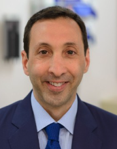 Steven M. Horwitz合作论文数Memorial Sloan Kettering Cancer Center59
 Jennifer R. Brown合作论文数CLL Center, Division of Hematologic Malignancies, Dana-Farber Cancer Institute;Harvard Medical School51
 Caron A. Jacobson合作论文数Dana-Farber Cancer Institute;Harvard Medical School48
 Arnold S. Freedman合作论文数Harvard Medical School;Department of Medical Oncology, Dana-Farber Cancer Institute;Brigham and Women's Hospital40
 Neha Mehta-Shah合作论文数39
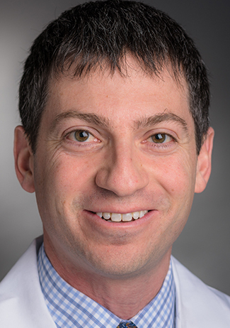 Matthew S. Davids合作论文数Division of Lymphoma, Dana-Farber Cancer Institute;Harvard Medical School;Brigham and Women's Hospital38
 Pier Luigi Zinzani合作论文数Department of Medical and Surgical Sciences, Università di Bologna36
 Robert Redd合作论文数Dana-Farber Cancer Institute35Barnes Jeffrey A合作论文数29
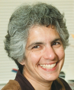 Donna Neuberg合作论文数Department of Biostatistics, Harvard T.H. Chan School of Public Health;Dana-Farber Cancer Institute26Austin I Kim合作论文数25Merryman Reid合作论文数25
 Nancy L. Bartlett合作论文数Divisions of Hematology & Oncology, Department of Medicine, Washington University School of Medicine in St. Louis25
 Francine M. Foss合作论文数Department of Internal Medicine, Yale School of Medicine, Yale University24Crombie Jennifer合作论文数24Odejide Oreofe O合作论文数24
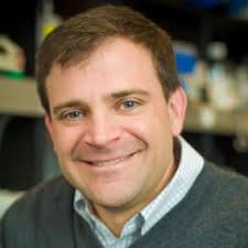 David Weinstock合作论文数Dana-Farber Cancer Institute;Harvard Medical School, Harvard University23
 Ephraim Hochberg合作论文数Cancer Center, Mass General Research Institute, Massachusetts General Hospital;Leuko;Harvard Medical School23
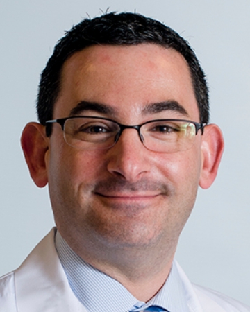 Jeremy Abramson合作论文数Jon and Jo Ann Hagler Center for Lymphoma, Massachusetts General Hospital Cancer Center;Harvard Medical School22
 Jasmine M. Zain合作论文数Division of Lymphoma, Department of Hematology & Hematopoietic Cell Transplantation, City of Hope;Toni Stephenson Lymphoma Center, City of Hope;Columbia University22
 Ranjana Advani合作论文数Division of Oncology, Department of Medicine, Stanford University;Stanford Cancer Institute22
 Youn H. Kim合作论文数Department of Dermatology, School of Medicine, Stanford University;Stanford Cancer Institute, Stanford University20
 Barbara Pro合作论文数Herbert Irving Comprehensive Cancer Center, Columbia University Irving Medical Center;Department of Medicine, Division of Hematology/Oncology, Vagelos College of Physicians and Surgeons, Columbia University19Samuel Yao-Ming Ng合作论文数18
 Haesook T. Kim合作论文数Department of Data Science, Dana-Farber Cancer Institute;Harvard School of Public Health;Department of Biostatistics and Computational Biology, Dana-Farber Cancer Institute18
 Ahmet Dogan合作论文数Memorial Sloan Kettering Cancer Center16
 Won Seog Kim合作论文数Division of Hematology-Oncology, Department of Medicine, Sungkyunkwan University School of Medicine16Jain Salvia合作论文数NYU Langone Medical Center, NYU Cancer Institute15
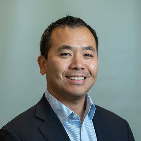 Yi-Bin Chen合作论文数Mass General Research Institute, Massachusetts General Hospital;Harvard Medical School15
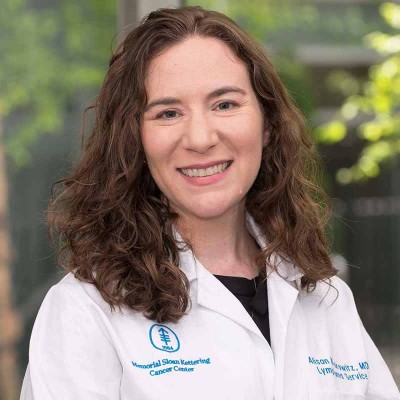 Alison J. Moskowitz合作论文数Memorial Sloan Kettering Cancer Center15Pierluigi Porcu合作论文数15Erin M Parry合作论文数Harvard Med Sch, Boston, MA USA14
 Owen A O'Connor合作论文数14
 Koji Izutsu合作论文数Graduate School of Medicine, University of Tokyo14
 Andre Henri Goy合作论文数John Theurer Cancer Center, Hackensack University Medical Center;Georgetown University;School of Medicine, Hackensack Meridian Health13
 Robert J. Soiffer合作论文数Department of Medical Oncology, Dana-Farber Cancer Institute;Division of Hematologic Malignancies, Dana-Farber Cancer Institute;Harvard Medical School13
 Andrei R. Shustov合作论文数School of Medicine, University of Washington;Fred Hutchinson Cancer Center13
 Theresa Davey合作论文数Mem Sloan Kettering Canc Ctr12
 Lubomir Sokol合作论文数Moffitt Cancer Center12
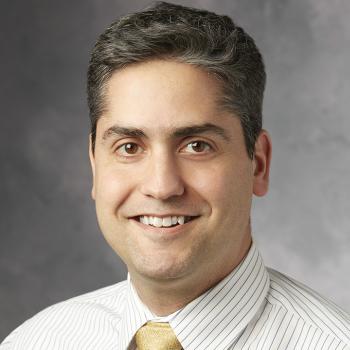 Michael Khodadoust合作论文数Medicine Department, Stanford University;Dermatology Department, Stanford University12Natasha Galasso合作论文数Mem Sloan Kettering Canc Ctr11
 Tanya Siddiqi合作论文数11
 Gritti Giuseppe合作论文数Hematology and Bone Marrow Transplant Units, A. O. Papa Giovanni XXIII11Herrera Alex F合作论文数Division of Hematologic Malignancies, Dana-Farber Cancer Institute11
 Scott J. Rodig合作论文数Department of Pathology, Brigham and Women's Hospital, Harvard Medical School;Tissue Biomarker Laboratory, Center for Immuno-Oncology, Dana-Farber Cancer Institute11Tak Takvorian合作论文数11




