Cell Painting images offer valuable insights into a cell's state and enable many biological applications, but publicly available arrayed datasets only include hundreds of genes perturbed. The JUMP Cell Painting Consortium perturbed roughly 75% of the protein-coding genome in human U-2 OS cells, generating a rich resource of single-cell images and extracted features. These profiles capture the phenotypic impacts of perturbing 15,243 human genes, including overexpressing 12,609 genes (using open reading frames) and knocking out 7,975 genes (using CRISPR-Cas9). Here we mitigated technical artifacts by rigorously evaluating data processing options and validated the dataset's robustness and biological relevance. Analysis of phenotypic profiles revealed previously undiscovered gene clusters and functional relationships, including those associated with mitochondrial function, cancer and neural processes. The JUMP Cell Painting genetic dataset is a valuable resource for exploring gene relationships and uncovering previously unknown functions.
In image-based profiling, software extracts thousands of morphological features of cells from multi-channel fluorescence microscopy images, yielding single-cell profiles that can be used for basic research and drug discovery. Powerful applications have been proven, including clustering chemical and genetic perturbations on the basis of their similar morphological impact, identifying disease phenotypes by observing differences in profiles between healthy and diseased cells and predicting assay outcomes by using machine learning, among many others. Here, we provide an updated protocol for the most popular assay for image-based profiling, Cell Painting. Introduced in 2013, it uses six stains imaged in five channels and labels eight diverse components of the cell: DNA, cytoplasmic RNA, nucleoli, actin, Golgi apparatus, plasma membrane, endoplasmic reticulum and mitochondria. The original protocol was updated in 2016 on the basis of several years’ experience running it at two sites, after optimizing it by visual stain quality. Here, we describe the work of the Joint Undertaking for Morphological Profiling Cell Painting Consortium, to improve upon the assay via quantitative optimization by measuring the assay’s ability to detect morphological phenotypes and group similar perturbations together. The assay gives very robust outputs despite various changes to the protocol, and two vendors’ dyes work equivalently well. We present Cell Painting version 3, in which some steps are simplified and several stain concentrations can be reduced, saving costs. Cell culture and image acquisition take 1–2 weeks for typically sized batches of ≤20 plates; feature extraction and data analysis take an additional 1–2 weeks. This protocol is an update to Nat. Protoc. 11, 1757–1774 (2016): https://doi.org/10.1038/nprot.2016.105 We provide an updated protocol for image-based profiling with Cell Painting. A detailed procedure, with standardized conditions for the assay, is presented, along with a comprehensive description of parameters to be considered when optimizing the assay.
Image-based profiling has emerged as a powerful technology for various steps in basic biological and pharmaceutical discovery, but the community has lacked a large, public reference set of data from chemical and genetic perturbations. Here we present data generated by the Joint Undertaking for Morphological Profiling (JUMP)-Cell Painting Consortium, a collaboration between 10 pharmaceutical companies, six supporting technology companies, and two non-profit partners. When completed, the dataset will contain images and profiles from the Cell Painting assay for over 116,750 unique compounds, over-expression of 12,602 genes, and knockout of 7,975 genes using CRISPR-Cas9, all in human osteosarcoma cells (U2OS). The dataset is estimated to be 115 TB in size and capturing 1.6 billion cells and their single-cell profiles. File quality control and upload is underway and will be completed over the coming months at the Cell Painting Gallery: https://registry.opendata.aws/cellpainting-gallery . A portal to visualize a subset of the data is available at https://phenaid.ardigen.com/jumpcpexplorer/ .
Some osteoblasts embed within bone matrix, change shape, and become dendrite-bearing osteocytes. The circuitry that drives dendrite formation during “osteocytogenesis” is poorly understood. Here we show that deletion of Sp7 in osteoblasts and osteocytes causes defects in osteocyte dendrites. Profiling of Sp7 target genes and binding sites reveals unexpected repurposing of this transcription factor to drive dendrite formation. Osteocrin is a Sp7 target gene that promotes osteocyte dendrite formation and rescues defects in Sp7-deficient mice. Single-cell RNA-sequencing demonstrates defects in osteocyte maturation in the absence of Sp7. Sp7-dependent osteocyte gene networks are associated with human skeletal diseases. Moreover, humans with a SP7 R316C mutation show defective osteocyte morphology. Sp7-dependent genes that mark osteocytes are enriched in neurons, highlighting shared features between osteocytic and neuronal connectivity. These findings reveal a role for Sp7 and its target gene Osteocrin in osteocytogenesis, revealing that pathways that control osteocyte development influence human bone diseases.
Drug repurposing has the advantage of identifying potential treatments on a shortened timescale. In response to the pandemic spread of SARS-CoV-2, we took advantage of a high-content screen of 3,713 compounds at different stages of clinical development to identify FDA-approved compounds that reduce mucin-1 (MUC1) protein abundance. Elevated MUC1 levels predict the development of acute lung injury (ALI) and acute respiratory distress syndrome (ARDS) and correlate with poor clinical outcomes. Our screen identifies fostamatinib (R788), an inhibitor of spleen tyrosine kinase (SYK) approved for the treatment of chronic immune thrombocytopenia, as a repurposing candidate for the treatment of ALI. In vivo, fostamatinib reduces MUC1 abundance in lung epithelial cells in a mouse model of ALI. In vitro, SYK inhibition by the active metabolite R406 promotes MUC1 removal from the cell surface. Our work suggests fostamatinib as a repurposing drug candidate for ALI.
 Maria Kost-Alimova合作论文数Broad Inst MIT & Harvard, Cambridge, MA 02142 USA5
 Beth A Cimini合作论文数Broad Inst MIT & Harvard, Imaging Platform, Cambridge, MA 02142 USA4Desiree Hernandez合作论文数3Briana Fritchman合作论文数3Lisa Miller合作论文数3Erin Weisbart合作论文数3Amy Goodale合作论文数Broad Inst MIT & Harvard, Cambridge, MA 02142 USA3
 Srinivas N. Chandrasekaran合作论文数Department of Biochemistry and Biophysics,;Program in Molecular and Cellular Biophysics, and;Department of Biochemistry and Biophysics,;Program in Molecular and Cellular Biophysics, and3
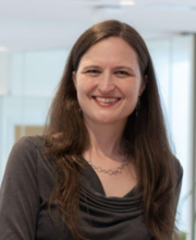 Anne E. Carpenter合作论文数Broad Institute3
 Shantanu Singh合作论文数Dept. of Computer Science and Engg., The Ohio State University3Tamara Gilbert合作论文数2Elizabeth Mouchet合作论文数2Amandine Thibaudeau合作论文数2Gisela Hormel合作论文数2Shan Yu合作论文数2Zahra Hanifehlou合作论文数2
 Peter Sommer合作论文数Ksilink2
 Adriana Borowa合作论文数Jagiellonian Univ, Fac Math & Comp Sci, 6 Lojasiewicza St, PL-30348 Krakow, Poland2Tiziana Monteverde合作论文数2Michał Warchoł合作论文数Ardigen, Cracow, Poland2Michelle Melanson合作论文数2Yu Han合作论文数2
 Lewis H Mervin合作论文数Univ Cambridge, Dept Chem, Ctr Mol Informat, Cambridge, England2
 Bolek Zapiec合作论文数Merck Healthcare KGaA2
 David C Logan合作论文数Institut de Recherche en Horticulture et Semences, University of Angers2
 Justin D Boyd合作论文数2Charles-Hugues Lardeau合作论文数CeMM Research Center for Molecular Medicine2Alix Eric合作论文数2
 Gnutt David合作论文数Department of Physical Chemistry II, Ruhr University Bochum2Garg Sakshi合作论文数2
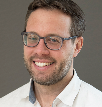 John Doench合作论文数Broad Institute of MIT and Harvard2
 David Root合作论文数Broad Institute2Hariharan Santosh合作论文数Sunnybrook Research Institute and Department of Biochemistry, University of Toronto2Martin Glynn合作论文数2Pilling James E合作论文数2McCarthy Nicola合作论文数2
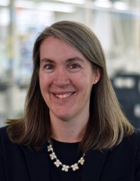 Susanne E. Swalley合作论文数2Arnaud Ogier合作论文数Image Mining Group, Institut Pasteur Korea2
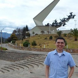 John E. Arevalo合作论文数Bioingenium Research Group, Universidad Nacional de Colombia2Guy B. Williams合作论文数Addenbrooke's Hospital, Cambridge University2Krzysztof Rataj合作论文数2Andrew J B Watts合作论文数1Zitong S. Chen合作论文数1Thierry Dorval合作论文数1Katherine A. Vernon合作论文数1
 Iffat Sumia Khader合作论文数1Jean Santos合作论文数1Alissa Campbell合作论文数1Melissa Bennion合作论文数1




