BACKGROUND:Transcatheter aortic valve replacement (TAVR) is a common treatment for aortic stenosis (AS). Although it is generally safe, its rising use has led to increased device failures and complications requiring surgery. Data on surgical explantation after TAVR remain limited. CASE PRESENTATION:An 85-year-old woman, 1-year post-TAVR, developed recurrent AS. Imaging revealed a mobile transvalvular mass and subvalvular pannus. Medical therapy failed, and because of stroke risk, she underwent TAVR explantation, subannular pannus resection, left ventricular outflow tract repair, and surgical aortic valve replacement. Histology confirmed leaflet thrombosis and valve deterioration. DISCUSSION:Surgical explantation after TAVR occurs in about 1% of cases. Among these, 47% require surgical aortic valve replacement, and 53% need additional procedures, often involving the aorta. Recognizing structural valve deterioration and its cause is critical. TAKE-HOME MESSAGE:As TAVR use rises, so will complications. Further research is needed to enhance management and outcomes.
Description of Case: An 81-year old-female with paroxysmal atrial fibrillation (AF) and severe aortic stenosis presented with AF with rapid ventricular response and volume overload. She had undergone an uncomplicated transcatheter aortic valve replacement (TAVR) 14 days prior with post procedure echocardiogram (TTE) demonstrating ejection fraction greater than 70%, a mean gradient across the TAVR valve of 11 mmHg, and mild mitral regurgitation (MR). Management included diuresis, rate control, and a failed cardioversion. TEE at the time of cardioversion, while tachycardic, demonstrated systolic anterior motion of the mitral valve (SAM) but no gradient across the aortic valve. She underwent atrioventricular (AV) junction ablation and placement of a left bundle branch area pacing (LBBAP) lead. Immediately post-procedure, she developed acute dyspnea and a new systolic murmur. Right heart catheterization revealed pulmonary artery pressure 90/30 mmHg and wedge pressure 39 mmHg with a prominent v-wave, and cardiac index of 1.6 L/min/m 2 . A simultaneous LV-AO pressure tracing demonstrated a gradient of 52 mmHg. TTE revealed a late-peaking doppler signal across the aortic valve with a peak velocity of 4 m/s, SAM, and severe MR. An Impella CP was placed for circulatory support. TTE revealed the LBBAP lead was adjacent to a 1.8 cm basal septal bulge (Figure 1). The next day, a transvenous RV apical pacing lead was placed, and simulatenous LV-AO pressure tracing revealed a peak-to-peak gradient of 45 mmHg during LBBAP, which immediately decreased to 5 mmHg with RV apical pacing (Figure 2). This led to a decrease in MR from severe to mild. Given the dynamic nature of the obstruction, alcohol septal ablation was performed. The Impella CP was removed and vasopressors were weaned. A permanent RV apical lead was added and used for pacing. The patient remained hemodynamically stable and was discharged to rehabilitation. Discussion: This case highlights a report of dynamic left ventricular outflow tract (LVOT) obstruction precipitated by LBBAP in a patient post-TAVR who previously did not have evidence of LVOT obstruction. Previously, RV apical pacing was a seldom used treatment for obstructive hypertrophic cardiomyopathy. Changing the pacing location to RV apical pacing instantly resolved the LVOT obstruction highlighting the need to be wary of pacing strategies in post TAVR patients who may have septal hypertrophy and may be at risk of LVOT obstruction secondary to LBBAP.
Background: Transcatheter aortic valve replacement (TAVR) has become the dominant treatment for aortic valve disease. While TAVR safety has improved over time, concern remains over the occurrence of cerebrovascular accidents (CVA) secondary to device placement, which is associated with increased morbidity and mortality. The Sentinel Cerebral Protection System (CPS) was developed to reduce the risk of embolic strokes associated with debris produced during TAVR. Studies evaluating Sentinel CPS efficacy have produced conflicting results, and there is little understanding of which patients are selected for device placement in “real-world” settings. With no existing guidelines on device use, the purpose of this study was to describe and compare the characteristics of patients who receive CPS with those who do not in a “real-world” setting of consecutive TAVR patients and evaluate its impact on postoperative complications, namely stroke. Methods: This was a single-center, retrospective study of all patients undergoing TAVR between July 1, 2019, and December 31, 2020. Patient demographics, baseline, and perioperative characteristics were collected prospectively using the Society of Thoracic Surgeons (STS)/American College of Cardiology (ACC) Transcatheter Valve Therapy (TVT) Registry and our institution’s TAVR database for analysis. Postoperative outcomes were assessed using primary endpoints of in-hospital/30-day stroke and the composite of death, stroke, and bleeding/vascular events at one-year. To adjust for baseline differences, a propensity score was developed including all factors that were different between groups, and Multivariate Cox Regression analysis was used to control for these differences. Patient follow-up was 97% complete at 12 months with 100% echocardiographic follow-up. Results: A total of 242 consecutive patients (57.9% male) were analyzed, with a mean age of 79.9 ± 9 years. Of these patients, 134 (55.4%) received the Sentinel CPS and 108 (44.6%) did not. Sentinel CPS patients were more likely to be male, not on dialysis, without prior CVA or pacemaker, had less severe chronic lung disease, and were lower operative risk compared to concurrent non-CPS patients. CPS patients were also found to have higher hemoglobin and albumin levels, lower creatinine, and were less likely to be on immunosuppressant therapy. The incidence of in-hospital/30-day stroke after TAVR did not differ between CPS and non-CPS patients (0.0% vs. 1.9%; p = 0.198). Unadjusted analyses at one-year showed a lower occurrence of the composite endpoint in CPS patients compared non-CPS patients (8.3% vs. 17.0%; p = 0.034). After adjustment, the hazard ratio (Adj HR) for the CPS group was no longer significantly associated with a lower composite endpoint (Adj HR = 0.609, 95% CI 0.244–1.523; p = 0.289). Both unadjusted (p = 0.233) and adjusted (p = 0.132) analyses showed no difference in the incidence of stroke at one-year. Conclusions: Our study demonstrates that in a “real-world” setting, the Sentinel CPS device is more likely to be used in healthier and less complex patients. In analyses adjusted for illness severity and patient complexity, CPS use did not have a significant effect on the incidence of in-hospital/30-day stroke or the composite endpoint of death, stroke, and bleeding/vascular events at one-year.
BackgroundWhile not available for clinical use in the United States, dedicated drug-coated balloons (DCB) are currently under investigation for the management of coronary in-stent restenosis (ISR). Peripheral drug-coated balloons (P-DCB) have been used off-label for coronary ISR. Further data regarding this practice are needed. We aimed to describe outcomes in patients who underwent off-label P-DCB angioplasty for coronary ISR.MethodsWe analyzed data on P-DCB angioplasty for coronary ISR at a single high-volume center between April 1, 2015, and December 30, 2017. Demographic and procedural details were collected, with systematic follow-up as clinically indicated.ResultsData from 31 patients treated with P-DCB angioplasty (mean age 68.0 ± 10.7 years) with coronary ISR (17 recurrent and 14 first time) were analyzed. Most patients presented with high-grade angina (81%) or myocardial infarction (13%). Treated ISR lesions were in native coronary arteries (68%), saphenous vein grafts (SVG, 23%), and the left internal mammary artery (10%). Diffuse intrastent ISR was common (69%) with a mean lesion length of 21.7 ± 12.4 mm. No postprocedural myocardial infarction occurred and 1 nonprocedural mortality occurred during index admission. At follow-up (median: 283, interquartile range [IQR]: 354 days), repeat angiography was performed in 19 patients (median: 212, IQR: 188 days), and 11 patients had target lesion recurrent ISR (Kaplan-Meier event-free survival estimate: 44.7%, 95% CI: 26.1-76.5%).ConclusionsIn the absence of availability of dedicated coronary DCB, treatment of coronary ISR using P-DCB angioplasty was feasible, although follow-up demonstrated continued risk for recurrent ISR in this high-risk population.
The goal of this review is to provide the reader with an overview of how to safely obtain large-bore access, descriptions of closure devices and techniques, and methods of identifying and troubleshooting common complications. Traditional methods of femoral access utilizing only anatomic landmarks should be supplemented with preprocedural imaging, ultrasound guidance, and micropuncture to ensure the adequacy of the femoral access for the intended procedure as well as to reduce the risk of complications. Closure of larger arteriotomies requires familiarity with both suture-based devices as well as collagen-based devices. A familiarity with dedicated closure devices is also necessary as each has its own learning curve as well as risks and benefits. Large-bore access carries increased risks of complications including bleeding, perforation, pseudoaneurysms, distal limb ischemia, and arteriovenous fistulas. Large-bore access is rapidly becoming a skill necessary for all interventional cardiologists. Due to the increased risks involved, familiarity with appropriate techniques and bailout strategies is critical.
We present a unique case of a paravalvular leak through a periannular channel around a bioprosthetic mitral valve. The role of multimodality imaging, in addition to novel technology, helped uncover the complex course of the jet, including its origin and direction, which translated into excellent procedural success. (Level of Difficulty: Advanced.).
HomeCirculation: Cardiovascular InterventionsVol. 13, No. 2Competency-Based Assessment of Interventional Cardiology Fellows’ Abilities in Intracoronary Physiology and Imaging Free AccessLetterPDF/EPUBAboutView PDFView EPUBSections ToolsAdd to favoritesDownload citationsTrack citationsPermissions ShareShare onFacebookTwitterLinked InMendeleyReddit Jump toFree AccessLetterPDF/EPUBCompetency-Based Assessment of Interventional Cardiology Fellows’ Abilities in Intracoronary Physiology and Imaging Erin Flattery, MD, Hussein M. Rahim, MD, Gregory Petrossian, BS, Evan Shlofmitz, DO, Fotis Gkargkoulas, MD, Mitsuaki Matsumura, BS, Ajay J. Kirtane, MD, SM, Sahil A. Parikh, MD, Manish A. Parikh, MD, Jeffrey W. Moses, MD, Dimitri Karmpaliotis, MD, PhD, Ori Ben-Yehuda, MD, Martin B. Leon, MD, Allen Jeremias, MD, Richard A. Shlofmitz, MD, Gregg W. Stone, MD, Akiko Maehara, MD, Ehtisham Mahmud, MD, Gary S. Mintz, MD and Ziad A. Ali, MD, DPhil Erin FlatteryErin Flattery NewYork-Presbyterian Hospital/Columbia University Medical Center (E.F., H.M.R., F.G., A.J.K., S.A.P., M.A.P., J.W.M., D.K., O.B.-Y., M.B.L., A.M., Z.A.A.). Cardiovascular Research Foundation, New York, NY (E.S., F.G., M.M., A.J.K., S.A.P., M.A.P., J.W.M., D.K., O.B.-Y., M.B.L., A.J., R.A.S., G.W.S., A.M., G.S.M., Z.A.A.). Search for more papers by this author , Hussein M. RahimHussein M. Rahim NewYork-Presbyterian Hospital/Columbia University Medical Center (E.F., H.M.R., F.G., A.J.K., S.A.P., M.A.P., J.W.M., D.K., O.B.-Y., M.B.L., A.M., Z.A.A.). Search for more papers by this author , Gregory PetrossianGregory Petrossian St Francis Hospital, Roslyn, NY (G.P., A.J., Z.A.A.). Search for more papers by this author , Evan ShlofmitzEvan Shlofmitz MedStar Washington Hospital Center, DC (E.S.). Search for more papers by this author , Fotis GkargkoulasFotis Gkargkoulas NewYork-Presbyterian Hospital/Columbia University Medical Center (E.F., H.M.R., F.G., A.J.K., S.A.P., M.A.P., J.W.M., D.K., O.B.-Y., M.B.L., A.M., Z.A.A.). Cardiovascular Research Foundation, New York, NY (E.S., F.G., M.M., A.J.K., S.A.P., M.A.P., J.W.M., D.K., O.B.-Y., M.B.L., A.J., R.A.S., G.W.S., A.M., G.S.M., Z.A.A.). Search for more papers by this author , Mitsuaki MatsumuraMitsuaki Matsumura Cardiovascular Research Foundation, New York, NY (E.S., F.G., M.M., A.J.K., S.A.P., M.A.P., J.W.M., D.K., O.B.-Y., M.B.L., A.J., R.A.S., G.W.S., A.M., G.S.M., Z.A.A.). Search for more papers by this author , Ajay J. KirtaneAjay J. Kirtane NewYork-Presbyterian Hospital/Columbia University Medical Center (E.F., H.M.R., F.G., A.J.K., S.A.P., M.A.P., J.W.M., D.K., O.B.-Y., M.B.L., A.M., Z.A.A.). Cardiovascular Research Foundation, New York, NY (E.S., F.G., M.M., A.J.K., S.A.P., M.A.P., J.W.M., D.K., O.B.-Y., M.B.L., A.J., R.A.S., G.W.S., A.M., G.S.M., Z.A.A.). Search for more papers by this author , Sahil A. ParikhSahil A. Parikh NewYork-Presbyterian Hospital/Columbia University Medical Center (E.F., H.M.R., F.G., A.J.K., S.A.P., M.A.P., J.W.M., D.K., O.B.-Y., M.B.L., A.M., Z.A.A.). Cardiovascular Research Foundation, New York, NY (E.S., F.G., M.M., A.J.K., S.A.P., M.A.P., J.W.M., D.K., O.B.-Y., M.B.L., A.J., R.A.S., G.W.S., A.M., G.S.M., Z.A.A.). Search for more papers by this author , Manish A. ParikhManish A. Parikh NewYork-Presbyterian Hospital/Columbia University Medical Center (E.F., H.M.R., F.G., A.J.K., S.A.P., M.A.P., J.W.M., D.K., O.B.-Y., M.B.L., A.M., Z.A.A.). Cardiovascular Research Foundation, New York, NY (E.S., F.G., M.M., A.J.K., S.A.P., M.A.P., J.W.M., D.K., O.B.-Y., M.B.L., A.J., R.A.S., G.W.S., A.M., G.S.M., Z.A.A.). Search for more papers by this author , Jeffrey W. MosesJeffrey W. Moses NewYork-Presbyterian Hospital/Columbia University Medical Center (E.F., H.M.R., F.G., A.J.K., S.A.P., M.A.P., J.W.M., D.K., O.B.-Y., M.B.L., A.M., Z.A.A.). Cardiovascular Research Foundation, New York, NY (E.S., F.G., M.M., A.J.K., S.A.P., M.A.P., J.W.M., D.K., O.B.-Y., M.B.L., A.J., R.A.S., G.W.S., A.M., G.S.M., Z.A.A.). Search for more papers by this author , Dimitri KarmpaliotisDimitri Karmpaliotis NewYork-Presbyterian Hospital/Columbia University Medical Center (E.F., H.M.R., F.G., A.J.K., S.A.P., M.A.P., J.W.M., D.K., O.B.-Y., M.B.L., A.M., Z.A.A.). Cardiovascular Research Foundation, New York, NY (E.S., F.G., M.M., A.J.K., S.A.P., M.A.P., J.W.M., D.K., O.B.-Y., M.B.L., A.J., R.A.S., G.W.S., A.M., G.S.M., Z.A.A.). Search for more papers by this author , Ori Ben-YehudaOri Ben-Yehuda NewYork-Presbyterian Hospital/Columbia University Medical Center (E.F., H.M.R., F.G., A.J.K., S.A.P., M.A.P., J.W.M., D.K., O.B.-Y., M.B.L., A.M., Z.A.A.). Cardiovascular Research Foundation, New York, NY (E.S., F.G., M.M., A.J.K., S.A.P., M.A.P., J.W.M., D.K., O.B.-Y., M.B.L., A.J., R.A.S., G.W.S., A.M., G.S.M., Z.A.A.). Search for more papers by this author , Martin B. LeonMartin B. Leon NewYork-Presbyterian Hospital/Columbia University Medical Center (E.F., H.M.R., F.G., A.J.K., S.A.P., M.A.P., J.W.M., D.K., O.B.-Y., M.B.L., A.M., Z.A.A.). Cardiovascular Research Foundation, New York, NY (E.S., F.G., M.M., A.J.K., S.A.P., M.A.P., J.W.M., D.K., O.B.-Y., M.B.L., A.J., R.A.S., G.W.S., A.M., G.S.M., Z.A.A.). Search for more papers by this author , Allen JeremiasAllen Jeremias St Francis Hospital, Roslyn, NY (G.P., A.J., Z.A.A.). Cardiovascular Research Foundation, New York, NY (E.S., F.G., M.M., A.J.K., S.A.P., M.A.P., J.W.M., D.K., O.B.-Y., M.B.L., A.J., R.A.S., G.W.S., A.M., G.S.M., Z.A.A.). Search for more papers by this author , Richard A. ShlofmitzRichard A. Shlofmitz Cardiovascular Research Foundation, New York, NY (E.S., F.G., M.M., A.J.K., S.A.P., M.A.P., J.W.M., D.K., O.B.-Y., M.B.L., A.J., R.A.S., G.W.S., A.M., G.S.M., Z.A.A.). Search for more papers by this author , Gregg W. StoneGregg W. Stone Cardiovascular Research Foundation, New York, NY (E.S., F.G., M.M., A.J.K., S.A.P., M.A.P., J.W.M., D.K., O.B.-Y., M.B.L., A.J., R.A.S., G.W.S., A.M., G.S.M., Z.A.A.). The Zena and Michael A. Wiener Cardiovascular Institute, Icahn School of Medicine at Mount Sinai, New York, NY (G.W.S.). Search for more papers by this author , Akiko MaeharaAkiko Maehara NewYork-Presbyterian Hospital/Columbia University Medical Center (E.F., H.M.R., F.G., A.J.K., S.A.P., M.A.P., J.W.M., D.K., O.B.-Y., M.B.L., A.M., Z.A.A.). Cardiovascular Research Foundation, New York, NY (E.S., F.G., M.M., A.J.K., S.A.P., M.A.P., J.W.M., D.K., O.B.-Y., M.B.L., A.J., R.A.S., G.W.S., A.M., G.S.M., Z.A.A.). Search for more papers by this author , Ehtisham MahmudEhtisham Mahmud Division of Cardiology, School of Medicine, University of California-San Diego (E.M.). Search for more papers by this author , Gary S. MintzGary S. Mintz Cardiovascular Research Foundation, New York, NY (E.S., F.G., M.M., A.J.K., S.A.P., M.A.P., J.W.M., D.K., O.B.-Y., M.B.L., A.J., R.A.S., G.W.S., A.M., G.S.M., Z.A.A.). Search for more papers by this author and Ziad A. AliZiad A. Ali Correspondence to: Ziad A. Ali, MD, DPhil, Columbia University Medical Center, Cardiovascular Research Foundation, 1700 Broadway, 9th Floor, New York, NY 10019. Email E-mail Address: [email protected] NewYork-Presbyterian Hospital/Columbia University Medical Center (E.F., H.M.R., F.G., A.J.K., S.A.P., M.A.P., J.W.M., D.K., O.B.-Y., M.B.L., A.M., Z.A.A.). St Francis Hospital, Roslyn, NY (G.P., A.J., Z.A.A.). Cardiovascular Research Foundation, New York, NY (E.S., F.G., M.M., A.J.K., S.A.P., M.A.P., J.W.M., D.K., O.B.-Y., M.B.L., A.J., R.A.S., G.W.S., A.M., G.S.M., Z.A.A.). Search for more papers by this author Originally published24 Jan 2020https://doi.org/10.1161/CIRCINTERVENTIONS.119.008760Circulation: Cardiovascular Interventions. 2020;13:e008760Although coronary angiography remains necessary for percutaneous coronary intervention, it provides limited information about lesion morphology, functional significance, and percutaneous coronary intervention results—limitations that are addressed by intravascular imaging and invasive physiology with demonstrated improved procedural and clinical outcomes.1,2 Nevertheless uptake remains low, especially among early career practitioners.3 Data regarding preparation of interventional cardiology fellows-in-training (IC-FIT) to use these modalities is scarce. We assessed IC-FIT perceptions regarding training and competency-based independence in invasive physiology, intravascular ultrasound (IVUS), and optical coherence tomography (OCT).An anonymous survey was distributed at the annual Cardiovascular Research Foundation Interventional Fellows Course in April 2018 and 2019 before the Imaging and Physiology session. Survey questions first assessed self-perceived training sufficiency using a multiple-choice scale that included expert, sufficient for clinical utility, rudimentary, or none. IC-FIT next assessed ability to perform specific core competencies independently, with assistance, or unable. Physiology competencies included device set-up and ability to identify aortic pressure dampening, distorted wave forms, and pressure drift. IVUS and OCT competencies included device set-up and ability to identify different plaque morphologies; reference segments for stent positioning, stent length, and diameter; minimal stent area; edge dissections; and malapposition. Student t test was used to compare differences between 2018 and 2019.Overall, 259 surveys were collected; complete responses were from 29% (74) IC-FIT. Others were general cardiology fellows or specialist imaging fellows. Demographic information was not collected in 2018; demographics obtained in 2019 showed that 77% (57) were from the United States and 74% (55) trained at University hospitals.Among IC-FIT, expert or sufficient training was initially reported by 95% (70) in invasive physiology, 82% (61) in IVUS, and 46% (34) in OCT (Figure [A]). Subsequently, when surveyed about specific core competencies involved in executing and interpreting these modalities (Figure [B and C]), only 57% (42) reported independence and preparedness for practice in all components of invasive physiology, 15% (11) in IVUS, and 18% (13) in OCT (Figure [D]). Only 7% (5) reported independence in all competencies of all modalities; 53% (39) report independence in all competencies of at least one, but not all modalities; and 40% (30) reported lack of competency-based independence in any modality (Figure [D]. There was no difference between surveys in April 2018 and April 2019 (data not shown).Download figureDownload PowerPointFigure. Competency-based assessment of interventional cardiology fellows’ abilities in intracoronary physiology and imaging. A, Percentage of interventional cardiology fellows-in-training (IC-FIT) reporting expert, sufficient, rudimentary, or no exposure in invasive physiology, intravascular ultrasound (IVUS), or optical coherence tomography (OCT) during training. B, Survey questions assessing perceived ability to perform specific competencies involved in executing and interpreting intravascular imaging (IVI) and physiology. C, Percentage of IC-FIT reporting perceived independence, need for assistance, or inability to practice each assessed competency in physiology, IVUS, and OCT. D, Percentage of IC-FIT perceived competency vs competency-based independence by modality. PCI indicates percutaneous coronary intervention.The results of our survey demonstrate that a significant portion of IC-FIT initially reports expert or sufficient experience in intracoronary physiology, IVUS, and OCT. However, among those who report such training, in fact, very few are independent in all components of performance and interpretation required for independent practice.There is a lack of emphasis on intravascular imaging and physiology in national educational guidelines. In 2017 American College of Graduate Medical Education IC fellowship program requirements, “competence in the performance of [intracoronary pressure monitoring… and intravascular ultrasound]” is a requirement without elaboration; OCT is not mentioned.4 In 2015 ACC-COCATS4 (American College of Cardiology - Core Cardiology Training Symposium 4), it is recommended that program faculty include persons competent in intravascular imaging and physiological assessment, but no modality is listed as a procedural skill to be obtained during training.5 This lack of an educational mandate may explain current training that is heterogeneous, dependent on institutional norms, and often inadequate or absent even in university hospitals.Our study was a small 2-year survey from a single educational conference with limited demographic data. Nevertheless, our survey demonstrated that IC-FIT are inadequately trained to perform and interpret intracoronary physiology, IVUS, and OCT independently after their training. The data, methods use in the analysis, and materials used to conduct this research will be made available to any researcher for the purpose of reproducing these results.DisclosuresEvan Shlofmitz is a consultant for Abbott Vascular, Opsens. Mitsuaki Matsumura is a consultant for Terumo Corporation. Dr Kirtane reports institutional funding to Columbia University and/or Cardiovascular Research; foundation from Medtronic, Boston Scientific, Abbott Vascular, Abiomed, CSI, CathWorks, Siemens, Philips, and ReCor Medical. Dr Parikh is a member of the advisory board to Abbott, Boston Scientific, Medtronic, and Philips. Dr Parikh reports speakers bureau for Abbott Vascular, Medtronic, Boston Scientific, CSI; also member of the advisory board to Abbott Vascular, Medtronic. Dr Karmpaliotis received honoraria from Abbott Vascular, Boston Scientific, Abiomed. Dr Leon received institutional grant support from Abbott, Boston Scientific, and Medtronic. Dr Jeremias received institutional grant support/consultant from Philips/Volcano and Abbott Vascular. Dr Stone reports speaker honoraria from Terumo, Novartis, and Amaranth; consultant to Shockwave, Valfix, TherOx, Reva, Vascular Dynamics, Robocath, Gore, Ablative Solutions, Matrizyme, Miracor, Neovasc, V-wave, Abiomed, Claret, Sirtex, Ancora, MAIA Pharmaceuticals, Spectrawave, Orchestra Biomed, Qool Therapeutics; equity/options in Qool Therapeutics, Cagent, Applied Therapeutics, Biostar family of funds, MedFocus family of funds, Spectrawave, Orchestra Biomed, Aria, and Ancora. Dr Maehara received grant support from Abbott Vascular and Boston Scientific, consultant for Conavi Medical Inc. Dr Mintz reports honoraria from Boston Scientific, Philips, and Terumo. Dr Ali received institutional research grants to Columbia University, Abbott, Cardiovascular Systems Inc; consultant of Abbott, Medtronic, Boston Scientific, Opsens, AstraZeneca. The other authors report no conflicts.FootnotesFor Sources of Funding and Disclosures, see page 2.Correspondence to: Ziad A. Ali, MD, DPhil, Columbia University Medical Center, Cardiovascular Research Foundation, 1700 Broadway, 9th Floor, New York, NY 10019. Email [email protected]eduReferences1. Buccheri S, Franchina G, Romano S, Puglisi S, Venuti G, D’Arrigo P, Francaviglia B, Scalia M, Condorelli A, Barbanti M, et al. Clinical outcomes following intravascular imaging-guided versus coronary angiography-guided percutaneous coronary intervention with stent implantation: a systematic review and bayesian network meta-analysis of 31 Studies and 17,882 patients.JACC Cardiovasc Interv. 2017; 10:2488–2498. doi: 10.1016/j.jcin.2017.08.051CrossrefMedlineGoogle Scholar2. Neumann FJ, Sousa-Uva M, Ahlsson A, Alfonso F, Banning AP, Benedetto U, Byrne RA, Collet JP, Falk V, Head SJet al; ESC Scientific Document Group. 2018 ESC/EACTS guidelines on myocardial revascularization.Eur Heart J. 2019; 40:87–165. doi: 10.1093/eurheartj/ehy394CrossrefMedlineGoogle Scholar3. Koskinas KC, Nakamura M, Räber L, Colleran R, Kadota K, Capodanno D, Wijns W, Akasaka T, Valgimigli M, Guagliumi G, et al. Current use of intracoronary imaging in interventional practice - results of a European Association of Percutaneous Cardiovascular Interventions (EAPCI) and Japanese Association of Cardiovascular Interventions and Therapeutics (CVIT) clinical practice survey.Circ J. 2018; 82:1360–1368. doi: 10.1253/circj.CJ-17-1144MedlineGoogle Scholar4. Accreditation Council for Graduate Medical Education (ACGME). ACGME Program Requirements for Graduate Medical Educationin Interventional Cardiology (Subspecialty of Internal Medicine).https://www.acgme.org/Portals/0/PFAssets/ProgramRequirements/152_InterventionalCardiology_2019_TCC.pdf. Published 2019. Accessed November 21, 2019.Google Scholar5. King SB, Babb JD, Bates ER, Crawford MH, Dangas GD, Voeltz MD, White CJ. COCATS 4 task force 10: Training in cardiac catheterization.J Am Coll Cardiol. 2015; 65:1844–1853. doi: 10.1016/j.jacc.2015.03.026CrossrefMedlineGoogle Scholar Previous Back to top Next FiguresReferencesRelatedDetailsCited By Jeremias A, Nijjer S, Davies J and DiMario C (2022) Physiologic Assessment and Guidance in the Cardiac Catheterization Laboratory Interventional Cardiology, 10.1002/9781119697367.ch7, (75-92), Online publication date: 3-Jun-2022. Shlofmitz E and Busch J (2022) Recognition of Drift: A Key to Success With Invasive Physiology, Cardiovascular Revascularization Medicine, 10.1016/j.carrev.2021.11.037, 35, (57-58), Online publication date: 1-Feb-2022. Gogia S, Edens M, Fall K, Petrossian G, Horst J, Jas Garcia J, Ozdemir D, Karimi Galougahi K, Karmpaliotis D, Kirtane A, Ben-Yehuda O, Maehara A, Mintz G and Ali Z (2022) Temporal trends of invasive physiologic assessment of coronary artery stenosis severity: insights from a quaternary care center in the United States, Coronary Artery Disease, 10.1097/MCA.0000000000001137, Publish Ahead of Print Saito Y, Kobayashi Y, Fujii K, Sonoda S, Tsujita K, Hibi K, Morino Y, Okura H, Ikari Y and Honye J (2021) Clinical expert consensus document on intravascular ultrasound from the Japanese Association of Cardiovascular Intervention and Therapeutics (2021), Cardiovascular Intervention and Therapeutics, 10.1007/s12928-021-00824-0, 37:1, (40-51), Online publication date: 1-Jan-2022. Sung J, Sharkawi M, Shah P, Croce K and Bergmark B (2021) Integrating Intracoronary Imaging into PCI Workflow and Catheterization Laboratory Culture, Current Cardiovascular Imaging Reports, 10.1007/s12410-021-09556-4, 14:6, Online publication date: 1-Jun-2021. Shah K and Cohen D (2021) Why Is Intravascular Ultrasound Guidance Underutilized in Percutaneous Coronary Intervention?: It Is Not “All About the Benjamins”, Circulation: Cardiovascular Quality and Outcomes, 14:5, Online publication date: 1-May-2021.Shlofmitz E, Ali Z, Maehara A, Mintz G, Shlofmitz R and Jeremias A (2020) Intravascular Imaging-Guided Percutaneous Coronary Intervention, Circulation: Cardiovascular Interventions, 13:12, Online publication date: 1-Dec-2020. Shlofmitz E, Khalid N and Hashim H (2020) Seeing Is Believing: Imaging-Guided Treatment of Calcified Lesions, Cardiovascular Revascularization Medicine, 10.1016/j.carrev.2020.07.004, 21:9, (1106-1107), Online publication date: 1-Sep-2020. Truesdell A, Khuddus M, Martinez S and Shlofmitz E (2020) Calcified Lesion Assessment and Intervention in Complex Percutaneous Coronary Intervention: Overview of Angioplasty, Atherectomy, and Lithotripsy, US Cardiology Review, 10.15420/usc.2020.16, 14 Mintz G (2020) Back to the Future, JACC: Cardiovascular Interventions, 10.1016/j.jcin.2020.04.042, 13:12, (1458-1459), Online publication date: 1-Jun-2020. Ali Z and Mintz G (2020) Intravascular Imaging, JACC: Case Reports, 10.1016/j.jaccas.2020.01.022, 2:3, (516-517), Online publication date: 1-Mar-2020. Rao S (2020) Proficiency Divide, Circulation: Cardiovascular Interventions, 13:2, Online publication date: 1-Feb-2020. February 2020Vol 13, Issue 2 Advertisement Article InformationMetrics © 2020 American Heart Association, Inc.https://doi.org/10.1161/CIRCINTERVENTIONS.119.008760PMID: 31973554 Originally publishedJanuary 24, 2020 Keywordscoronary angiographytomographypercutaneous coronary interventionstentscardiologyPDF download Advertisement SubjectsImagingOptical Coherence Tomography (OCT)Ultrasound
This review offers a summary of the current evidence on management of patients with combined AS and CAD. We discuss epidemiology, physiology, considerations about why and when CAD should be treated in this patient population, and the natural history of these diseases, all with particular attention to the impact and role of transcatheter aortic valve replacement (TAVR) in the management of AS.
Muhammed A. Rahim, Gregory Petrossian, Madison Edens, Nathaniel Abittan, Hussein Rahim and Ziad A. Ali, Penn State College of Medicine, Hershey, Pennsylvania, Albany Medical College, Albany, New York, Cardiovascular Research Foundation, Department of Medicine, Icahn School of Medicine at Mount Sinai, Mount Sinai Hospital, New York, Department of Medicine, Jacobi Medical Center, Albert Einstein College of Medicine, Bronx, Department of Cardiology, New York-Presbyterian Hospital/Columbia University Irving Medical Center, New York, New York, USA
Background Suprasternal access is an alternative access strategy for transcatheter aortic valve replacement (TAVR) where the innominate artery is cannulated from an incision above the sternal notch. To date, suprasternal access has never been compared with transfemoral TAVR. Thus, we sought to assess safety, feasibility, and early clinical outcomes between suprasternal and transfemoral access for patients undergoing TAVR. Methods and Results We evaluated patients from 2 institutional prospective, observational registries containing 1348 patients. Patients were selected in a 2:1 ratio (transfemoral:suprasternal) on the basis of propensity score matching. The primary outcome was in‐hospital mortality, and secondary outcomes included the incidence of ischemic stroke, major bleeding, vascular injury, left bundle‐branch block, and permanent pacemaker implantation at 30‐day follow‐up. Propensity score matching identified 89 patients undergoing suprasternal TAVR and 159 patients undergoing transfemoral TAVR suitable for analysis. There was no significant difference between suprasternal TAVR and transfemoral TAVR with respect to in‐hospital mortality (1.1% versus 0.6%; odds ratio [OR], 1.80; 95% CI, 0.11–29.06; P=0.680). No patients in either cohort suffered an ischemic stroke. The incidence of major bleeding (2.2% versus 2.5%; OR, 0.89; 95% CI, 0.16–4.96; P=0.895) and vascular injury (1.1% versus 1.9%; OR, 0.59; 95% CI, 0.06–5.77; P=0.651) did not differ significantly. The frequency of left bundle‐branch block (9.4% versus 15.8%; OR, 0.56; 95% CI, 0.24–1.30; P=0.177) and permanent pacemaker implantation (11.2% versus 5.9%; OR, 2.01; 95% CI, 0.75–5.45; P=0.169) were not statistically significantly different. Conclusions Suprasternal TAVR was safe and achieved promising short‐term clinical outcomes when compared with transfemoral TAVR. Future studies seeking to identify the optimal alternative access site should evaluate suprasternal TAVR access alongside other substitutes for transfemoral TAVR.
One of the primary reasons for under-use of optical coherence tomography (OCT) is the need for administration of radiocontrast to clear blood from the imaging field. The additional radiocontrast required to acquire OCT images is undesirable, particularly in patients with chronic kidney disease.
Optical coherence tomography (OCT) is underutilized partly due to the need for contrast flushing. Saline flush is an alternative without renal risk, but image quality and vascular dimensions have not been evaluated in vivo. Coronary arteries from 20 patients were imaged sequentially with contrast
Patients with chronic kidney disease are at elevated risk for adverse events after traditional coronary angiography and percutaneous intervention with contrast media. The case presented in this report highlights the potential benefits of zero-contrast multivessel percutaneous coronary intervention in a patient presenting with a non–ST-segment elevation acute coronary syndrome. (Level of Difficulty: Beginner.)
 Martin Leon合作论文数Center for Interventional Vascular Therapy, Columbia University Medical Center15
 Akiko Maehara合作论文数Seymour, Paul and Gloria Milstein Division of Cardiology, Department of Medicine, Columbia University;Intravascular Imaging Core Laboratory, CRF Clinical Trials Center;MRI Core Laboratory, CRF Clinical Trials Center14
 Gregg Stone合作论文数The Zena and Michael A. Wiener Cardiovascular Institute, Icahn School of Medicine at Mount Sinai12
 Gary Mintz合作论文数Cardiovascular Research Foundation11
 Ori Ben-Yehuda合作论文数Sulpizio Cardiovascular Institute, University of California, San Diego10
 Ayyaz Ali合作论文数10Ziad Ali合作论文数9Erin Flattery合作论文数8
 Evan Shlofmitz合作论文数St. Francis Hospital – The Heart Center8Dimitri Karmpaliotis合作论文数8
 Ankita Gore合作论文数Rutgers Robert Wood Johnson Medical School7
 Jeffrey W Moses合作论文数Division of Cardiology, Department of Medice, Columbia University7Allen Jeremias合作论文数6Nadira Hamid合作论文数6
 Mitsuaki Matsumura合作论文数MRI Core Laboratories, Cardiovascular Research Foundation6Shmuel Chen合作论文数Hadassah Ein Kerem Medical Center, Hebrew University6Torsten Vahl合作论文数6Ajay Jayant Kirtane合作论文数5
 Amisha rameshbhai Patel合作论文数P G Dept Biosci, Sardar Patel Univ5Khalique Omar K合作论文数Heart Valve Center, Columbia University Medical Center5Vivian Ng合作论文数5
 Rebecca T. Hahn合作论文数Seymour, Paul and Gloria Milstein Division of Cardiology, Department of Medicine, Columbia University Irving Medical Center5Richard A. Shlofmitz合作论文数5
 Yaser Nemshah合作论文数4Susheel Kodali合作论文数4Aaron Crowley合作论文数Genesis Research4Anjali Sharma合作论文数4
 Lauren S Ranard合作论文数Irving Medical Center, Columbia University4Cahill Thomas J合作论文数4Tayal Rajiv合作论文数4
 Tamim Nazif合作论文数4
 Isaac George合作论文数Department of Surgery;Division of Cardiothoracic;Columbia University;College of Physicians4Sahil A. Parikh合作论文数3Fotios Gkargkoulas合作论文数3Russell J Crew合作论文数3Fotis Gkargkoulas合作论文数3Kim Francis合作论文数3Gregory Petrossian合作论文数3
 Habib Jabagi合作论文数Address correspondence to Dr Jabagi, University of Ottawa Heart Institute3
 Sumit Mohan合作论文数Department of Medicine and Epidemiology, Mailman School of Public Health, Columbia University3Geoffrey Dube合作论文数3
 Mahesh Madhavan合作论文数Columbia University Irving Medical Center;Garnet Health Medical Center3
 David J. Cohen合作论文数University of Missouri-Kansas City3Konigstein Maayan合作论文数3
 Khady Fall合作论文数3
 Lloyd E. Ratner合作论文数Department of Surgery, Columbia University Irving Medical Center3
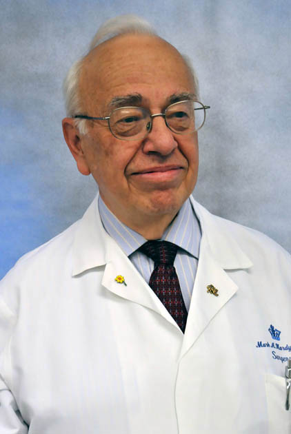 Mark A. Hardy合作论文数Department of Surgery, New York Presbyterian Hospital3
 Ajay Kirtane合作论文数Center for Interventional Vascular Therapy, Columbia University3




