Nitrogen-fixing eukaryotes were not believed to exist in nature until the recent discovery of a N2-fixing organelle, or nitroplast, in the marine microalga Braarudosphaera bigelowii. This nitroplast (formerly known as UCYN-A2) has long been recognized as key cyanobacterial contributor to global oceanic N2 fixation. However, how this novel organelle is integrated and regulated within the architecture of a eukaryotic cell remains unclear. Here, we combine multiscale volumetric imaging with cryo-electron tomography to resolve the native architecture, cellular integration, and diel remodeling of the nitroplast in cultured and environmental cells. We find that the nitroplast occupies up to 10% of the cell volume and exhibits close interfaces with multiple host organelles through membrane contact sites, while integration of this metabolically demanding compartment does not disrupt global scaling of host organelles. Interestingly, the chloroplast-to-nitroplast volume ratio is conserved across distinct life stages. Cryo-electron tomography reveals that the nitroplast retains a reinforced four-layer cyanobacterial envelope and is additionally surrounded by two host-derived layers that remodel across the day-night cycle. During daytime N2 fixation, these host-derived barriers become locally discontinuous and the organelle interface becomes enriched with two distinct vesicle populations. Our findings suggest that dynamic control of organelle accessibility through transient membrane gating represents a fundamental strategy by which eukaryotic cells could domesticate new endosymbiotic functions during early organellogenesis.
Radiolaria are unicellular marine organisms (protists) that have been drifting in oceanic plankton for hundreds of millions of years. These mineral architects can build extraordinarily complex skeletons, which fascinated and puzzled naturalists observing water samples through rudimentary microscopes. In the 19th century, the discovery and study of Radiolaria are associated with scientific voyages and human adventures. Naturalists who studied medicine and anatomy in European universities were captivated by the morphology of Radiolaria and expressed a profound wanderlust to collect them in the ocean. These intrepid and workaholic adventurers devoted their restless lives to studying microscopic life, while also actively engaging in teaching and sharing their observations and hypotheses with students. This article aims to retrace the discovery of Radiolaria through the lives of prominent naturalists and marine biologists, primarily Christian Gottfried Ehrenberg, Thomas Henry Huxley, Johannes Müller, and Ernst Haeckel. It also highlights the intellectual and geographic influences that shaped their research, including figures such as Johannes Wolfgang von Goethe and Alexander von Humboldt, as well as places like Jena, Helgoland, Villefranche-sur-Mer and Italy, which served as sampling locations and sources of romantic and artistic inspiration. Pioneering work on Radiolaria played a central role in shaping several emerging concepts (e.g., cell theory, individuality) and fields (e.g., taxonomy, evolution of morphology, symbiosis). The discovery of Radiolaria therefore reveals that even the most elusive marine microorganisms can deeply transform our understanding of life.
Light availability plays a central role in shaping the photophysiology and energy metabolism of photosymbiotic organisms such as reef-building corals. Although light varies greatly within coral colonies, the effects of this spatial heterogeneity on the subcellular organization and energy storage of symbiotic algae (Symbiodiniaceae) remain poorly understood. Here, we combined microscale measurements of light and oxygen across both light-exposed upper regions and shaded basal regions of a Favites abdita colony with three-dimensional cellular imaging using focused ion beam scanning electron microscopy. Our multiscale approach revealed subcellular heterogeneity among symbiont populations, suggesting different cell-cycle stages and physiological states across a spatial stratification in the coral. Subcellular morphometrics revealed that symbiont cells at the top of the colony were twice as voluminous as those at the shaded base, despite similar plastid volume occupancy. Compared to symbionts at the top of the colony, symbionts in the basal region accumulated nearly three times more starch relative to their cell volume. These findings show that light gradients within coral colonies shape symbiont morphology and energy storage patterns, with important implications for coral stress tolerance and resilience.
Symbiosis between a host and intracellular eukaryotic microalgae is a widespread life strategy in aquatic ecosystems. This partnership is considered to be mainly energized by the supply of photosynthetically derived carbon energy from microalgal symbionts. A major question is whether microalgae increase their photosynthetic production and decrease carbon storage in order to maximize carbon translocation to their host. By combining three-dimensional subcellular imaging and physiological analyses, we show that the chloroplast and CO2-fixing pyrenoid of the microalga Micractinium conductrix significantly expands during symbiosis within their host (the ciliate Paramecium bursaria) compared to the free-living stage. This is accompanied by a threefold higher quantity of Rubisco enzymes, 16-fold higher carbon fixation rate per algal cell and upregulation of several Carbon Concentrating Mechanism-related genes. Time-resolved subcellular quantitative imaging revealed that photosynthetically fixed carbon is first allocated to starch during the day, with five times higher production in symbiosis. Nearly half of the carbon stored in starch is consumed overnight while some is converted into lipid droplets, which are 20-fold more voluminous in symbiotic microalgae. We also show that carbon is transferred to the host and potentially respired by the high density of surrounding host mitochondria. Yet, high starch and lipid content in symbiotic microalgae suggest a moderate carbon export to the host relative to the high primary productivity. Overall, this study provides an original view of the subcellular remodeling and dynamics of carbon metabolism of microalgae inside a host, and opens new questions on the mechanisms of the source-sink relationship in aquatic photosymbiosis.
The ability to reproduce is a key process for the perpetuation of organisms. Along the evolution of protist reproductive strategies, the molecular machinery of sexual recombination is estimated to have been inherited from the last eukaryotic common ancestor (LECA). Unraveling the sexual cycles of free-living protists remains challenging, given the enigmatic roles of many uncultivated life stages. For the planktonic group of Acantharia (Radiolaria), a hypothetical sexual cycle has been proposed since the late 19th century, including a gamete-like stage, referred to as swarmers. In order to investigate the sexual nature of acantharian reproductive stages, we compared transcriptomes of various acantharian life stages. Our results show distinct functional profiles for reproductive and vegetative stages, while revealing the expression of the gamete fusion genes, HAP2/GCS1 and KAR5-GEX1-BMB in swarmers. Annotation of differentially expressed life stage-specific genes, also highlighted putative meiosis-related functions among pre-swarmer and swarmer stages, while suggesting the existence of a putative zygotic stage. This original life stage-specific genetic data is coherent with morphological evidence supporting the acantharian sexual cycle, with swarmers acting as gametes. Moreover, it paves the way for a deeper understanding of radiolarian cell biology and ecology at a single-cell scale.
Phaeocystales, comprising the genus Phaeocystis and an uncharacterized sister lineage, are nanoplanktonic haptophytes widespread in the global ocean. Several species form mucilaginous colonies and influence key biogeochemical cycles, yet their underlying diversity and ecological strategies remain underexplored. Here, we present new genomic data from 13 strains, including three high-quality reference genomes (N50 > 30 kbp), and integrate previous metagenome-assembled genomes to resolve a robust phylogeny. Divergence timing of P. antarctica aligns with Miocene cooling and Southern Ocean isolation. Genomic traits reveal metabolic flexibility, including mixotrophic nitrogen acquisition in temperate waters and gene expansions linked to polar nutrient adaptation. Concordantly, transcriptomic comparisons between temperate and polar Phaeocystis suggest Southern Ocean populations experience iron and B12 limitation. We also identify signatures of horizontal gene transfer and endogenous giant virus/virophage insertions. Together, these findings highlight Phaeocystales as an ecologically versatile and geographically widespread lineage shaped by evolutionary innovation and adaptation to contrasting environmental stressors.
Marine alveolates (MALVs) are diverse, primarily parasitic micro-eukaryotes that significantly impact marine ecosystems. The life cycles of most MALVs remain elusive and the role of sexual reproduction in these organisms is a key question that may determine their ecological success. In this study we focus on a widespread dinoflagellate parasite of bloom-forming dinoflagellates, Amoebophrya . After infection, we identified two distinct spores, differing in size, ultrastructure, swimming behavior, lifespan, gene expression, and metabolite composition. The smaller spores serve as infectious propagules, equipped with an apical complex for host invasion. They exhibit a distinct, shorter, and straighter swimming pattern, likely optimized for an extended lifespan while enhancing dispersion and chance for host encounters. Transcriptomic analysis reveals that these smaller spores are primed for efficient protein synthesis upon initiating a new infection. Conversely, the larger spores cannot infect new hosts and are characterized by the expression of meiotic genes, underscoring their sexual nature. They have a shorter lifespan, exhibit more tortuous movement, along display condensed chromosomes, signaling readiness for mating. Interestingly, infected hosts already express meiotic genes, and a single infected host only produces progeny of the same spore type, suggesting that cell fate is determined prior to spore release. Our study provides one of the first formal demonstrations of a sexually specialized cell in MALVs. Isolating compatible strains for cross-breeding and understanding how environmental conditions favor each reproductive route are the next key questions for elucidating the ecological success of MALVs in marine waters. Significance Statement Marine alveolates (MALVs) are ecologically significant parasites that impact carbon cycling, causing major disease outbreaks affecting fisheries and aquaculture, and influencing the dynamics of harmful algal blooms. Despite their diversity and wide host range, much of our knowledge comes from environmental DNA, leaving important aspects of their biology, such as their life cycles, largely unknown. This study provides the first evidence of sexual reproduction in MALVs, linking spore polymorphism to infective or sexual routes. This discovery is crucial as sexual reproduction increases genetic diversity and adaptability, aiding MALVs’ resilience in changing environments. Understanding MALVs’ reproductive strategies deepens our insight into their ecological roles and their broader impact on marine ecosystems. ### Competing Interest Statement The authors have declared no competing interest.
In oceanic plankton, various hosts are capable of engulfing and temporarily integrating microalgae (photosymbiosis) or just their photosynthetic plastids (kleptoplastidy) from the environment. These cellular interactions have been hypothesized to be representative of evolutionary steps in plastid acquisition in eukaryotes, but the underlying mechanisms are not fully understood. Here, we studied a polar kleptoplastidic dinoflagellate, which is known to steal plastids of the microalga Phaeocystis antarctica. We tracked the morphology and activity of stolen plastids over several months by combining multimodal subcellular imaging and photophysiology. Upon integration inside a host vacuole, the volume of plastids and pyrenoids significantly increased, and photosynthetic activity was boosted. This may be supported by the retention of a 50-fold larger algal nucleus for ∼1 week. Once the algal nucleus was lost, there was a decrease in plastid volume and photosynthesis, but nucleus- and plastid-encoded photosystem subunits were still detected. Carbon fixation and transfer to the host were also maintained after >2 months. We also showed that the algal mitochondrion was stolen and retained for several months, transforming into an extensive network interacting with plastids. This highlights a complex strategy in plankton along the continuum of plastid symbioses, where both plastids and mitochondria of a microalga are hijacked by a host for several months without the algal nucleus. This association, which we found to be widely distributed in polar regions, suggests that plastid-mitochondrion interaction may have played a role in the evolution of plastid acquisition and opens new questions about host control and organelle maintenance.
Symbiosis between eukaryotic microalgae and heterotrophic hosts is a widespread, phylogenetically convergent, and ecologically important phenomenon in aquatic ecosystems. Partners include taxonomically diverse microalgae interacting with multicellular or unicellular hosts in marine or freshwater environments. While progress has been made recently, there are still major knowledge gaps on the microenvironmental conditions of microalgae in hospite (e.g. nutrient and CO2 availability), the algal carbon metabolism (production and storage), and the cellular mechanisms of carbohydrate export to the host. This review aims to provide current knowledge on the physiology and metabolism of symbiotic microalgae, to highlight whether there are commonalities across different photosymbioses, and to identify new approaches and technologies for disentangling photosymbiotic interactions at relevant temporal and spatial scales.
Marine planktonic Radiolaria harboring symbiotic microalgae are ubiquitous in the oceans and abundant in oligotrophic areas. In these low-nutrient environments, they are among the most important primary producers. Systematic studies of radiolarian biology are limited because radiolaria are non-culturable and prone to damage during sampling. To obtain insight into the mechanistic basis of radiolarian photosymbiosis, we address here the metabolic contributions of the partners to the performance of the holobiont. Therefore, we describe the metabolic inventory of two highly abundant photosymbiotic radiolaria-colony-forming Collodaria and single-celled Acantharia and compare their metabolomes to metabolomes of respective free-living algae. Most of the metabolites detected in the symbiosis are not present in the free-living algae, suggesting a significant transformation of symbionts' metabolites by the host. The metabolites identified in the holobiont and the free-living algae encompass molecules of primary metabolism and a number of osmolytes, including dimethylsulfoniopropionate. Mass spectrometry imaging revealed the presence of dimethylsulfoniopropionate in the symbionts and host cells, indicating that the algae provide osmolytic protection to the host. Furthermore, our findings suggest a possible dependence of Collodaria on symbiotic vitamin B3. Distinctive differences in phospholipid composition between free-living and symbiotic stages indicate that the algal cell membrane may undergo rearrangement in the symbiosis. Our results demonstrate a strong interdependence and rewiring of the algal metabolism underlying radiolaria-microalgae photosymbioses.
Symbiosis is everywhere, and “we have never been individuals”[[1][1], [2][2]]. In animal-microbe symbioses, established symbionts are often thought to be confined to a specific cellular or tissue niche[[3][3]–[7][4]] and generally lose their motile appendages such as flagella[[8][5]–[15][6]]. However, whether the loss of motile appendages necessarily implies immobility within the animal host remains an open conundrum. Here, we present the discovery of long-range, host actin-driven symbiont mobility in a dinoflagellate-acoel worm symbiosis. Using long-term tracking, fluorescence, and electron microscopy, we find that dinoflagellate symbionts ( Amphidinium sp. , 10-20 µm in size) travel throughout an extensive network of thin host cells ( ∼ 200 nm in regions without symbionts) in Waminoa sp. acoel worms, which are part of a deep-diverging bilaterian lineage[[16][7]–[18][8]]. Although FIB-SEM-based 3D reconstruction shows symbionts still retain both flagella, we uncover that it is host actin machinery that plays a primary role in overcoming large drag forces under confinement to achieve mobility throughout the worm at surprisingly high velocities (around 1 µm/s ). Long term in-toto imaging further reveals diel rhythms and spatiotemporal regulation of symbionts during regeneration. Our findings show the presence of host-mediated mobility in animal-microbe symbioses, which suggests the existence of previously overlooked regulatory processes in holobionts’ maintenance of dynamic homeostasis. ### Competing Interest Statement The authors have declared no competing interest. NSF Center for Cellular Construction Woods Institute for the Environment Gordon and Betty Moore Foundation, https://ror.org/006wxqw41 Schmidt Futures Innovation Fellowship [1]: #ref-1 [2]: #ref-2 [3]: #ref-3 [4]: #ref-7 [5]: #ref-8 [6]: #ref-15 [7]: #ref-16 [8]: #ref-18
Light availability plays a central role in shaping the photophysiology and energy metabolism of photosymbiotic organisms such as reef-building corals. Although light varies greatly within coral colonies, the effects of this spatial heterogeneity on the subcellular organization and energy storage of symbiotic algae (Symbiodiniaceae) remain poorly understood. Here, we combined microscale measurements of light and oxygen across both light-exposed upper regions and shaded basal regions of a Favites abdita colony with three-dimensional subcellular imaging using Focused Ion Beam Scanning Electron Microscopy (FIB-SEM). Our multi-scale approach revealed subcellular heterogeneity among symbiont populations, suggesting different cell cycle stages and physiological states across a spatial stratification in the coral. Subcellular morphometrics revealed that symbiont cells at the top of the colony were twice more voluminous than those at the shaded base with similar plastid volume occupancy. Compared to symbionts at the top of the colony, symbionts in the basal region accumulated nearly three times more starch relative to their cell volume. These findings show that light gradients within coral colonies shape symbiont morphology and energy storage patterns, with important implications for coral stress tolerance and resilience. ### Competing Interest Statement The authors have declared no competing interest. Gordon and Betty Moore Foundation, GBMF9325, GBMF9206, GBMF11532 NSF-DBI, 2316391 NSF-BSF, 2149925 LabEx GRAL, ANR-10-LABX-49-01 CBHEUR-GS, ANR-17-EURE-0003 SymbiOCEAN, 101088661
The plastids of photosynthetic organisms on land are predominantly “primary plastids,” derived from an ancient endosymbiosis of a cyanobacterium. Conversely, the plastids of marine photosynthetic organisms were mostly gained through subsequent endosymbioses of photosynthetic eukaryotes generating so-called “complex plastids.” The plastids of the major eukaryotic lineages—cryptophytes, haptophytes, ochrophytes, dinoflagellates, and apicomplexans—were posited to derive from a single secondary endosymbiosis of a red alga in the “chromalveloate” hypothesis. Subsequent phylogenetic resolution of eukaryotes has shown that separate events of plastid acquisition must have occurred to account for this distribution of plastids. However, the number of such events and the donor organisms for the new plastid endosymbioses are still not resolved. A perceived bottleneck of endosymbiotic plastid gain is the development of protein targeting from the hosts into the new plastids, and this supposition has often driven hypotheses toward minimizing the number of plastid-gain events to explain plastid distribution in eukaryotes. But how plastid-protein-targeting is established for new endosymbionts is often unclear, which makes it difficult to assess the likelihood of plastid transfers between lineages. Here, we show that Kareniaceae dinoflagellates, which possess complex plastids known to be derived from haptophytes, acquired all the necessary protein import machinery from these haptophytes. Furthermore, cryo-electron tomography revealed that no additional membranes were added to the Kareniaceae complex plastid during serial endosymbiosis, suggesting that the haptophyte-derived import processes were sufficient. Our analyses suggest that complex red plastids are preadapted for horizontal transmission, potentially explaining their widespread distribution in algal diversity.
Cryptophytes are abundant and ubiquitous microalgae that constitute a major plastid source for kleptoplastidic ciliates and dinoflagellates. Despite their ecological significance, the understanding of their light preferences and photophysiology remains limited. Here, we provide a comprehensive study of the response of the haploid strain Teleaulax amphioxeia (Cr10EHU) to varying light irradiance. This strain is capable of growing under a wide range of irradiance levels, notably by finely tuning the different pigments bound to the membrane light-harvesting proteins. Analysis of the luminal phycoerythrin content revealed remarkable flexibility, with phycoerythrin emerging as a pivotal protein facilitating acclimation to varying light levels. Detailed ultrastructure examinations unveiled that this adaptability was supported by the synthesis of large thylakoidal vesicles, likely enhancing the capture of green photons efficiently under low light, a phenomenon previously undocumented. Teleaulax amphioxeia Cr10EHU effectively regulated light utilization by using a cryptophyte state transition-like process, with a larger amplitude observed under high growth irradiance. Furthermore, our results revealed the establishment of growth irradiance-dependent non-photochemical quenching of fluorescence, likely inducing the dissipation of excess light. This study underscores the particularities and the significant photoadaptability of the plastid of the haploid form of T. amphioxeia. It constitutes a comprehensive photophysiological characterization of the Cr10EHU strain that paves the way for future studies of the kleptoplastidy process.
Once the work at the microscope is done, biological discoveries rely heavily on proper downstream analysis. This often amounts to first segmenting the biological objects of interest in the image before performing a quantitative analysis. Deep-learning (DL) is nowadays ubiquitous in such segmentation tasks. However, DL can be cumbersome to apply, as it often requires large amount of manual labeling to produce ground-truth data, and expert knowledge to train the models from scratch. Nonetheless, the performance of large foundation models, although trained on natural images, are improving on scientific images with every new model released. They, however, require either manual prompting or tedious post-processing to selectively segment the biological objects of interest. Classical machine learning algorithms, such as random forest classifiers, on the other hand, are well-established, easy to train, and often yield results of sufficient quality for downstream processing tasks, hence their continued popularity. Unfortunately, they are limited to objects with distinct, well-defined textures compared to their environment. This generally limits their usefulness to structures easy to recognize. Here, we present FeatureForest, an open-source tool that leverages the feature embeddings of large foundation models to train a random forest classifier, thereby providing users with a rapid way of semantically segmenting complex images using only a few labeling strokes. We demonstrate the improvement in performance over a variety of datasets, including large and complex volumetric electron microscopy stacks. Our implementation is available in napari, currently integrates four foundation models, and can easily be extended to any new model once they become available. ### Competing Interest Statement The authors have declared no competing interest.
Electron microscopy and chemical imaging are now essential in life science to access the interior of a cell and unveil its structural and chemical landscape at nanoscale. Because of the high vacuum conditions required to visualize subcellular structures, organisms need to be fixed, dehydrated, and sectioned/milled. For fixation, the use of aldehydes at room temperature alters the ultrastructure and the chemistry of the cell creating major osmotic changes. Therefore, rapid freezing methods such as high-pressure freezing superiors in preserving native-state cell structure and chemistry at a fast rate (a scale of milliseconds compared with minutes for chemical fixation). Here, we present the workflow starting from rapid freezing (high pressure freezing) and ending with resin embedding (using freeze substitution) in order to prepare biological samples (cells or tissues) for 2D or 3D electron microscopy (Transmission/Scanning Electron microscopy, and FIB-SEM: Focused Ion Beam Scanning Electron Microscope) and chemical imaging.
Metabolic exchange is one of the foundations of symbiotic associations between organisms and is a driving force in evolution. In the ocean, photosymbiosis between heterotrophic hosts and microalgae is powered by photosynthesis and relies on the transfer of organic carbon to the host (e.g. sugars). Yet, the identity of transferred carbohydrates as well as the molecular mechanisms that drive this exchange remain largely unknown, especially in unicellular photosymbioses that are widespread in the open ocean. Combining genomics, single-holobiont transcriptomics, and environmental metatranscriptomics, we revealed the transportome of the marine microalga Phaeocystis in symbiosis within acantharia, with a focus on sugar transporters. At the genomic level, the sugar transportome of Phaeocystis is comparable to non-symbiotic haptophytes. By contrast, we found significant remodeling of the expression of the transportome in symbiotic microalgae compared to the free-living stage. More particularly, 36% of sugar transporter genes were differentially expressed. Several of them, such as GLUTs, TPTs, and aquaporins, with glucose, triose-phosphate sugars, and glycerol as potential substrates, were upregulated at the holobiont and community level. We also showed that algal sugar transporter genes exhibit distinct temporal expression patterns during the day. This reprogramed transportome indicates that symbiosis has a major impact on sugar fluxes within and outside the algal cell, and highlights the complexity and the dynamics of metabolic exchanges between partners. This study improves our understanding of the molecular players of the metabolic connectivity underlying the ecological success of planktonic photosymbiosis and paves the way for more studies on transporters across photosymbiotic models.
As an innate property of life, the ability to reproduce is a key process for the perpetuation of organisms. Along the evolution of protist reproductive strategies, the molecular machinery of sexual recombination is estimated to have been inherited from the last eukaryotic common ancestor (LECA). Nevertheless, unraveling the sexual cycles of extant free-living protist lineages remains challenging, given the enigmatic roles of many uncultivated life stages. Among the uncultivated planktonic group of Acantharia (Radiolaria), a hypothetical sexual cycle has been proposed since the late 19th century, including the existence of a gamete-like life stage of undetermined ploidy, referred to as swarmers. In order to investigate the sexual nature of acantharian reproductive stages, we conducted single-cell transcriptomic analysis across various acantharian life stages. Our results show distinct functional profiles for reproductive and vegetative life stages, while revealing the expression of the reference eukaryotic genes involved in gamete fusion, HAP2/GCS1 and GEX1-KAR5, in swarmers and pre-swarmer stages. Annotation of differentially expressed life stage-specific genes, also highlights putative meiosis-related functions among swarmers, while suggesting the existence of a potential swarmer/vegetative intermediate stage expressing putative growth-related genes. This original life stage-specific genetic data is coherent with morphological evidence supporting the existence of an acantharian sexual cycle, with swarmers acting as gametes. Moreover, it paves the way for a deeper understanding of radiolarian cell biology and ecology at a single-cell scale. ### Competing Interest Statement The authors have declared no competing interest.
Gephyrocapsa huxleyi is a prevalent, bloom-forming phytoplankton species in the oceans. It exhibits a complex haplodiplontic life cycle, featuring a diploid-calcified phase, a haploid phase and a third 'decoupled' phase produced during viral infection. Decoupled cells display a haploid-like phenotype, but are diploid. Here, we investigated the fate of decoupled cells during culture observations and we compared the transcriptome profiles and the cellular ultrastructure of the three life cycle cell types. We found that decoupled cells can revert to the calcified form in the absence of viral pressure, revealing the ability of G. huxleyi to modulate cell differentiation as a function of external conditions. Ultrastructural analyses showed distinct nuclear organization with variations in chromatin volume. Transcriptomic analyses revealed gene expression patterns specific to each life phase. These included multiple regulatory functions in chromatin remodeling, broader epigenetic mechanisms and life cycling, likely contributing to cell differentiation. Finally, analyses of available host-virus transcriptomes support life cycle transition during viral infection. This study provides cellular and molecular foundations for nuclear remodeling and cell differentiation in coccolithophores and the identification of gene markers for studying coccolithophore life cycles in natural populations.
 Fabrice Not合作论文数UMR 7127 CNRS, INSU et Université Pierre et Marie Curie25
 Colomban de Vargas合作论文数CNRS, Stn Biol Roscoff22
 Benoit Gallet合作论文数Institut de Biologie Structurale UMR5075 (, CEA/CNRS/Université Joseph Fourier),19
 Sarah Romac合作论文数Sorbonne Université, CNRS, Station Biologique de Roscoff16
 Ian Probert合作论文数CNRS, Université Pierre et Marie Curie15
 Frédéric Mahé合作论文数13
 Stéphane Audic合作论文数Institut de Microbiologie de la Méditerranée (IM2, IFR-88), Aix-Marseille University13
 Chevalier Fabien合作论文数12
 Lucie Bittner合作论文数UMR CNRS 7138 Systématique, Adaptation, Evolution, Muséum National d'Histoire Naturelle12
 Dunthorn Micah合作论文数Dept Ecol, Univ Kaiserslautern11
 Jan Pawłowski合作论文数Department of Genetics and Evolution, University of Geneva;Laboratory of Paleoceanography, Marine Ecology Department, Institute of Oceanology Polish Academy of Sciences11
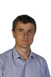 Yannick Schwab合作论文数Imaging Center, Université de Strasbourg BP. 1014210
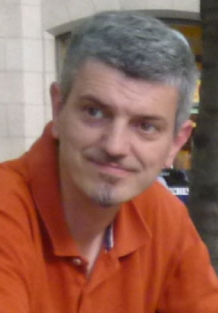 Giovanni Finazzi合作论文数Cell & Plant Physiology Laboratory9
 Pierre-Henri Jouneau合作论文数Univ Grenoble Alpes9
 Patrick Wincker合作论文数Centre National de Recherche en Génomique Humaine;University of Paris-Saclay9
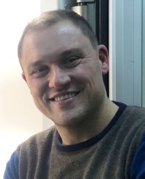 Raffaele Siano合作论文数TIMP, Stazione Zoologica Anton Dohrn9
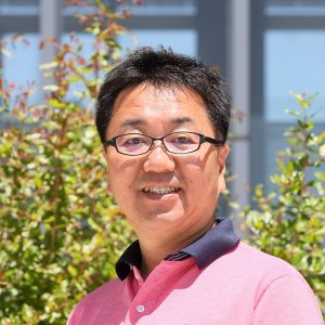 Hiroyuki Ogata (緒方博之)合作论文数Bioinformatics Center, Institute for Chemical Research, Kyoto University8
 Ramiro Logares合作论文数Department of Marine Biology and Oceanography, Institut de Ciències del Mar, Consejo Superior de Investigaciones Científicas8
 Adriana Zingone合作论文数Section of Integrated Marine Ecology, Stazione Zoologica Anton Dohrn, Napoli8
 Thorsten Stoeck合作论文数Department of Ecology, Technische Universität Kaiserslautern, Kaiserslautern, Germany8Sébastien Colin合作论文数8
 Charlotte Le Kieffre合作论文数Cell & Plant Physiol Lab, Univ Grenoble Alpes7
 Uwizeye Clarisse合作论文数Cell & Plant Physiology Laboratory, University of Grenoble Alpes, CNRS, CEA, INRA, 38054 Grenoble Cedex 9, France7
 Nicolas Henry合作论文数CNRS, Sorbonne Univ7
 Niculina Musat合作论文数UFZ Helmholtz Ctr Environm Res, Dept Isotope Biogeochem, Permoserstr 15, D-04318 Leipzig, Germany7
 Schieber Nicole L合作论文数European Mol Biol Lab, Heidelberg, Germany7
 David Bass合作论文数7Daniel P. Yee合作论文数6
 Cedric Berney合作论文数6Richard Christen合作论文数6Stryhanyuk Hryhoriy合作论文数6
 Ramon Massana合作论文数Instituto de Ciencias del Mar-CSIC6
 Wiebe H. C. F. Kooistra合作论文数Smithsonian Tropical Research Institute6
 Bente Edvardsen合作论文数Microbial Evolution Research Group (MERG),, University of Oslo6Margaux Carmichael合作论文数5
 Noan Le Bescot合作论文数5Giulia Veronesi合作论文数5
 Shinichi Sunagawa合作论文数Department of Biology, Eidgenossische Technische Hochschule Zurich5
 Daniel Vaulot合作论文数GEEK Lab, Asian School of the Environment, Nanyang Technological University5Falconet Denis合作论文数5Remi Tucoulou合作论文数5
 Jean-Michel Claverie合作论文数Structural and Genomic Information Laboratory, School of Medicine, Aix-Marseille University5
 Laure Guillou合作论文数Centre for Environment5
 Enrique Lara合作论文数WSL Wetlands Research Group, Swiss Federal Research Institute5Christophe Boutte合作论文数5
 John R. Dolan合作论文数Marine Microbial Ecology & Biogeochemistry Group, Laboratoire d'Oceanographie de Villefranche, CNRS & Universite Paris VI;Laboratoire d'oceanographie de Villefranche, Sorbonne Universite5
 Gabriel Gorsky合作论文数5Gilles Curien合作论文数5Rachel M Templin合作论文数4




