We evaluate deconvolution methods, which infer levels of immune infiltration from bulk expression of tumor samples, through a community-wide DREAM Challenge. We assess six published and 22 community-contributed methods using in vitro and in silico transcriptional profiles of admixed cancer and healthy immune cells. Several published methods predict most cell types well, though they either were not trained to evaluate all functional CD8+ T cell states or do so with low accuracy. Several community-contributed methods address this gap, including a deep learning-based approach, whose strong performance establishes the applicability of this paradigm to deconvolution. Despite being developed largely using immune cells from healthy tissues, deconvolution methods predict levels of tumor-derived immune cells well. Our admixed and purified transcriptional profiles will be a valuable resource for developing deconvolution methods, including in response to common challenges we observe across methods, such as sensitive identification of functional CD4+ T cell states. Deconvolution methods infer levels of immune infiltration from bulk expression of tumour samples. Here, authors assess 6 published and 22 community-contributed methods via a DREAM Challenge using in vitro and in silico transcriptional profiles of admixed cancer and healthy immune cells.
AbstractCancers evolve not only through the acquisition and clonal transmission of somatic mutations but also by epigenetic mechanisms that modify cell phenotype. Here, we use histology-guided and spatial transcriptomics to characterize hepatoblastoma, a childhood liver cancer that exhibits significant histologic and proliferative heterogeneity despite clonal activating mutations in the Wnt/β-catenin pathway. Highly proliferative regions with embryonal histology show high expression of Wnt target genes, the embryonic biliary transcription factor SOX4, and striking focal expression of the growth factor FGF19. In patient-derived tumoroids with constitutive Wnt activation, FGF19 is a required growth signal for FGF19-negative cells. Indeed, some tumoroids contain subsets of cells that endogenously express FGF19, downstream of Wnt/β-catenin and SOX4. Thus, the embryonic biliary lineage program cooperates with stabilized nuclear β-catenin, inducing FGF19 as a paracrine growth signal that promotes tumor cell proliferation, together with active Wnt signaling. In this pediatric cancer presumed to originate from a multipotent hepatobiliary progenitor, lineage-driven heterogeneity results in a functional growth advantage, a non-genetic mechanism whereby developmental lineage programs influence tumor evolution.
Aptamers incorporating chemically modified bases can achieve superior affinity and specificity compared to natural aptamers, but their characterization remains a labor-intensive, low-throughput task. Here, we describe the "non-natural aptamer array" (N2A2) system, in which a minimally modified Illumina MiSeq instrument is used for the high-throughput generation and characterization of large libraries of base-modified DNA aptamer candidates based on both target binding and specificity. We first demonstrate the capability to screen multiple different base modifications to identify the optimal chemistry for high-affinity target binding. We next use N2A2 to generate aptamers that can maintain excellent specificity even in complex samples, with equally strong target affinity in both buffer and diluted human serum. For both aptamers, affinity was formally calculated with gold-standard binding assays. Given that N2A2 requires only minor mechanical modifications to the MiSeq, we believe that N2A2 offers a broadly accessible tool for generating high-quality affinity reagents for diverse applications.
Despite its rapidly increased availability for the study of complex tissue, single cell RNA sequencing remains prohibitively expensive for large studies. Here, we present a protocol using oligonucleotide barcoding for the tagging and pooling of multiple samples from healing wounds, which are among the most challenging tissue types for this application. We describe steps to generate skin wounds in mice, followed by tissue harvest and oligonucleotide barcoding. This protocol is also applicable to other species including rats, pigs, and humans.For complete details on the use and execution of this protocol, please refer to Stoeckius et al. (2018),1 Galiano et al. (2004),2 and Mascharak et al. (2022).3
Chronic rhinosinusitis (CRS) is a common inflammatory disease of the sinonasal cavity that affects millions of individuals worldwide. The complex pathophysiology of CRS remains poorly understood, with emerging evidence implicating the orchestration between diverse immune and epithelial cell types in disease progression. We applied single-cell RNA sequencing (scRNA-seq) and spatial transcriptomics to both dissociated and intact, freshly isolated sinonasal human tissues to investigate the cellular and molecular heterogeneity of CRS with and without nasal polyp formation compared to non-CRS control samples. Our findings reveal a mechanism for macrophage-eosinophil recruitment into the nasal mucosa, systematic dysregulation of CD4+ and CD8+ T cells, and enrichment of mast cell populations to the upper airway tissues with intricate interactions between mast cells and CD4 T cells. Additionally, we identify immune-epithelial interactions and dysregulation, particularly involving understudied basal progenitor cells and Tuft chemosensory cells. We further describe a distinct basal cell differential trajectory in CRS patients with nasal polyps (NP), and link it to NP formation through immune-epithelial remodeling. By harnessing stringent patient tissue selection and advanced technologies, our study unveils novel aspects of CRS pathophysiology, and sheds light onto both intricate immune and epithelial cell interactions within the disrupted CRS tissue microenvironment and promising targets for therapeutic intervention. These findings expand upon existing knowledge of nasal inflammation and provide a comprehensive resource towards understanding the cellular and molecular mechanisms underlying this uniquely complex disease entity, and beyond.
The supplementary data include one table and 5 figures. Table S1 presents cabazitaxel cytotoxic activity in the human breast cancer cell line MCF-7, uterine sarcoma cell line MES-SA, and ovarian cancer cell lines ES-2, MES-OV, and OVCAR-3. Figure S1 presents the molecular structure of cabazitaxel and cytotoxicity curves for three taxanes in MCF-7 cells. Figure S2 shows an immunoblot of drug transporter proteins in parental and drug resistant variants of MCF-7. Figure S3 shown rhodamine123 and BODIPY-paclitaxel accumulation by flow cytometry in parental and resistant variants. Figure S4 shows polymerized versus soluble tubulin in parental vs resistant variants. Figure S5 shows expression of apoptotic regulators.
Abstract Autologous T cells genetically engineered to express a chimeric antigen receptor (CAR) targeting CD19 and/or CD22 have achieved high complete response rates in patients with hematologic malignancies, but >50% of patients progress following therapy. Here, we sought to understand key T cell intrinsic factors impacting efficacy: CAR T cell expansion, persistence, and homing to the tumor. Using an endogenous T cell receptor (TCR) sequence as a ‘barcode’, we followed individual T cell clonotypes at the single-cell level from pre-manufacture apheresis and infusion products to tumor-involved lymph node and blood at peak and late expansion in 22 adult patients with relapsed or refractory large B cell lymphoma (LBCL) or acute lymphoblastic leukemia (ALL) treated with axicabtagene ciloleucel, an FDA-approved CD19-CAR T cell immunotherapy, or bispecific CD19/CD22 CAR T cells on an investigator-initiated trial (NCT03233854). The resulting CAR T cell atlas comprises matched transcriptome (scRNA-seq) and surface protein expression (CITE-seq) for 846,344 cells from 97 samples, with 215,045 unique TCR clonotypes identified, including 8,747 clonotypes that could be traced across 2+ timepoints in CAR mRNA+ cells. This atlas enabled us to ask: “What were the phenotypes of ‘successful’ CAR T cell clonotypes with optimal homing, expansion, and persistence properties at the time of infusion or pre-manufacture apheresis?” We found that successful T cell clonotypes at apheresis had juvenile features, including IL7R expression. Conversely, successful clonotypes in the infusion product had elevated interferon pathway activity and effector signatures, including GZMB expression. Further, we built a cell-cell interactome using all live cells from on-treatment biopsies and identified a set of 149 specific ligand-receptor pairs significantly enriched in patients who progressed. Finally, we defined dynamics of TCR clonotypes with predicted specificities for viral and self-antigens. These analyses pinpoint the identities of source T cells and infusion CAR T cells with properties impacting efficacy, and also identify ligand-receptor pairs that could be modulated to enhance CAR T cell response in the tumor at the genetic or pharmacological level. This work was supported in part by the Parker Institute for Cancer Immunotherapy, California Institute for Regenerative Medicine, Kite Pharma, and Stanford Cancer Institute. Citation Format: Zinaida Good, Mark P. Hamilton, Jay Y. Spiegel, Sreevidya Kurra, Moksha H. Desai, Snehit Prabhu, Shin-Heng Chiou, Christine Y. Yeh, Yiyun Chen, Eric Yang, Michael G. Ozawa, Fang Wu, Matthew J. Frank, Lori Muffly, Gursharan K. Claire, Juliana Craig, Maria I. Iglesias, Sushma Bharadwaj, Katherine A. Kong, Dhananjay Wagh, John Coller, Mark M. Davis, Sylvia K. Plevritis, Bita Sahaf, David B. Miklos, Crystal L. Mackall. Lineage tracing of CAR T cells in patients with B cell malignancies [abstract]. In: Proceedings of the American Association for Cancer Research Annual Meeting 2023; Part 1 (Regular and Invited Abstracts); 2023 Apr 14-19; Orlando, FL. Philadelphia (PA): AACR; Cancer Res 2023;83(7_Suppl):Abstract nr 1128.
Atherosclerosis is an inflammatory process resulting in the deposition of cholesterol and cellular debris, narrowing of the vessel lumen and clot formation. Characterization of the morphology and vulnerability of the lesion is essential for effective clinical management. Here, near-infrared auto-photoacoustic (NIRAPA) imaging is shown to detect plaque components and, when combined with ultrasound imaging, to differentiate stable and vulnerable plaque. In an ex vivo study of photoacoustic imaging of excised plaque from 25 patients, 88.2% sensitivity and 71.4% specificity were achieved using a clinically-relevant protocol. In order to determine the origin of the NIRAPA signal, immunohistochemistry, spatial transcriptomics and spatial proteomics were co-registered with imaging and applied to adjacent plaque sections. The highest NIRAPA signal was spatially correlated with bilirubin and associated blood-based residue and with the cytoplasmic contents of inflammatory macrophages bearing CD74, HLA-DR, CD14 and CD163 markers. In summary, we establish the potential to apply the NIRAPA-ultrasound imaging combination to detect vulnerable carotid plaque and a methodology for fusing molecular imaging with spatial transcriptomic and proteomic methods.
Cancer-associated fibroblasts (CAFs) are integral to the solid tumor microenvironment. CAFs were once thought to be a relatively uniform population of matrix-producing cells, but single-cell RNA sequencing has revealed diverse CAF phenotypes. Here, we further probed CAF heterogeneity with a comprehensive multiomics approach. Using paired, same-cell chromatin accessibility and transcriptome analysis, we provided an integrated analysis of CAF subpopulations over a complex spatial transcriptomic and proteomic landscape to identify three superclusters: steady state-like (SSL), mechanoresponsive (MR), and immunomodulatory (IM) CAFs. These superclusters are recapitulated across multiple tissue types and species. Selective disruption of underlying mechanical force or immune checkpoint inhibition therapy results in shifts in CAF subpopulation distributions and affected tumor growth. As such, the balance among CAF superclusters may have considerable translational implications. Collectively, this research expands our understanding of CAF biology, identifying regulatory pathways in CAF differentiation and elucidating therapeutic targets in a species- and tumor-agnostic manner.
Approximately 60% of patients with large B cell lymphoma treated with chimeric antigen receptor (CAR) T cell therapies targeting CD19 experience disease progression, and neurotoxicity remains a challenge. Biomarkers associated with resistance and toxicity are limited. In this study, single-cell proteomic profiling of circulating CAR T cells in 32 patients treated with CD19-CAR identified that CD4+Helios+ CAR T cells on day 7 after infusion are associated with progressive disease and less severe neurotoxicity. Deep profiling demonstrated that this population is non-clonal and manifests hallmark features of T regulatory (TReg) cells. Validation cohort analysis upheld the link between higher CAR TReg cells with clinical progression and less severe neurotoxicity. A model combining expansion of this subset with lactate dehydrogenase levels, as a surrogate for tumor burden, was superior for predicting durable clinical response compared to models relying on each feature alone. These data credential CAR TReg cell expansion as a novel biomarker of response and toxicity after CAR T cell therapy and raise the prospect that this subset may regulate CAR T cell responses in humans. Single-cell proteomic profiling of circulating CAR T cells in patients treated with CD19-CAR shows that CD4+Helios+ CAR T cells on day 7 after infusion are associated with progressive disease and less severe neurotoxicity.
Autologous T cells genetically engineered to express a chimeric antigen receptor targeting CD19 (CD19-CAR) have achieved high complete response rates in patients with hematologic malignancies, but >50% of patients progress following therapy. Here, we sought to understand key T-cell intrinsic factors impacting efficacy, namely CAR T-cell expansion, persistence, and homing to the tumor. Using an approach called reverse fate mapping, we followed individual T-cell clones at the single-cell level from pre-manufacture apheresis to the infusion product, tumor-involved lymph node, and blood at peak and late expansion in 12 adult patients with relapsed or refractory large B-cell lymphoma treated with axicabtagene ciloleucel, an FDA-approved CD19-CAR T-cell immunotherapy. The resulting CAR T-cell atlas comprises matched transcriptome (scRNA-seq) and surface protein expression (CITE-seq) for 322,028 cells from 44 samples, with 119,397 unique T-cell receptor (TCR) clonotypes identified. This atlas enabled us to ask questions like: “What were the phenotypes of the most successful CAR T-cell clones at the time of infusion or pre-manufacture apheresis?” We found that T-cell clonotypes with juvenile features at apheresis, including IL7R expression, were the most successful at expansion to higher frequencies in the infusion product, while clones with effector gene expression programs, such as those encoding perforin and granzymes, contracted between apheresis and product. Conversely, it was GZMK-expressing T cells in pre-manufacture apheresis that were dominant in the tumor early following CAR T-cell infusion. Further, T-cell clonotypes with active effector programs at infusion dominated at peak expansion. Finally, we defined active expression modules and pathways in the infusion product for CAR T-cell clones that homed to the tumor or became dominant at late expansion. These analyses pinpoint the molecular mechanisms that could be modulated to rationally steer CAR T-cell differentiation trajectories at the genetic or pharmacological level. This work was supported in part by the Parker Institute for Cancer Immunotherapy, California Institute for Regenerative Medicine, Kite Pharma, and Stanford Cancer Institute. Citation Format: Zinaida Good, Mark P. Hamilton, Jay Y. Spiegel, Sreevidya Kurra, Moksha Desai, Snehit Prabhu, Eric Yang, Michael G. Ozawa, Paul J. Hanson, Fang Wu, Matthew J. Frank, John H. Baird, Lori Muffly, Gursharan K. Claire, Juliana Craig, Katherine A. Kong, Dhananjay Wagh, John Coller, Sylvia K. Plevritis, Bita Sahaf, David B. Miklos, Crystal L. Mackall. Reverse fate mapping of CD19-targeted CAR T cells in patients with large B-cell lymphoma [abstract]. In: Proceedings of the American Association for Cancer Research Annual Meeting 2022; 2022 Apr 8-13. Philadelphia (PA): AACR; Cancer Res 2022;82(12_Suppl):Abstract nr 3603.
Genomic and epigenetic changes are known to be the underlying mechanism of cancer development. Assessing the genomic (mutation) and epigenetic (methylation) changes has become increasingly important for cancer detection and treatment selection, especially in the field of cfDNA-based liquid biopsy. Targeted Next-Generation Sequencing (NGS) enables in-depth analysis of specific genomic regions that is essential for the clinical application of liquid biopsy. However, current targeted NGS assays are complicated, lengthy, and inefficient. Two major hurdles of existing targeted NGS methods make them impractical for the clinical setting. First, there is no comprehensive, end to end, kit solution available for targeted methylation sequencing (TMS) from sample to sequencing ready library. Second, the low yield of cfDNA from clinical blood samples presents a major challenge for multi-modal analyses on one sample. Thus, an assay that is capable of both methylation and mutation analysis would be optimal for clinical research and future diagnostic assays.Here, we report the performance of Point-n-Seq, a kit solution that can provide in-depth DNA analysis with highly flexible and customizable focused panels to enable both methylation and mutation analysis without sample splitting. Custom panels of 10 to 1000s of markers can be designed with >99% first-pass success rate. We conducted both performance validation and multi-center, multi-operator, reproducibility studies. With spike-in titration of cancer cell-line gDNA with known mutation and methylation profile, we achieved a reliable detection level down to 0.003% of tumor DNA with a linear relationship between the measured and expected fractions.In a pilot study, we designed a colorectal cancer (CRC) TMS panel covering 560 methylation markers and a mutation panel with >350 hotspot mutations in 22 genes. From only 1ml of plasma from late stage CRC patients, we detected cancer-specific methylation signals in all samples tested, and hotspot mutations with clinical implications. Next, a tumor-informed mutation panel covering approximately 100 personalized mutations for each patient was compared to the tumor-independent CRC methylation panel to analyze 23 stage I and II CRC patient plasma samples. The initial results showed that tumor-independent TMS assay achieved a comparable or superior limit of detection compared to the personalized tumor-informed approach. Moreover, to further improve assay sensitivity, metrics for cfDNA size can be integrated into the analysis of the same workflow without sample splitting. The Point-n-Seq kit, with a highly streamlined workflow and dual methylation and mutation analysis, provides a highly sensitive and robust non-invasive method for cancer detection, monitoring and treatment guidance. Citation Format: Grace Q. Zhao, Yun Bao, Heng Wang, Vanita S. Natu, Jayashree Joshi, Yingmin Wang, Elena Arzumanyan, Wanping Wang, Jianmin Wang, John Coller, Shengrong Lin. Dual methylation and mutation analysis from a single input of plasma cfDNA without sample splitting [abstract]. In: Proceedings of the American Association for Cancer Research Annual Meeting 2021; 2021 Apr 10-15 and May 17-21. Philadelphia (PA): AACR; Cancer Res 2021;81(13_Suppl):Abstract nr LB059.
In the skin, tissue injury results in fibrosis in the form of scars composed of dense extracellular matrix deposited by fibroblasts. The therapeutic goal of regenerative wound healing has remained elusive, in part because principles of fibroblast programming and adaptive response to injury remain incompletely understood. Here, we present a multimodal -omics platform for the comprehensive study of cell populations in complex tissue, which has allowed us to characterize the cells involved in wound healing across both time and space. We employ a stented wound model that recapitulates human tissue repair kinetics and multiple Rainbow transgenic lines to precisely track fibroblast fate during the physiologic response to skin injury. Through integrated analysis of single cell chromatin landscapes and gene expression states, coupled with spatial transcriptomic profiling, we are able to impute fibroblast epigenomes with temporospatial resolution. This has allowed us to reveal potential mechanisms controlling fibroblast fate during migration, proliferation, and differentiation following skin injury, and thereby reexamine the canonical phases of wound healing. These findings have broad implications for the study of tissue repair in complex organ systems.
3043 Background: Assessing the genomic and epigenomic changes on plasma cell-free DNA (cfDNA) using next-generation sequencing (NGS) has become increasingly important for cancer detection and treatment selection guidance. However, two major hurdles of existing targeted NGS methods make them impractical for the clinical setting. First, there is no comprehensive, end to end, kit solution available for targeted methylation sequencing (TMS), let alone one that analyzes both mutation and methylation information in one assay. Second, the low yield of cfDNA from clinical blood samples presents a major challenge for conducting multi-omic analysis. Thus, an assay that is capable of both genomic and epigenomic analysis would be advantageous for clinical research and future diagnostic assays. Methods: Here, we report the performance of Point-n-SeqTM dual analysis, a kit solution that can provide in-depth DNA analysis with highly flexible and customizable focused panels to enable both genomic and epigenomic analysis without sample splitting. With custom panels of tens to thousands of markers designed with > 99% first-pass success rate, we conducted both performance validation and multi-center, multi-operator, reproducibility studies. Using spike-in titration of cancer cell-line gDNA with known mutation and methylation profiles, Point-n-Seq assay achieved a reliable detection level down to 0.003% of tumor DNA with a linear relationship between the measured and expected fractions. Benchmarked with conventional targeted sequencing and methylation sequencing, Point-n-Seq solution also demonstrated improved performance, speed and shortened hands-on time. Results: In a pilot clinical study, a colorectal cancer (CRC) TMS panel covering 560 methylation markers and a mutation panel with > 350 hotspot mutations in 22 genes were used in the dual assay. Using 1ml of plasma from late-stage CRC patients, cancer-specific methylation signals were detected in all samples tested, and oncogenic mutations. In an early-stage cohort (33 stage I/II CRC patient ), comparison of the analysis between tumor-informed, personalized-mutation panels (̃100 private SNVs) for each patient and the tumor-independent CRC methylation panels were conducted. The initial results showed that tumor-independent TMS assay achieved a comparable detection compared to the personalized tumor-informed approach. Moreover, cfDNA size information (fragmentome) is also integrated into the analysis of the same Point-n-Seq workflow to improve the assay sensitivity. Conclusions: Point-n-Seq dual analysis is poised to advance both research and clinical applications of early cancer detection, minimal residual disease (MRD), and monitoring.
Protein phosphorylation is the most common mechanism of regulating protein function. With an expanding phosphoproteome of known functional pathways, much interest now turns towards targeted quantitative analyses based on candidates earlier identified in discovery screens for detailed comparison of differential phosphorylation and linking with genomic evidence of mutagenesis. Even complex tissue samples can be probed selectively for phosphopeptides of interest by devoting instrument sensitivity and sampling speed to a subset of relevant targets for peptide quantification. Here, we screen for phosphoproteins in the AKT/mTOR pathway using a new targeted sample prep approach (SureQuant). This approach provides a simple, robust method to multiplex immunoprecipitation and mass spec sample prep for multiple phosphopeptides simultaneously: this particular kit is used to identify and quantify 30 unique peptides from 10 phosphorylated proteins and can be used in conjunction with genomic analyses. This panel covers significant proteins throughout the AKT-mTOR signaling pathway from human clinical samples. Using this approach, we enriched for ∼300 proteins and ∼1k peptides from both cell lysates and tissues in untargeted runs. Most of the 10 AKT/mTOR targets were successfully identified from frozen tissue and lysates. Identification of a couple of targets in FFPE samples is encouraging and an indication of sensitivity even in these challenging samples. Quantitation of these targets is being evaluated in ongoing efforts. With this technology, we identify peptide targets from both cell lysates and frozen tissues, and additionally demonstrate viability for some of these target phosphoproteins in FFPE tissue, laying the foundation for connecting these observations with genetic screening. This new tool adds a critical 'next step' for phosphoproteomics approaches previously limited to qualitative screening and is portable to analogous protein targets in other research areas.
Children have elevated fever risk 1 to 2 weeks after the first dose of a measles-containing vaccine (MCV), which is likely affected by genetic, immunologic, and clinical factors. Fever after MCV is associated with febrile seizures, though may also be associated with higher measles antibody titers. This exploratory study investigated genetic and immunologic associations with a fever after MCV. Concurrent with a randomized Phase 3 clinical trial of 12-15-month-olds who received their first measles-mumps-rubella (MMR) vaccine in which parents recorded post-vaccination temperatures daily, we consented a subset to collect additional blood and performed human leukocyte antigens (HLA) typing. Association between fever 5-12 days after MMR ("MMR-associated") and HLA type was assessed using logistic regression. We compared 42-day post-vaccination geometric mean titers (GMT) to measles between children who did and did not have fever using a t-test. We enrolled 86 children and performed HLA typing on 82; 13 (15.1%) had MMR-associated fever. Logistic regressions identified associations between MMR-associated fever and HLA Class I loci A-29:02 (P = .036), B-57:01 (P = .018), C-06:02 (P = .006), C-14:02 (P = .022), and Class II loci DRB1-15 (P = .045). However, Bonferroni's adjustment for multiple comparisons suggests that these associations could have been due to chance. Ninety-eight percent of children had protective antibody titers to measles; however, GMT was higher among those with fever compared with children without fever (P = .006). Fever after the measles vaccine correlated with genetic factors and higher immune response. This study suggests a possible genetic susceptibility to MMR-associated fever.
Coronavirus disease 2019 (COVID-19) represents a global crisis, yet major knowledge gaps remain about human immunity to severe acute respiratory syndrome coronavirus 2 (SARS-CoV-2). We analyzed immune responses in 76 COVID-19 patients and 69 healthy individuals from Hong Kong and Atlanta, Georgia, United States. In the peripheral blood mononuclear cells (PBMCs) of COVID-19 patients, we observed reduced expression of human leukocyte antigen class DR (HLA-DR) and proinflammatory cytokines by myeloid cells as well as impaired mammalian target of rapamycin (mTOR) signaling and interferon-α (IFN-α) production by plasmacytoid dendritic cells. By contrast, we detected enhanced plasma levels of inflammatory mediators-including EN-RAGE, TNFSF14, and oncostatin M-which correlated with disease severity and increased bacterial products in plasma. Single-cell transcriptomics revealed a lack of type I IFNs, reduced HLA-DR in the myeloid cells of patients with severe COVID-19, and transient expression of IFN-stimulated genes. This was consistent with bulk PBMC transcriptomics and transient, low IFN-α levels in plasma during infection. These results reveal mechanisms and potential therapeutic targets for COVID-19.
In response to various stimuli, vascular smooth muscle cells (SMCs) can de-differentiate, proliferate and migrate in a process known as phenotypic modulation. However, the phenotype of modulated SMCs in vivo during atherosclerosis and the influence of this process on coronary artery disease (CAD) risk have not been clearly established. Using single-cell RNA sequencing, we comprehensively characterized the transcriptomic phenotype of modulated SMCs in vivo in atherosclerotic lesions of both mouse and human arteries and found that these cells transform into unique fibroblast-like cells, termed ‘fibromyocytes’, rather than into a classical macrophage phenotype. SMC-specific knockout of TCF21 —a causal CAD gene—markedly inhibited SMC phenotypic modulation in mice, leading to the presence of fewer fibromyocytes within lesions as well as within the protective fibrous cap of the lesions. Moreover, TCF21 expression was strongly associated with SMC phenotypic modulation in diseased human coronary arteries, and higher levels of TCF21 expression were associated with decreased CAD risk in human CAD-relevant tissues. These results establish a protective role for both TCF21 and SMC phenotypic modulation in this disease.
 Malcolm C. Veidenheimer合作论文数61Patricia L. Roberts合作论文数50
 John J. Murray合作论文数Department of Colon and Rectal Surgery Lahey Clinic Medical Center 41 Mall Road 01805 Burlington Massachusetts50David J. Schoetz合作论文数47Marvin L. Corman合作论文数19Wagh Dhananjay合作论文数18
 Peter Marcello合作论文数Colon & Rectal Surgery, Lahey Hospital & Medical Center;Tufts University School of Medicine15Lawrence C. Rusin合作论文数14David J Schoetz合作论文数14Richard C. Barrett合作论文数11Yash P. Sangwan合作论文数9Vanita Natu合作论文数8
 Robert Madoff合作论文数Department of Surgery, University of Minnesota Medical School6
 Steven Wexner合作论文数Ellen Leifer Shulman and Steven Shulman Digestive Disease Center, Cleveland Clinic Florida;Department of Colorectal Surgery, Cleveland Clinic Florida6
 Ghislain Devroede合作论文数5
 Tracy L. Hull合作论文数Department of Colorectal Surgery, Cleveland Clinic Foundation5Anders Mellgren合作论文数5
 David Miklos合作论文数Blood and Marrow Transplantation & Cellular Therapy Division, School of Medicine, Stanford University;Stanford Cancer Institute, School of Medicine, Stanford University5
 Bita Sahaf合作论文数Bone Marrow Transplantation5
 Hou Luqia合作论文数Center for Arrhythmia Research, University of Michigan4Vida Shokoohi合作论文数4James O. Keck合作论文数4
 Ngan F. Huang合作论文数Department of Cardiothoracic Surgery, Stanford University;Veterans Affairs Palo Alto Health Care System;Stanford Cardiovascular Institute, Stanford University4Melanie Triboulet合作论文数3Gursharan K. Claire合作论文数3Josephine Y Lee合作论文数3Evelyn Kidess-Sigal合作论文数3Mary Oster合作论文数3Sreevidya Kurra合作论文数3Valeria Giangarra合作论文数3Grace Q. Zhao合作论文数3Jerald Sapida合作论文数3Trieu Nguyen合作论文数3Ramen Kundu合作论文数3Luz Montesclaros合作论文数3
 Shamik Mascharak合作论文数Stanford Univ, Sch Med, Stanford, CA 94305 USA3Robyn Fong合作论文数Department of Cardiothoracic Surgery, Stanford University, Stanford, Calif3
 Dominic Henn合作论文数Stanford Univ, Stanford, CA 94305 USA3Yun Bao合作论文数3Shengrong Lin合作论文数3Jay Y. Spiegel合作论文数Department of Medicine, University of Miami Health System3
 Kellen Chen合作论文数Stanford Univ, Surg, Stanford, CA 94305 USA3
 Ameen Salahudeen合作论文数Division of Hematology and Oncology, Department of Medicine, College of Medicine, University of Illinois at Chicago3Matthew Frank合作论文数Medicine Department, Stanford University3
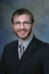 Hamilton Mark P合作论文数Department of Molecular and Cellular Biology, Baylor College of Medicine3
 Deshka Foster合作论文数Dept Surg, Stanford Univ3
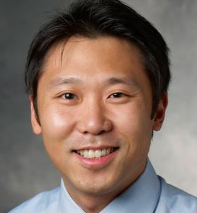 Juyong Brian Kim合作论文数Department of Medicine, Stanford University3
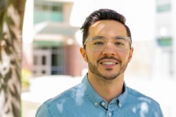 Sharifi Osman合作论文数Department of Biological Sciences, California State University3Kong Katherine A合作论文数3




