It is well known that exercise training (TRN) and ischemic preconditioning (IPC) reduce cardiomyocyte (CM) death following ischemia and reperfusion (IR), but it is unknown if protection can be elicited by moderate exercise TRN or if the effects of TRN and IPC summate to generate greater protection than either protocol alone. To address these questions, we used wheel running, a moderate-intensity paradigm, to TRN male and female rats, and two IPC protocols. Hearts were studied ex vivo with regional ischemia (RI) produced by occluding the left anterior descending coronary artery for 30 min followed by 3 h reperfusion. IPC consisted of a 5-min period of global ischemia and either 5 (PC5) or 10 (PC10) min reperfusion before the initiation of index RI. We found that wheel running and PC10, but not PC5, reduced CM death compared with sedentary, with the TRN effect greater in males. TRN, but not IPC, showed functional protection by reducing the loss of left ventricular developed pressure. Surprisingly, the TRN and PC10 protection from cell death was not additive, suggesting that these modalities converge on the same pathway. We also showed that TRN significantly improved mitochondrial respiration and ΔΨm repolarization during oxidative phosphorylation after IR when compared with SED rats; the improved bioenergetics was associated with the increased prosurvival kinase, hexokinase II (HKII), translocation to mitochondria, which was mediated by increased total Akt and the phosphorylated Akt. We conclude that TRN-induced cardioprotection is mediated, in part, by preservation of mitochondrial bioenergetics likely mediated via the Akt-pAkt-HKII signaling pathway.NEW & NOTEWORTHY Our study produced the novel findings that wheel running in rats, a form of moderate-intensity continuous training (MICT), can provide protection against IR injury as evidenced by a reduction in cardiomyocyte death following 3 h reperfusion and that the protection generated by MICT and IPC is not additive. Importantly, we demonstrate for the first time that MICT-induced cardioprotection is associated with preservations of mitochondrial bioenergetics likely by the Akt-pAkt-HKII signaling pathway.
INTRODUCTION:A previous study found that following out-of-hospital cardiac arrest (OHCA), 67% of out-of-hospital 12-lead electrocardiograms (ECGs) diagnostic for ST-segment elevation myocardial infarction (STEMI) changed to non-STEMI on repeat emergency department (ED) ECG. Here we evaluated associations with resolution of STEMI on ED ECG. METHODS:In this secondary analysis of a previous retrospective study, adults (≥18 years) with return of spontaneous circulation (ROSC) following OHCA, at least 1 out-of-hospital and ED ECG and transport to the study hospital were entered. We analyzed variables suspected of influencing ischemic changes on ECG including arrest characteristics, treatment interventions, resuscitation duration, and out-of-hospital and ED ECG acquisition times. RESULTS:Forty-nine of 176 patients entered had out-of-hospital ECGs diagnostic for STEMI, and 33/49 (67%) had resolved STEMI upon ED evaluation. Shorter resuscitation time (13 [interquartile range 5-18] vs 21 [14-28] minutes), p = 0.007), less epinephrine (3 [1-4] vs 5 [2-10] milligrams, p = 0.018), lower incidence of norepinephrine (5/33 (15%) vs 11/16 (69%), p ≤ 0.001), less time from ROSC to out-of-hospital ECG acquisition (5.5 [1-8] vs 8.5 [7-14] minutes, p = 0.044), and more time between out-of-hospital and ED ECG acquisition (34 [25-52] vs 21 [14-27] minutes, p = 0.001) were associated with resolution of out-of-hospital STEMI on ED evaluation. More defibrillations were associated with increased ischemia on ED ECG for patients with non-STEMI out-of-hospital ECGs. CONCLUSION:ROSC patients with STEMI on out-of-hospital ECG commonly resolve in the ED (67%). These identified associations may better inform clinical decision making. Post-ROSC out-of-hospital 12-lead ECGs should be repeated on arrival in the ED.
Exposure to diagnostic and therapeutic radiation introduces risks for development of diseases later in life by causing DNA damage in cells. Currently, there is no clinical method for determining exposure risk caused by radiation toxicity to DNA. Cell-free DNA (cfDNA), a marker of DNA damage, is currently used to assess risk for long-term effects following organ transplantation, surgery and inflammation. The goal of our proposed study is to develop cfDNA as an early biomarker for assessing risk for cardiovascular disease and cancer from radiation exposure so that strategies to mitigate the damaging effects of medical radiation can be assessed. Hearts from male and female WAG/RijCmcr rats (n = 6-10/group) were exposed to increasing doses of X-radiation (50 mGy and 3.5 Gy). Blood was collected prior to and after (15 minutes-96 hours) irradiation, and cell-free plasma was prepared. Primers and probes were designed for quantitative analysis of sequences of mitochondria (12S rRNA) and nuclear (Gapdh) cfDNA present in rat plasma using quantitative reverse transcription polymerase chain reaction (RT-qPCR). Exposure of hearts to radiation increased nuclear and mitochondrial cfDNA in a dose-dependent manner. Three point five grays from X-radiation increase cfDNA for Gapdh in plasma after 1 hour with a 15.8-fold increase (P < 0.001) after 6 hours. The earliest time nuclear and mitochondrial cfDNA increases were detected in plasma was at 60 minutes following exposure to 3.5 Gy. cfDNA has potential to advance as a biomarker of exposure to medical doses of radiation in patients.
Introduction: The space environment astronauts experience during space missions consists of multiple environmental challenges, including microgravity. In this study, we assessed the behavioral and cognitive performances of male Fisher rats 2 months after sham irradiation or total body irradiation with photons in the absence or presence of simulated microgravity. We analyzed the plasma collected 9 months after sham irradiation or total body irradiation for distinct alterations in metabolic pathways and to determine whether changes to metabolic measures were associated with specific behavioral and cognitive measures.Methods: A total of 344 male Fischer rats were irradiated with photons (6 MeV; 3, 8, or 10 Gy) in the absence or presence of simulated weightlessness achieved using hindlimb unloading (HU). To identify potential plasma biomarkers of photon radiation exposure or the HU condition for behavioral or cognitive performance, we performed regression analyses.Results: The behavioral effects of HU on activity levels in an open field, measures of anxiety in an elevated plus maze, and anhedonia in the M&M consumption test were more pronounced than those of photon irradiation. Phenylalanine, tyrosine, and tryptophan metabolism, and phenylalanine metabolism and biosynthesis showed very strong pathway changes, following photon irradiation and HU in animals irradiated with 3 Gy. Here, 29 out of 101 plasma metabolites were associated with 1 out of 13 behavioral measures. In the absence of HU, 22 metabolites were related to behavioral and cognitive measures. In HU animals that were sham-irradiated or irradiated with 8 Gy, one metabolite was related to behavioral and cognitive measures. In HU animals irradiated with 3 Gy, six metabolites were related to behavioral and cognitive measures.Discussion: These data suggest that it will be possible to develop stable plasma biomarkers of behavioral and cognitive performance, following environmental challenges like HU and radiation exposure.
IntroductionEffects and interactions between different spaceflight stressors are expected to be experienced by crew on missions when exposed to microgravity and galactic cosmic rays (GCRs). One of the limitations of previous studies on simulated weightlessness using hindlimb unloading (HU) is that a control HU condition was not included.MethodsWe characterized the behavioral performance of male Fischer rats 2 months after sham or total body irradiation with a simplified 5-ion 6-mixed-beam exposure representative of GCRs in the absence or presence of HU. Six months later, the plasma, hippocampus, and cortex were processed to determine whether the behavioral effects were associated with long-term alterations in the metabolic pathways.ResultsIn the open field without and with objects, interactions were observed for radiation × HU. In the plasma of animals that were not under the HU or control HU condition, the riboflavin metabolic pathway was affected most for sham irradiation vs. 0.75 Gy exposure. Analysis of the effects of control HU on plasma in the sham-irradiated animals showed that the alanine, aspartate, glutamate, riboflavin, and glutamine metabolisms as well as arginine biosynthesis were affected. The effects of control HU on the hippocampus in the sham-irradiated animals showed that the phenylalanine, tyrosine, and tryptophan pathway was affected the most. Analysis of effects of 0.75 Gy irradiation on the cortex of control HU animals showed that the glutamine and glutamate metabolic pathway was affected similar to the hippocampus, while the riboflavin pathway was affected in animals that were not under the control HU condition. The effects of control HU on the cortex in sham-irradiated animals showed that the riboflavin metabolic pathway was affected. Animals receiving 0.75 Gy of irradiation showed impaired glutamine and glutamate metabolic pathway, whereas animals receiving 1.5 Gy of irradiation showed impaired riboflavin metabolic pathways. A total of 21 plasma metabolites were correlated with the behavioral measures, indicating that plasma and brain biomarkers associated with behavioral performance are dependent on the environmental conditions experienced.DiscussionPhenylalanine, tyrosine, and tryptophan metabolism as well as phenylalanine and tryptophan as plasma metabolites are biomarkers that can be considered for spaceflight as they were revealed in both Fischer and WAG/Rij rats exposed to simGCRsim and/or HU.
Radiation therapy (RT) plays an integral role in treating thoracic cancers, despite the risk of radiation-induced cardiotoxicity. We hypothesize that our newly developed magnetic resonance imaging (MRI)-based contractility index (ContractiX) is a sensitive marker for early detection of RT-induced cardiotoxicity in a preclinical rat model of thoracic cancer RT. Adult salt-sensitive rats received image-guided heart RT and were imaged with MRI at 8 weeks and 10 weeks post-RT or sham. The MRI exam included cine and tagging sequences to measure left-ventricular ejection fraction (LVEF), mass, myocardial strain, and ContractiX. Furthermore, ventricular torsion, diastolic strain rate, and mechanical dyssynchrony were measured. Statistical analyses were performed between the sham, 8 weeks post-RT, and 10 weeks post-RT MRI parameters. The results showed that both LVEF and myocardial mass increased post-RT. Peak systolic strain and ContractiX significantly decreased post-RT, with a more relative reduction in ContractiX compared to strain. ContractiX showed an inverse nonlinear relationship with LVEF and continuously decreased with time post-RT. While early diastolic strain rate and mechanical dyssynchrony significantly changed post-RT, ventricular torsion changes were not significant post-RT. In conclusion, ContractiX measured via non-contrast MRI is a sensitive early marker for the detection of subclinical cardiac dysfunction post-RT, and it is superior to other MRI cardiac measures.
Heart disease is a significant adverse event caused by radiotherapy for some cancers. Identifying the origins of radiogenic heart disease will allow therapies to be developed. Previous studies showed non-targeted effects manifest as fibrosis in the non-irradiated heart after 120 days following targeted X-irradiation of the kidneys with 10 Gy in WAG/RijCmcr rats. To demonstrate the involvement of T cells in driving pathophysiological responses in the out-of-field heart, and to characterize the timing of immune cell engagement, we created and validated a T cell knock downrat on the WAG genetic backgrou nd. Irradiation of the kidneys with 10 Gy of X-rays in wild-type rats resulted in infiltration of T cells, natural killer cells, and macrophages after 120 days, and none of these after 40 days, suggesting immune cell engagement is a late response. The radiation nephropathy and cardiac fibrosis that resulted in these animals after 120 days was significantly decreased in irradiated T cell depleted rats. We conclude that T cells function as an effector cell in communicating signals from the irradiated kidneys which cause pathologic remodeling of non-targeted heart.
Long-duration space exploratory missions to the Earth's moon and the planet Mars are actively being planned. Such missions will require humans to live for prolonged periods beyond low earth orbit where astronauts will be continuously exposed to high energy galactic cosmic rays (GCRs). A major unknown is the potential impact of GCRs on the risks of developing degenerative cardiovascular disease, which is a concern to NASA. A ground-based rat model has been used to provide a detailed characterization of the risk of long-term cardiovascular disease from components of GCRs at radiation doses relevant to future human missions beyond low earth orbit. Six month old male WAG/RijCmcr rats were irradiated at a ground-based charged particle accelerator facility with high energy ion beams broadly representative of GCRs: protons, silicon and iron. Irradiation was given either as a single ion beam or as a combination of three ion beams. For the doses used, the single ion beam studies did not show any significant changes in the known cardiac risk factors and no evidence of cardiovascular disease could be demonstrated. In the three ion beam study, the total cholesterol levels in the circulation increased modestly over the 270 day follow up period, and inflammatory cytokines were also increased, transiently, 30 days after irradiation. Perivascular cardiac collagen content, systolic blood pressure and the number of macrophages found in the kidney and in the heart were each increased 270 days after irradiation with 1.5 Gy of the three ion beam grouping. These findings provide evidence for a cardiac vascular pathology and indicate a possible threshold dose for perivascular cardiac fibrosis and increased systemic systolic blood pressure for complex radiation fields during the 9 month follow up period. The development of perivascular cardiac fibrosis and increased systemic systolic blood pressure occurred at a physical dose of the three ion beam grouping (1.5 Gy) that was much lower than that required to show similar outcomes in earlier studies with the same rat strain exposed to photons. Further studies with longer follow up periods may help determine whether humans exposed to lower, mission-relevant doses of GCRs will develop radiation-induced heart disease.
In the original publication [...].
A limitation of simulated space radiation studies is that radiation exposure is not the only environmental challenge astronauts face during missions. Therefore, we characterized behavioral and cognitive performance of male WAG/Rij rats 3 months after sham-irradiation or total body irradiation with a simplified 5-ion mixed beam exposure in the absence or presence of simulated weightlessness using hindlimb unloading (HU) alone. Six months following behavioral and cognitive testing or 9 months following sham-irradiation or total body irradiation, plasma and brain tissues (hippocampus and cortex) were processed to determine whether the behavioral and cognitive effects were associated with long-term alterations in metabolic pathways in plasma and brain. Sham HU, but not irradiated HU, rats were impaired in spatial habituation learning. Rats irradiated with 1.5 Gy showed increased depressive-like behaviors. This was seen in the absence but not presence of HU. Thus, HU has differential effects in sham-irradiated and irradiated animals and specific behavioral measures are associated with plasma levels of distinct metabolites 6 months later. The combined effects of HU and radiation on metabolic pathways in plasma and brain illustrate the complex interaction of environmental stressors and highlights the importance of assessing these interactions.
A primary objective of the National Aeronautics and Space Administration (NASA) is expansion of humankind’s presence outside low-Earth orbit, culminating in permanent interplanetary travel and habitation. Having no inherent means of physiological detection or protection against ionizing radiation, humans incur capricious risk when journeying beyond low-Earth orbit for long periods. NASA has made large investments to analyze pathologies from space radiation exposure, emphasizing the importance of characterizing radiation’s physiological effects. Because natural evolution would require many generations to confer resistance against space radiation, immediately pragmatic approaches should be considered. Volitional evolution, defined as humans steering their own heredity, may inevitably retrofit the genome to mitigate resultant pathologies from space radiation exposure. Recently, uniquely radioprotective genes have been identified, conferring local or systemic radiotolerance when overexpressed in vitro and in vivo. Aiding in this process, the CRISPR/Cas9 technique is an inexpensive and reproducible instrument capable of making limited additions and deletions to the genome. Although cohorts can be identified and engineered to protect against radiation, alternative and supplemental strategies should be seriously considered. Advanced propulsion and mild synthetic torpor are perhaps the most likely to be integrated. Interfacing artificial intelligence with genetic engineering using predefined boundary conditions may enable the computational modeling of otherwise overly complex biological networks. The ethical context and boundaries of introducing genetically pioneered humans are considered.
Introduction: We evaluated the incidence of change in serial 12-lead electrocardiogram (ECG) diagnostic classifications in patients resuscitated from out-of-hospital (OH) cardiac arrest (OHCA) comparing OH to emergency department (ED) ECGs. Methods: This retrospective case series included: 1) adults (> 18 years old), 2) resuscitated from OHCA, 3) > 1 OH and 1 ED ECG/patient, and 4) emergency medical services (EMS) transport to the study hospital. OH and ED ECGs were classified as: 1) STEMI (ST-segment Elevation Myocardial Infarction), 2) Ischemic, and 3) Non-ischemic. Two ED physicians and one cardiologist independently classified all ECGs, then generated a consensus opinion classification for each ECG based on American Heart Association's 2018 Expert Consensus criteria. The most ischemic OH ECG classification was compared with the last ED ECG classification. Results: From 7/27/12 to 7/18/19, 176 patients were entered with a mean age of 61.2 +/- 16.6 years; 102/176 (58%) were male. Overall, 504 OH and ED 12-lead ECGs were acquired (2.9 ECGs/patient). ECG classification inter-rater reliability kappa score was 0.63 +/- 0.02 (substantial agreement). Overall, 86/176 (49%) changed ECG classification from the OH to ED setting; 69/86 (80%) of these ECGs changed from more to less ischemic classifications. Of 49 OH STEMI ECG classifications, 33/49 (67%) changed to a less ischemic (non-STEMI) ED ECG classification. Conclusions: Change in 12-lead ECG classification from OH to ED setting in patients resuscitated from OHCA was common (49%). The OH STEMI classification changed to a less ischemic (non-STEMI) ED classification in 67% of cases.
Cardiac disease is a frequent and significant adverse event associated with radiotherapy for cancer. Identifying the underlying mechanism responsible for radiation injury to the heart will allow interventions to be developed. In the present study, we tested if local kidney irradiation results in remodeling of the shielded, nontargeted heart. One kidney, two kidneys, or the total body of male WAG and Dahl SS rats were irradiated with 10 Gy of X-rays. Local kidney irradiation resulted in systemic hypertension, increased BUN, infiltration of T lymphocytes, natural killer cells, and macrophages into the renal cortex and medulla, and renal fibrosis. Local irradiation of kidneys in WAG rats resulted in remodeling in the nontargeted heart after 120 days, manifested by perivascular fibrosis and increased interventricular septal thickness, but was not seen in Dahl SS rats due to a high baseline level of fibrosis in the sham-irradiated animals. Genetic depletion of T cells mitigated the nephropathy after local kidney irradiation, indicating a role for the immune system in mediating this outcome. Local kidney irradiation resulted in a cascade of pro-inflammatory cytokines and low-molecular weight metabolites into the circulation associated with transmission of signals resulting in pathologic remodeling in the nontargeted heart. A new model is proposed whereby radiation-induced cardiac remodeling in susceptible animals is indirect, with lower hemi body organs such as the kidney exporting factors into the circulation that cause remodeling outside of the irradiated field in the shielded, nontargeted heart. This nontargeted effect appears to be mediated, in part, by the immune system.
Radiotherapy with sparsely ionizing photons is a cornerstone of successful cancer treatment. Age at time of exposure to radiation is known to influence biological outcomes for many end points. The effect of dose and age at exposure upon the occurrence of radiogenic cardiovascular disease is poorly understood. The goal of this work was to determine the response of maleWAG/RijCmcr rats at 6 months of age to gamma rays, and at 6 months or 6 weeks of age to X rays, using clinically relevant biomarkers of cardiovascular disease and kidney injury. Overall, there were significant radiation-induced effects on the levels of bicarbonate (P=0.0016), creatinine (P=0.0002), calcium (P = 0.0009), triglycerides (P = 0.0269) and blood urea nitrogen, albumin, protein, AST, alkaline phosphatase, total cholesterol and HDL (all P < 0.0001). Of those variables with a significant radiation-dose effect, there were significant modifications by age at time of exposure for bicarbonate (P = 0.0033), creatinine (P = 0.0015), AST (P = 0.0040), total cholesterol (P = 0.0006) and blood urea nitrogen, calcium, albumin, protein, alkaline phosphatase and HDL (all P < 0.0001). Cardiac perivascular collagen content was significantly increased in rats that were 8.0 Gy X-ray irradiated at 6 weeks of age (P < 0.047) but not at 6 months of age. While systemic blood pressure was elevated in both cohorts after 8.0 Gy X-ray irradiation (compared to agematched sham-irradiated controls), the magnitude of the increase above baseline was greater in the younger rats (P < 0.05). These findings indicate that dose and age at time of irradiation determine the timeline and severity of cardiac and renal injury.
Rationale: A strong association has emerged between the gut microbiome and atherosclerotic disease. Our recent data suggest Lactobacillus plantarum 299v (Lp299v) supplementation reduces infarct size in male rats. Limited human data are available on the impact of Lp299v on the vasculature. Objective: To determine whether oral Lp299v supplementation improves vascular endothelial function and reduces systemic inflammation in humans with stable coronary artery disease (CAD). Methods and Results: Twenty men with stable CAD consumed a drink containing Lp299v (20 billion CFU) once daily for 6 weeks. After a 4-week washout, subjects were given an option of additionally participating in a 10-day study of oral liquid vancomycin (250 mg QID). Vascular endothelial function was measured by brachial artery flow-mediated dilation. Before and after Lp299v, plasma short-chain fatty acids, trimethylamine oxide, and adipokine levels were measured. Additional plasma samples underwent unbiased metabolomic analyses using liquid chromatography/mass spectroscopy. 16S rRNA sequencing was used to determine changes of the stool microbiome. Arterioles from patients with CAD were obtained, and endothelium-dependent vasodilation was measured by video microscopy after intraluminal incubation with plasma from Lp299v study subjects. Lp299v supplementation improved brachial flow-mediated dilation ( P =0.008) without significant changes in plasma cholesterol profiles, fasting glucose, or body mass index. Vancomycin did not impact flow-mediated dilation. Lp299v supplementation decreased circulating levels of IL (interleukin)-8 ( P =0.01), IL-12 ( P =0.02), and leptin ( P =0.0007) but did not significantly change plasma trimethylamine oxide concentrations ( P =0.27). Plasma propionate ( P =0.004) increased, whereas acetate levels decreased ( P =0.03). Post-Lp299v plasma improved endothelium-dependent vasodilation in resistance arteries from patients with CAD ( P =0.02).16S rRNA analysis showed the Lactobacillus genus was enriched in postprobiotic stool samples without other changes. Conclusions: Lp299v improved vascular endothelial function and decreased systemic inflammation in men with CAD, independent of changes in traditional risk factors and trimethylamine oxide. Circulating gut-derived metabolites likely account for these improvements and merit further study. Clinical Trial Registration: URL: http://www.clinicaltrials.gov . Unique identifier: NCT01952834.
Radiation is a cornerstone of successful cancer treatment, with one-half to two-thirds of all patients receiving radiotherapy. Survivors of cancer treated with radiation are at increased risk for cardiovascular disease (CVD). Understanding how radiation causes CVD will allow development of novel therapies. Irradiation of the lower hemi body, but not the upper hemi body, with 10 Gy in rats increases risk factors for CVD and results in cardiac fibrosis quantitatively similar to total body irradiation, suggesting radiation injury to the heart can be indirect. Simvastatin, an inhibitor of liver cholesterol synthesis administered continuously to rats (10 mg/kg/day) after 10 Gy lower hemi body irradiation mitigated against increased blood cholesterol and cardiac fibrosis. These findings indicate simvastatin limits transmission of a signal from the lower hemi body that decreases risk for and occurrence of CVD independent of any direct exposure of the heart to radiation. Bivariate examination of 3,607 patients following therapeutic lower hemi body irradiation using Chi-square, Wilcoxon rank-sum and t-tests was used to examine risk factors for CVD in patients diagnosed with congestive heart failure, myocardial infarction, atrial fibrillation, and cardiomyopathy before 80 years of age. We found that 47.4% of patients age 70-80 developed CVD compared to 29.7% who received simvastatin (p= < 0.001, n = 293 and 361, respectively). Patients who were male, overweight, smokers, and had a diagnosis of chronic kidney disease and diabetes also had significantly higher risk of CVD. Race and hypertension were not indicative of increased risk for CVD. These clinical findings, taken together with the results from our animal studies, support a new research paradigm where radiation-induced heart disease can be indirect, with abdominal organs exporting factors that cause CVD. Simvastatin can be developed to mitigate and treat CVD after therapeutic radiation.
Intestinal microbiota determine severity of myocardial infarction in rats. We determined whether low molecular weight metabolites derived from intestinal microbiota and transported to the systemic circulation are linked to severity of myocardial infarction. Plasma from rats treated for seven days with the non-absorbed antibiotic vancomycin or a mixture of streptomycin, neomycin, polymyxin B and bacitracin was analyzed using mass spectrometry-based metabolite profiling platforms. Antibiotic-induced changes in the abundance of individual groups of intestinal microbiota dramatically altered the host's metabolism. Hierarchical clustering of dissimilarities separated the levels of 284 identified metabolites from treated vs. untreated rats; 193 were altered by the antibiotic treatments with a tendency towards decreased metabolite levels. Catabolism of the aromatic amino acids phenylalanine, tryptophan and tyrosine was the most affected pathway comprising 33 affected metabolites. Both antibiotic treatments decreased the severity of an induced myocardial infarction in vivo by 27% and 29%, respectively. We then determined whether microbial metabolites of the amino acids phenylalanine, tryptophan and tyrosine were linked to decreased severity of myocardial infarction. Vancomycin-treated rats were administered amino acid metabolites prior to ischemia/reperfusion studies. Oral or intravenous pretreatment of rats with these amino acid metabolites abolished the decrease in infarct size conferred by vancomycin. Inhibition of JAK-2 (AG-490, 10 μM), Src kinase (PP1, 20 μM), Akt/PI3 kinase (Wortmannin, 100 nM), p44/42 MAPK (PD98059, 10 μM), p38 MAPK (SB203580, 10 μM), or KATP channels (glibenclamide, 3 μM) abolished cardioprotection by vancomycin, indicating microbial metabolites are interacting with cell surface receptors to transduce their signals through Src kinase, cell survival pathways and KATP channels. These inhibitors have no effect on myocardial infarct size in untreated rats. This study links gut microbiota metabolites to severity of myocardial infarction and may provide future opportunities for novel diagnostic tests and interventions for the prevention of cardiovascular disease.
Objective: Recombinant Human Erythropoietin (rhEPO) is strongly inferred to protect the cardiomyocytes from the reperfusion injury and our aim is to elucidate the cardioprotective effect and the exact mechanism behind the cardioprotection. Methods: Neonatal rat cardiomyocytes (NCM) exposed to Hypoxia/Reperfusion (H/R) with or without pretreatment using various concentrations of rhEPO. To determine the cell viability-MTT assay, Acridine orange and Ethidium Bromide (Ao/EtBr) staining was performed. To determine the reactive oxygen species (ROS) and mitochondrial membrane potential (Δψm), Dichlorofluorescein diacetate (DCF-DA) and Rhodamine-123 was used. To determine the signaling pathways Western blot analysis of pAkt, pp38 MAPK, cytochrome-c were performed. Results: rhEPO was found to reduce the cell death by stabilizing ROS significantly, Δψm, cytochrome c release, and caspase-3. rhEPO, increases the phosphorylation of p38 MAPK, Akt and BAD compared to H/R. Further myocytes blocked with Wortmannin (WT), and SB203580 showed increased caspase-3 activity. Conclusion: Hence we conclude from this study that rhEPO regulated the factors involved in reperfusion injury through modulation of Akt and p38 MAPK pathways.
Introduction: There is a significant unmet clinical need to identify novel therapies to prevent and treat the progression of cardiovascular disease. This requires identification of additional contributory processes to the pathogenesis of heart disease so that mechanism-based interventions may be developed. Hypothesis: Our objective was to translate the recently discovered mechanistic link in rats between changes in the intestinal microbiota and the pathogenesis of cardiovascular disease to humans. Methods: Twenty male adult patients with stable coronary heart disease on stable doses of vasoactive medications and HMG-CoA reductase inhibitors consumed a probiotic drink containing Lactobacillus plantarum 299v (3 oz. serving containing 20 billion colony forming units) once a day for 42 days. Vascular endothelial function was measured at the beginning and end of the study period by brachial flow-mediated dilation. Results: The probiotic improved brachial artery flow mediated dilation, a surrogate marker of cardiovascular risk, by 33% from 3.55 + 1.96% to 4.73 + 2.33% with a trend towards decreased LDL-cholesterol by 8% from 96 + 11 mg/dl to 88 + 10 mg/dl. Probiotic therapy decreased circulating levels of the inflammatory cytokines IL-8 by 24% (17±2 to 13±1 pg/ml) and IL-12 by 20% (56±7 to 45±6 pg/ml). Whole microbiome analysis based on abundance of 2,206 taxa using hierarchical clustering revealed no separation of the overall intestinal microbial population between pre- and post-probiotic treatment samples. Bacterial genus richness ranged from 334 to 530 and archaeal ranged from 8 to 25, with no significant differences between pre- and post-probiotic samples. Lactobacillus and Bacillus species were enriched in post-probiotic samples. Lactobacillus plantarum 299v levels were elevated in post-probiotic samples, confirming patient compliance. Conclusions: Our discovery of a relationship between Lactobacillus plantarum 299v, improved vascular endothelial function and decreased inflammation indicates intestinal microbiota may be developed as therapies to prevent and treat the progression of cardiovascular disease.
 Garrett Gross合作论文数Department of Pharmacology and Toxicology, Medical College of Wisconsin31
 Balaraman Kalyanaraman合作论文数Department of Biophysics, Medical College of Wisconsin25
 Jidong Su合作论文数24KA PRITCHARD合作论文数20
 Yang Shi合作论文数National Institutes of Health18
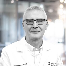 Eugene A. Konorev合作论文数Biophysics Research Institute, Medical College of Wisconsin15Richard A. Komorowski合作论文数15
 Nicholas N Kipshidze合作论文数Lenox Hill Heart and Vascular Institute of New York15Gordon N. Olinger合作论文数13Anna Hsu合作论文数13
 Marek Lenarczyk合作论文数Department of Radiation Protection and Radiobiology, National Institute of Hygiene13Js Tweddell合作论文数12Joy Joseph合作论文数Med Coll Wisconsin11David J. Hearse合作论文数11Parvaneh Rafiee合作论文数11
 Jennifer L Strande合作论文数10LE BOERBOOM合作论文数9Kronenberg Amy合作论文数9John E. Moulder合作论文数8Victor Nikolaychik合作论文数8
 Zeljko J. Bosnjak合作论文数Medical College of Wisconsin8
 John W Hopewell合作论文数University of Oxford NHS Trust7
 Stephen J. Sugden合作论文数7
 Nita H. Salzman合作论文数Department of Pediatrics and Microbiology & Immunology, Medical College of Wisconsin6
 Howard J. Jacob合作论文数AbbVie;MedRedi Solutions, LLP.6Vy Lam合作论文数6Xiangping Fu合作论文数6Raymond Q. Migrino合作论文数6
 Brian L Fish合作论文数6Er Jacobs合作论文数5SJ CONTNEY合作论文数5Mh Keelan合作论文数5P Holman合作论文数5H Sahota合作论文数5J Su合作论文数5G.R. Bullock合作论文数5
 Anuradha Dhanasekaran合作论文数5Wc Hutchins合作论文数4Dl Zhu合作论文数4Xiangrong Kong合作论文数4
 James Tweddell合作论文数4Alexander M Borg合作论文数4
 David L. Mattson合作论文数Department of Physiology, Medical College of Georgia, Augusta University4
 Widlansky Michael E合作论文数Div Cardiovasc Med, Med Coll Wisconsin4Devendran Ajay合作论文数4Alfred C. Nicolosi合作论文数4
 William Hutchins合作论文数4Marylou Mäder合作论文数4




