Background:Long non-coding RNAs (lncRNAs) have emerged as key regulators of tumor biology, however, thus far none have translated to cancer therapies. The lncRNA MALAT1 is overexpressed in more than 20 cancers, including breast cancer and has been shown to function via various mechanisms in a context-dependent manner, in 2D cell lines and mouse models. However, its functional role and therapeutic potential have not been evaluated in clinically relevant patient-derived models. Methods:We investigated the therapeutic potential of a MALAT1-targeting antisense oligonucleotide (ASO) for breast cancer, using clinically relevant 3D human patient-derived organoids (PDOs) and PDO-xenograft (PDO-X) models. We systematically evaluated the efficiency of MALAT1-targeting ASOs using a biobank of 28 PDO models. Using three independent PDO-X models of triple negative breast cancer (TNBC), we targeted MALAT1 in vivo to study its impact on transcription, alternative splicing, stromal remodeling and metastasis. Results:Across PDO-X models, MALAT1 depletion reproducibly drove widespread alternative splicing changes across all event types, particularly intron retention events, accompanied by modest gene expression alterations. Differentially spliced transcripts were enriched for targets of shared cancer-associated transcription factors, and MALAT1 knockdown altered the relative abundance of previously unannotated splicing isoforms. Beyond tumor-intrinsic effects, tumor-specific MALAT1 depletion induced a consistent reduction in macrophage-associated gene signatures and reduced lung metastatic burden. Conclusions:Our data define MALAT1's multifaceted role in TNBC, coordinating alternative splicing, transcriptional fine-tuning, tumor-stroma crosstalk, and metastatic progression. Our study provides strong preclinical evidence supporting MALAT1-targeted ASO therapy and establishes PDO-X models as a clinically relevant platform for functional interrogation of TNBC therapies.
Diffuse midline gliomas (DMGs) are a deadly class of pediatric high-grade brain cancers. Approximately 80% of pontine DMGs feature a dominant, somatic, heterozygous point mutation in the non-canonical histone H3.3-coding gene H3-3A. This dominant-negative mutation replaces lysine 27 with methionine (K27M) and prevents global K27 di- and tri-methylation of all wild-type histone H3 proteins. We aimed to target the H3.3K27M onco-histone pre-mRNA with splice-switching antisense oligonucleotides (ASOs) designed to promote skipping of H3-3A exon 2, as this constitutive exon comprises both the K27M mutation and the natural in-frame start codon of the gene. The lead ASO identified in a systematic screen specifically induced H3-3A exon 2 skipping, did not affect expression or splicing of the paralog gene H3-3B—which also encodes histone H3.3—and restored global H3K27me3 marks in patient-derived DMG cells grown as neurospheres. Furthermore, the lead ASO reduced proliferation and extended survival in a patient-derived orthotopic xenograft tumor mouse model. Our results show the potential of exon-skipping ASOs targeting H3-3A exon 2 as a therapeutic option for H3.3K27M-altered DMG. More generally, they exemplify the strategy of using ASOs to induce skipping of a constitutive exon to effectively achieve gene downregulation.
Lysyl oxidases crosslink type I collagen to promote fibrosis and cancer progression in mouse mammary tumor models. Pancreatic ductal adenocarcinomas (PDACs) are highly fibrotic and contain abundant type I collagen with elevated expression of lysyl oxidases. Indeed, inhibition of lysyl oxidases constitute an attractive anti-tumor therapeutic strategy, with several reported preclinical studies demonstrating efficacy at reducing PDAC fibrosis and progression. Yet, lysyl oxidase was first described as an anti-oncogene through its effect of directly suppressing cell transformation by mutant Ras which is present in around 90% of human pancreatic tumors. These prior studies highlight the dual functions, anti-ras and pro-fibrotic, of lysyl oxidases in pancreatic cancer. As a result, clinical trials targeting lysyl oxidase in cancers have demonstrated limited efficacy. Here we examined the effects of perturbation of lysyl oxidase activity or expression using syngeneic orthotopic transplantation models expressing mutant Ras and intravital imaging. Unexpectedly, genetic or pharmacological inhibition of lysyl oxidases increased invasion along collagen fibers and distant metastasis. Furthermore, inhibition of lysyl oxidases promoted focal adhesion kinase (FAK) activity which was required for metastasis. We found that mutant Kras status dictated lysyl oxidase-mediated suppression on FAK signaling in both mouse and human pancreatic cancer cells. These results suggest that the effect of lysyl oxidase on metastasis are dependent on signaling from Ras and FAK. These results strongly caution against inhibiting lysyl oxidases for cancers driven by mutant Ras. Lijuan Sun, Jean Albrengues, John E. Wilkinson, Sarah L. Dallas, Valerie M. Weaver, Mikala Egeblad, Mario A. Shields. Lysyl oxidases suppress pancreatic cancer progression by inhibiting focal adhesion kinase signaling [abstract]. In: Proceedings of the American Association for Cancer Research Annual Meeting 2025; Part 1 (Regular Abstracts); 2025 Apr 25-30; Chicago, IL. Philadelphia (PA): AACR; Cancer Res 2025;85(8_Suppl_1):Abstract nr 138.
The role of driver gene mutations in sustaining tumor growth at metastatic sites is poorly understood. SMAD4 inactivation is a paradigm of such mutations and a hallmark of pancreatic ductal adenocarcinoma (PDAC). To determine whether metastatic tumors are dependent on SMAD4 inactivation, we developed a mouse model of PDAC that enables spatiotemporal control of Smad4 expression. While Smad4 inactivation in the premalignant pancreas facilitated the formation of primary tumors, Smad4 reactivation in metastatic disease suppressed liver metastases but promoted lung metastases. These divergent effects were underpinned by organ-biased differences in the tumor cells' chromatin state that emerged in the premalignant pancreas and were distinguished by the dominance of KLF4 versus RUNX1 transcription factors. Our results show how epigenetic states favored by the organ of residence can influence the output of driver mutations in metastatic tumors, which has implications for interpreting tumor genetics and therapeutically targeting metastatic disease.
Figure S1. Elevated SRSF1 induces pancreatitis and is associated with its duration. Figure S2. Elevated SRSF1 predicts poor PDAC clinical prognosis and is associated with PDAC progression. Figure S3. SRSF1 accelerates KRASG12D-mediated PDAC tumorigenesis. Figure S4. SRSF1 loss delays KRASG12D-mediated PDAC initiation. Figure S5. Opposite patterns of pathway enrichment by KRASG12D mutation versus SRSF1 upregulation. Figure S6. Recombination and activation of KrasG12D allele in pancreas cells from KC mice. Figure S7. KSC and KPSC mice exhibit a hyper-activated MAPK pathway, suppression of which diminished SRSF1-induced pancreatitis. Figure S8. Decreased SRSF1 expression perturbs MAPK signaling. Figure S9. MYC promotes SRSF1 expression in PDAC. Figure S10. SRSF1 regulates IL1R1 alternative splicing and expression. Figure S11. Knockout of Il1r1 rescues SRSF1-induced pancreatitis. Table S1. Oligonucleotides for PCR and short hairpin RNAs.
Context-specific epigenetic dependencies, shaped by chromatin remodeling can create exploitable vulnerabilities for cancer therapies that are unique to tissue types and cellular identities. Here, we show that loss of BPTF (Bromodomain PHD Finger Transcription Factor), a core component of the NURF (Nucleosome Remodeling Factor) complex, results in the emergence of estrogen-responsive, tamoxifen-sensitive, Estrogen Receptor alpha (ERα) positive mammary tumors without altering cancer cell state and tumor pathology. Elevated ERα levels in BPTFKO mammary tumor cells are linked with decreased TGF-β activity and limited metastatic spread of mammary tumor cells to the lungs. Loss of ERα is sufficient to restore TGF-β activity and the metastatic potential in BPTFKO tumors. These findings highlight a mechanism through which BPTF regulates tumor development and progression in mammary epithelial cells, offering insights into the interplay between chromatin remodeling, estrogen signaling, and their resultant adjuvant therapeutic potential in breast cancer.
Exposure to pathogens throughout a lifetime influences immunity and organ function. Here, we explore how the systemic host-response to bacterial urinary tract infection (UTI) induces tissue-specific alterations to the mammary gland. Utilizing a combination of histological tissue analysis, single cell transcriptomics, and flow cytometry, we identify that mammary tissue from UTI-bearing mice displays collagen deposition, enlarged ductal structures, ductal hyperplasia with atypical epithelial transcriptomes and altered immune composition. Bacterial cells are absent in the mammary tissue and blood of UTI-bearing mice, therefore, alterations to the distal mammary tissue are mediated by the systemic host response to local infection. Furthermore, broad spectrum antibiotic treatment resolves the infection and restores mammary cellular and tissue homeostasis. Systemically, unresolved UTI correlates with increased plasma levels of the metalloproteinase inhibitor, TIMP1, which controls extracellular matrix remodeling and neutrophil function. Treatment of nulliparous and post-lactation UTI-bearing female mice with a TIMP1 neutralizing antibody, restores mammary tissue normal homeostasis, thus providing evidence for a link between the systemic host response during UTI and mammary gland alterations. Urinary tract infections (UTIs) can elicit systemic host-responses. Here the authors report that, in a mouse model, unresolved UTI is associated with alterations of the mammary tissue, including collagen deposition and hyperplasia.
Abstract Chromatin remodeling controls transcriptional programs that drive mammary epithelial lineage commitment and cellular differentiation. This process is also important for the regulation of highly dynamic cellular states during mammary tumorigenesis, and perturbing the chromatin landscape of cancer cells can halt cellular and molecular mechanisms of mammary cancer development and progression. However, this paradigm assumes that all cancer cells that develop in the mammary tissue bear the same chromatin dependencies, and rely on similar programs throughout cancer progression. Here, we show that molecular dependencies regulated by the epigenetic factor BPTF control tumor development and progression across distinct mammary cancer cell states. With the utilization of classical models of mammary tumorigenesis, single cell transcriptional profiling, and organoid cultures, we demonstrate that BPTF loss results in the development of mammary tumors with a longer latency of ER•+ expression and tamoxifen sensitivity and the inhibition of lung metastasis development. While gain of ER• expression was associated with gain of luminal markers in Krt5+ BPTF KO mammary epithelial cells (MECs), metastasis-like signatures were significantly perturbed in cells with defined luminal fate. In addition, we detected a partial rescue of metastatic features via the activation of transcriptional programs controlled by TGF-•, thus indicating that BPTF sits atop a canonical metastatic pathway. Collectively, our studies illustrate how BPTF-specific chromatin remodeling inhibition induces phenotypes that differentially affect tumor development and progression, while allowing for the development of a murine model of ER•+ mammary tumorigenesis. Citation Format: Chen Chen, Michael F. Ciccone, Marygrace c. Trousdell, Deeptiman Chatterjee, Yixin Zhao, Steven M. Lewis, Dhivya Anandan, John E. Wilkinson, William C. K. Pomerantz, Adam Siepel, Camila O. dos Santos. Loss of BPTF restores estrogen-regulated programs and suppress metastatic development of mammary tumors [abstract]. In: Proceedings of the AACR Special Conference in Cancer Research: Advances in Breast Cancer Research; 2023 Oct 19-22; San Diego, California. Philadelphia (PA): AACR; Cancer Res 2024;84(3 Suppl_1):Abstract nr A079.
Abstract Background: Pancreatic ductal adenocarcinoma (PDAC) is highly resistant to therapy. Stromal cancer-associated fibroblasts (CAFs) play a key role in promoting tumor progression and chemoresistance, by creating a fibrotic microenvironment that impedes drug access and feeds PDAC cells nutrients and pro-tumor signals. CRO-67 (Patent PCT/AU2023/050505) is a novel drug developed with Noxopharm Ltd using a rational medicinal chemistry design to improve bioavailability of chromans which have potent anti-cancer activity. Using our patient-derived PDAC tumor in a dish model (explants; maintain multicellular architecture and fibrosis) we previously showed that CRO-67 has both anti-tumor and CAF reprogramming capacity in 4 patient explants [Cancer Res (2022) 82 (22_Supplement):C073]. Aims: 1) To expand evaluation of CRO-67 in additional patient-derived PDAC tumor explants given PDAC heterogeneity. 2) Assess the effect of CRO-67 on PDAC cell and CAF function. 3) Validate the therapeutic potential of CRO-67 on PDAC growth in vivo. Methods: 1) PDAC tumor samples collected from 7 patients undergoing pancreatic resection. Tumor explants (1–2mm diameter) were cultured on gelatin sponges, treated with CRO-67 (0–50μg/mL) every 3 days and fixed on day 12. Therapeutic response assessed by immunohistochemistry for cytokeratin (PDAC cells), α-smooth muscle actin (CAFs), bromodeoxyuridine (proliferation) and TUNEL (cell death). 2) PDAC cells (MiaPaCa-2) and patient-derived CAFs were treated with CRO-67 (1.5μM) for 48h before proliferation analysis (IncuCYTE S3) and for 24h before apoptosis (Annexin V/DAPI staining; flow cytometry) and cell cycle (DAPI staining; flow cytometry) analysis. 3) Subcutaneous PDAC (BxPC3 human cells) tumors in mice were treated with CRO-67 (2.5mg/kg Intraperitoneally, twice a day) for 21 days and tumor volume measured (calipers). Results: 1) CRO-67 treatment decreased tumor and CAF cell frequency in 6/6 (no quantifiable tumor in patient 7 explants) and 7/7 patient PDAC explants, respectively. It also decreased explant cell proliferation and increased cell death versus controls. 2) CRO-67 completely abolished proliferation of MiaPaCa-2 and reduced the average growth rate of CAFs by 81.3±8.8% (p=0.0025; n=5), increased apoptosis in MiaPaCa-2 by 455.7±21.4% (p=0.003; n=3) and in CAFs by 172.6±13.1% (p=0.0067; n=5) and increased the fraction of cells in G2/M cell cycle phase by 163.8±12.8% (p<0.0001; n=3) in MiaPaCa-2 and by 74.6±8.6% (p=0.0015; n=5) in CAFs versus controls. 3) CRO-67 reduced PDAC tumors in vivo by 56.7±6.6% (p=0.0013; n=9 mice/group) versus controls. Conclusions: CRO-67: 1) reduced PDAC cells and CAFs in human PDAC explants via reduced proliferation and increased death; 2) reduced proliferation of PDAC cells and CAFs in vitro, increased apoptosis and disrupted cell cycle progression through G2/M phase; 3) reduced PDAC tumor growth in vivo. Implication: We predict CRO-67 is a potential ‘dual cell’ therapy with direct anti-tumor effects and CAF reprogramming capacity, warranting further investigation in vivo. Citation Format: Keilah Garcia Netto, Shannon Chiang, John Kokkinos, Koroush S. Haghighi, Aparna Raina, Omali Pitiyarachchi, Janet Youkhana, Quach Truong, Daniel Wenholz, Xiang Li, Olivier Laczka, Naresh Kumar, John Wilkinson, David Goldstein, George Sharbeen, Phoebe A. Phillips. CRO67 has therapeutic potential against pancreatic tumor cells and cancer associated fibroblasts [abstract]. In: Proceedings of the AACR Special Conference in Cancer Research: Pancreatic Cancer; 2023 Sep 27-30; Boston, Massachusetts. Philadelphia (PA): AACR; Cancer Res 2024;84(2 Suppl):Abstract nr B062.
Men taking antioxidant vitamin E supplements have increased prostate cancer (PC) risk. However, whether pro-oxidants protect from PC remained unclear. In this work, we show that a pro-oxidant vitamin K precursor [menadione sodium bisulfite (MSB)] suppresses PC progression in mice, killing cells through an oxidative cell death: MSB antagonizes the essential class III phosphatidylinositol (PI) 3-kinase VPS34—the regulator of endosome identity and sorting—through oxidation of key cysteines, pointing to a redox checkpoint in sorting. Testing MSB in a myotubular myopathy model that is driven by loss of MTM1 —the phosphatase antagonist of VPS34—we show that dietary MSB improved muscle histology and function and extended life span. These findings enhance our understanding of pro-oxidant selectivity and show how definition of the pathways they impinge on can give rise to unexpected therapeutic opportunities.
Abstract Improved preclinical tools are urgently needed to translate new brain cancer treatments. Patient-derived brain tumor explant organoids (GBOs) offer promise for studying tumor cells in a relevant human tumor microenvironment and predicting patient responses to therapy. However, generating GBOs is time-consuming, costly, and technically challenging, hindering the creation of comprehensive biobanks covering the spectrum of brain tumor heterogeneity. This work presents a semi-automated method for producing GBOs, involving tumor tissue processing, size selection, and same-day cryopreservation, reducing generation time to less than 1 hour. We established a biobank of 24 GBOs from 33 samples (11 males and 13 females) with primary (n=13, GBO Yield (GY)>90%) and recurrent (n=5, GY>90%) glioblastomas, high-grade gliomas (HGG, n=2, GY≤50%), and low-grade gliomas (LGG, n=2 with GY>90%, n=2 with GY≤50%). Using GBO size and propidium iodide as readouts, we used this biobank to compare anticancer activities of novel idronoxil-conjugated benzopyran compounds (NX786, NX904/904E1) against Bortezomib (100% cell death reference). NX786 and NX904/904E1 reduced GBO growth (≥50%) in all primary glioblastoma and one (of two) recurrent glioblastoma GBOs with milder effects in LGG GBOs. Two (of four) primary glioblastoma, one (of two) recurrent glioblastoma, and one LGG GBOs exhibited significant cell death (>60%) in response to NX904/904E1. In contrast, NX786 induced cell death in one (of three) primary glioblastoma (34% cell death) and one (of two) recurrent glioblastoma (71% cell death) GBOs but not in LGG GBOs, together showcasing varied responses across different brain tumors. Highly-passaged GBOs lacking the non-malignant tumor microenvironment were more susceptible to these treatments, underlining the tumor microenvironment’s critical role in responses to anticancer agents. We provide a new method for efficient processing and cryopreserving GBOs, enabling the establishment of large biobanks for patient-specific preclinical drug testing across brain tumor subgroups in a clinically relevant human 3D model.
Abstract Dysregulated RNA splicing factors have been widely reported in tumorigenesis, but their involvement in pancreatic ductal adenocarcinoma (PDAC) is not well understood. We found that two proteins involved in different stages of spliceosome assembly contribute to PDAC tumorigenesis by regulating alternative splicing, either through direct or indirect interaction with RNA. First, we showed that the splicing factor SRSF1—an RNA-binding protein involved in early spliceosome assembly—can induce pancreatitis and accelerate KRASG12D-mediated PDAC by activating the MAPK pathway. Moreover, the expression of SRSF1 decreased in morphologically normal pancreatic cells expressing KRASG12D to maintain cellular homeostasis. Second, we found that breast cancer amplified sequence 2 (BCAS2, also known as SPF27)—a spliceosome scaffold protein associated with conformational changes—also contributes to PDAC progression. Although BCAS2 does not bind RNA directly, it can regulate alternative splicing events in a sequence-specific manner by promoting the usage of specific 5’ splice sites. BCAS2 promotes the inclusion of exon 21, which has a weak 5' splice site, in the SOS Ras/Rac Guanine Nucleotide Exchange Factor 1 (SOS1) pre-mRNA, resulting in a more oncogenic isoform. These studies not only improve our understanding of the oncogenic role of aberrant alternative splicing, but also provide insights into the mechanisms of alternative-splicing regulation at different stages of spliceosome assembly. Citation Format: Ledong Wan, Kuan-Ting Lin, Mohammad A. Rahman, Yuma Ishigami, Alexander J. Kral, Dillon Voss, Zhikai Wang, Mads A. Jensen, John E. Wilkinson, Youngkyu Park, David A. Tuveson, Adrian R. Krainer. Dysregulated spliceosomal components promote pancreatic cancer progression [abstract]. In: Proceedings of the AACR Special Conference in Cancer Research: Pancreatic Cancer; 2023 Sep 27-30; Boston, Massachusetts. Philadelphia (PA): AACR; Cancer Res 2024;84(2 Suppl):Abstract nr C093.
Driver gene mutations can increase the metastatic potential of the primary tumor1-3, but their role in sustaining tumor growth at metastatic sites is poorly understood. A paradigm of such mutations is inactivation of SMAD4 - a transcriptional effector of TGFβ signaling - which is a hallmark of multiple gastrointestinal malignancies4,5. SMAD4 inactivation mediates TGFβ's remarkable anti- to pro-tumorigenic switch during cancer progression and can thus influence both tumor initiation and metastasis6-14. To determine whether metastatic tumors remain dependent on SMAD4 inactivation, we developed a mouse model of pancreatic ductal adenocarcinoma (PDAC) that enables Smad4 depletion in the pre-malignant pancreas and subsequent Smad4 reactivation in established metastases. As expected, Smad4 inactivation facilitated the formation of primary tumors that eventually colonized the liver and lungs. By contrast, Smad4 reactivation in metastatic disease had strikingly opposite effects depending on the tumor's organ of residence: suppression of liver metastases and promotion of lung metastases. Integrative multiomic analysis revealed organ-specific differences in the tumor cells' epigenomic state, whereby the liver and lungs harbored chromatin programs respectively dominated by the KLF and RUNX developmental transcription factors, with Klf4 depletion being sufficient to reverse Smad4's tumor-suppressive activity in liver metastases. Our results show how epigenetic states favored by the organ of residence can influence the function of driver genes in metastatic tumors. This organ-specific gene-chromatin interplay invites consideration of anatomical site in the interpretation of tumor genetics, with implications for the therapeutic targeting of metastatic disease.
Metastatic gastric carcinoma is a highly lethal cancer that responds poorly to conventional and molecularly targeted therapies. Despite its clinical relevance, the mechanisms underlying the behavior and therapeutic response of this disease are poorly understood owing, in part, to a paucity of tractable models. Here we developed methods to somatically introduce different oncogenic lesions directly into the murine gastric epithelium. Genotypic configurations observed in patients produced metastatic gastric cancers that recapitulated the histological, molecular and clinical features of all nonviral molecular subtypes of the human disease. Applying this platform to both wild-type and immunodeficient mice revealed previously unappreciated links between the genotype, organotropism and immune surveillance of metastatic cells, which produced distinct patterns of metastasis that were mirrored in patients. Our results establish a highly portable platform for generating autochthonous cancer models with flexible genotypes and host backgrounds, which can unravel mechanisms of gastric tumorigenesis or test new therapeutic concepts.
Supplementary Figure S6. Ar expression in primary and metastatic tumors. (A) IHC analysis of the Ar status in RapidCaP lesions shows no significant Ar staining (left panel) while most prostate glands show strong nuclear Ar staining. (B) The wild type and mutant prostates from Probasin-Cre animals show strong Ar staining.
Supplementary Figure S7. Pathway to Pten/ Trp53 deficient prostate metastasis and therapy resistance. Pten-loss triggers PIP3 signaling to activate AKT, which prompts activation of the p53/ p21/ p16 tumor suppressors and senescence arrest. Suppression and loss of p53 in this context results in cell proliferation and Il6 secretion. Il6 signals both auto and paracrine to activate Myc via the Jak/Stat pathway. Myc induces the Phlpp2 phosphatase which creates a negative feedback loop by dampening Akt activation. Metastases (and castration-resistant tumors) select for increased Myc expression (and gene amplification, and Akt inactivation, demonstrating that Myc supersedes the need for Akt in lethal prostate cancer.
 Lloyd C. Trotman合作论文数25Herzka Tali合作论文数Cold Spring Harbor, Cold Spring Harbor Laboratory23
 Brian D. Robinson合作论文数Weill Cornell Medical College, Cornell University;NewYork-Presbyterian Hospital22
 Mireia Castillo-Martin合作论文数Icahn Sch Med Mt Sinai, Dept Pathol, New York, NY 10029 USA22Hyejin Cho合作论文数21
 Scott W. Lowe合作论文数Howard Hughes Medical Institute;Sloan Kettering Institute, Memorial Sloan Kettering Cancer Center;Geoffrey Beene Cancer Research Center, Memorial Sloan Kettering Cancer Center;Gerstner Sloan Kettering Graduate School of Biomedical Sciences;Graduate School of Medical Sciences, Cornell University17
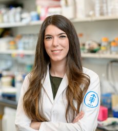 Direna Alonso-Curbelo合作论文数Mem Sloan Kettering Canc Ctr, Canc Biol & Genet Program, 1275 York Ave, New York, NY 10021 USA16
 Dawid Nowak合作论文数Graduate School of Medical Sciences, Weill Cornell Medicine;Nowak Lab, Sandra and Edward Meyer Cancer Center, Weill Cornell Medical College16Kaitlin Watrud合作论文数15
 Lukas Dow合作论文数Cell & Developmental Biology, Weill Cornell Graduate School of Medical Sciences, Cornell University15Muhan Chen合作论文数13
 Victoria Wang合作论文数12Daniel V. DeMarco合作论文数11David Kleinman合作论文数11Teng Han合作论文数Dept Med, Sandra & Edward Meyer Canc Ctr, Weill Cornell Med11Emma M Schatoff合作论文数Weill Cornell Med, Dept Med, Sandra & Edward Meyer Canc Ctr, New York, NY 10021 USA11Wu Zheng合作论文数11
 Christof Fellmann合作论文数11
 Tumas Beinortas合作论文数Faculty of Medicine, Vilnius University11Ding David合作论文数Cold Spring Harbor, Cold Spring Harbor Laboratory11
 Adrian Krainer合作论文数Cold Spring Harbor Laboratory11
 Mikala Egeblad合作论文数Departments of Cell Biology and Oncology, School of Medicine, Johns Hopkins University11
 Maria Paz Zafra合作论文数Centro de Investigaciones Biológicas, CSIC11
 Serif Senturk合作论文数Faculty of Science, Bilkent University11James E Bradner合作论文数10
 David Tuveson合作论文数Tuveson Laboratory, Cold Spring Harbor Laboratory;Cancer Center, Cold Spring Harbor Laboratory10
 Jun Qi合作论文数Dana-Farber Cancer Institute10Raffaella Sordella合作论文数10Sukanya Goswami合作论文数9Youngkyu Park合作论文数Cold Spring Harbor Laboratory9Marie J Parsons合作论文数Sandra & Edward Meyer Canc Ctr, Weill Cornell Med9
 Camila Oresco Dos Santos合作论文数9
 Carlos Cordon-Cardo合作论文数Department of Pathology, Molecular and Cell-Based Medicine, Icahn School of Medicine at Mount Sinai9María Fernández合作论文数9
 Richard A. Miller合作论文数Department of Pathology, University of Michigan;Paul Glenn Center for Biology of Aging, University of Michigan9
 Suzy Torti合作论文数9
 Richard Koche合作论文数Ctr Epigenet Res, Mem Sloan Kettering Canc Ctr9
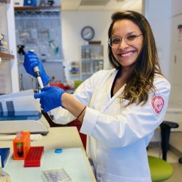 Alyna Katti合作论文数Dept Med, Sandra & Edward Meyer Canc Ctr, Weill Cornell Med8Elena Piskounova合作论文数Biochemistry & Structural Bioligy, Weill Cornell Graduate School of Medical Sciences, Cornell University8Frank M. Torti合作论文数8Sha Tian合作论文数7
 David L. Spector合作论文数Cold Spring Harbor Laboratory7Timour Baslan合作论文数6John P Morris合作论文数Mem Sloan Kettering Canc Ctr, Canc Biol & Genet Program, New York, NY 10065 USA6
 Michael F Ciccone合作论文数6
 Francisco M Barriga合作论文数6
 Vakoc Christopher R合作论文数Cold Spring Harbor Lab6
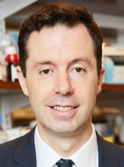 Olivier Elemento合作论文数Englander Institute for Precision Medicine, Weill Cornell Medical College;Institute for Computational Biomedicine , Weill Cornell Medical College;Clinical & Translational Science Center, Weill Cornell Medical College;Department of Physiology and Biophysics, Weill Cornell Medical College;Laboratory of Cancer Systems Biology, Weill Cornell Medical College;Institute for Computational Medicine, Weill Cornell Medical College;Meyer Cancer Center, Weill Cornell Medical College6




