Certain environmental toxins and chemotherapeutics are nucleic acid-damaging agents, causing adducts in DNA and RNA. While most of these adducts occur in RNA, the consequences of RNA damage are largely unexplored. Here, we demonstrate that nuclear RNA damage can result in loss of genome integrity in human cells. Specifically, we show that YTHDC1 regulates alkylation damage responses with the THO complex (THOC). In addition to its established binding to N6-methyladenosine (m6A), YTHDC1 binds to chemically induced N1-methyladenosine (m1A). Without YTHDC1, cells have greater alkylation damage sensitivity and increased DNA breaks, which are rescued by an RNA-specific dealkylase. These RNA-damage-induced DNA breaks (RDIBs) depend on R-loop formation, which is converted to DNA breaks by the XPG nuclease. Strikingly, in the absence of YTHDC1 or THOC, a nuclear RNA m1A methyltransferase is sufficient to induce DNA breaks. Our results provide mechanistic insight into how damaged RNAs can impact genomic integrity.
R-loops are RNA–DNA-hybrid-containing nucleic acids with important cellular roles. Deregulation of R-loop dynamics can lead to DNA damage and genome instability1, which has been linked to the action of endonucleases such as XPG2–4. However, the mechanisms and cellular consequences of such processing have remained unclear. Here we identify a new population of RNA–DNA hybrids in the cytoplasm that are R-loop-processing products. When nuclear R-loops were perturbed by depleting the RNA–DNA helicase senataxin (SETX) or the breast cancer gene BRCA1 (refs. 5–7), we observed XPG- and XPF-dependent cytoplasmic hybrid formation. We identify their source as a subset of stable, overlapping nuclear hybrids with a specific nucleotide signature. Cytoplasmic hybrids bind to the pattern recognition receptors cGAS and TLR3 (ref. 8), activating IRF3 and inducing apoptosis. Excised hybrids and an R-loop-induced innate immune response were also observed in SETX-mutated cells from patients with ataxia oculomotor apraxia type 2 (ref. 9) and in BRCA1-mutated cancer cells10. These findings establish RNA–DNA hybrids as immunogenic species that aberrantly accumulate in the cytoplasm after R-loop processing, linking R-loop accumulation to cell death through the innate immune response. Aberrant R-loop processing and subsequent innate immune activation may contribute to many diseases, such as neurodegeneration and cancer. RNA–DNA hybrids are immunogenic species that can aberrantly accumulate in the cytoplasm after R-loop processing, linking R-loop accumulation to cell death through the innate immune response.
The pre-mRNA life cycle requires intron processing; yet, how intron-processing defects influence splicing and gene expression is unclear. Here, we find that TTDN1/MPLKIP, which is encoded by a gene implicated in non-photosensitive trichothiodystrophy (NP-TTD), functionally links intron lariat processing to spliceosomal function. The conserved TTDN1 C-terminal region directly binds lariat debranching enzyme DBR1, whereas its N-terminal intrinsically disordered region (IDR) binds the intron-binding complex (IBC). TTDN1 loss, or a mutated IDR, causes significant intron lariat accumulation, as well as splicing and gene expression defects, mirroring phenotypes observed in NP-TTD patient cells. A Ttdn1-deficient mouse model recapitulates intron-processing defects and certain neurodevelopmental phenotypes seen in NP-TTD. Fusing DBR1 to the TTDN1 IDR is sufficient to recruit DBR1 to the IBC and circumvents the functional requirement for TTDN1. Collectively, our findings link RNA lariat processing with splicing outcomes by revealing the molecular function of TTDN1.
Supplementary Data from SMYD3 Impedes Small Cell Lung Cancer Sensitivity to Alkylation Damage through RNF113A Methylation–Phosphorylation Cross-talk
Although transcription is an essential cellular process, it is paradoxically also a well-recognized cause of genomic instability. R-loops, non-B DNA structures formed when nascent RNA hybridizes to DNA to displace the non-template strand as single-stranded DNA (ssDNA), are partially responsible for this instability. Yet, recent work has begun to elucidate regulatory roles for R-loops in maintaining the genome. In this review, we discuss the cellular contexts in which R-loops contribute to genomic instability, particularly during DNA replication and double-strand break (DSB) repair. We also summarize the evidence that R-loops participate as an intermediate during repair and may influence pathway choice to preserve genomic integrity. Finally, we discuss the immunogenic potential of R-loops and highlight their links to disease should they become pathogenic.
Summary The pre-mRNA life cycle requires intron processing; yet, how intron processing defects influence splicing and gene expression is unclear. Here, we find TTDN1, which is frequently mutated in non-photosensitive trichothiodystrophy (NP-TTD), functionally links intron lariat processing to the spliceosome. The conserved TTDN1 C-terminal region directly binds lariat debranching enzyme DBR1, while its N-terminal intrinsically disordered region (IDR) binds the intron binding complex (IBC). The IDR forms condensates in vitro and is needed for IBC interaction. TTDN1 loss causes significant intron lariat accumulation, as well as splicing and gene expression defects, mirroring phenotypes observed in NP-TTD patient cells. Ttdn1 Δ/Δ mice recapitulate intron processing defects and neurodevelopmental phenotypes seen in NP-TTD. A DBR1-IDR fusion recruits DBR1 to the IBC and circumvents the requirement for TTDN1, indicating this tethering role as its major molecular function. Collectively, our findings unveil key functional connections between lariat processing, splicing outcomes, and NP-TTD molecular pathology.
R-loops are three-stranded nucleic acid structures with both physiological and pathological roles in cells. R-loop imaging generally relies on detection of the RNA-DNA hybrid component of these structures using the S9.6 antibody. We show that the use of this antibody for imaging can be problematic because it readily binds to double-stranded RNA (dsRNA) in vitro and in vivo, giving rise to nonspecific signal. In contrast, purified, catalytically inactive human RNase H1 tagged with GFP (GFP-dRNH1) is a more specific reagent for imaging RNA-DNA hybrids. GFP-dRNH1 binds strongly to RNA-DNA hybrids but not to dsRNA oligonucleotides in fixed human cells and is not susceptible to binding endogenous RNA. Furthermore, we demonstrate that purified GFP-dRNH1 can be applied to fixed cells to detect hybrids after their induction, thereby bypassing the need for cell line engineering. GFP-dRNH1 therefore promises to be a versatile tool for imaging and quantifying RNA-DNA hybrids under a wide range of conditions.
Central to genotoxic responses is their ability to sense highly specific signals to activate the appropriate repair response. We previously reported that the activation of the ASCC-ALKBH3 repair pathway is exquisitely specific to alkylation damage in human cells, Yet the mechanistic basis for the selectivity of this pathway was not immediately obvious. Here, we demonstrate that RNA but not DNA alkylation is the initiating signal for this process. Aberrantly methylated RNA is sufficient to recruit ASCC, while an RNA dealkylase suppresses ASCC recruitment during chemical alkylation. In turn, recruitment of ASCC during alkylation damage, which is mediated by the E3 ubiquitin ligase RNF113A, suppresses transcription and R-loop formation. We further show that alkylated pre-mRNA is sufficient to activate RNF113A E3 ligase in vitro in a manner dependent on its RNA binding Zn-finger domain. Together, our work identifies an unexpected role for RNA damage in eliciting a specific response to genotoxins.
The response to DNA damage intersects with many other physiological processes in the cell, such as DNA replication, chromatin remodeling, and the cell cycle. Certain damaging lesions, such as UV-induced pyrimidine dimers, also strongly block RNA polymerases, necessitating the coordination of the repair mechanism with remodeling of the elongating transcriptional machinery, in a process called transcription-coupled nucleotide excision repair (TC-NER). This pathway is typically not thought to be engaged with smaller lesions such as base alkylation. However, recent work has uncovered the potential for shared molecular components between the cellular response to alkylation and UV damage. Here, we review our current understanding of the alkylation damage response and its impacts on RNA biogenesis. We give particular attention to the Activating Signal Cointegrator Complex (ASCC), which plays important roles in the transcriptional response during UV damage as well as alkylation damage reversal, and intersects with trichothiodystrophy, an inherited disease associated with TC-NER.
Cross-presentation allows for the presentation of exogenous viral and tumor antigens to CD8+ T cells by XCR1+ classical dendritic cells (cDC1s). Previously, the role of cross-presentation in cytotoxic T cell responses in vivo was unclear due to the lack of mouse models specifically deficient in cross-presentation. We developed a CRISPR-Cas9 screen to find novel regulators of cross-presentation and found that the protein WDFY4 is essential for cross-presentation of cell-associated antigens but not for direct presentation or presentation on MHCII. WDFY4-deficient mice had normal cDC1 development and were able to survive infections with Toxoplasma gondii through production of IL-12. However, WDFY4-deficient mice failed to prime CD8+ T cells to viral antigens and were completely unable to control immunogenic tumors. WDFY4 interacts with proteins related to vesicular formation and trafficking such as clathrin and HSP90ab1 and localizes near the cell surface, suggesting its involvement in vesicular targeting after antigen uptake during cross-presentation.
During the process of cross-presentation, viral or tumor-derived antigens are presented to CD8+ T cells by Batf3-dependent CD8α+/XCR1+ classical dendritic cells (cDC1s). We designed a functional CRISPR screen for previously unknown regulators of cross-presentation, and identified the BEACH domain-containing protein WDFY4 as essential for cross-presentation of cell-associated antigens by cDC1s in mice. However, WDFY4 was not required for major histocompatibility complex class II presentation, nor for cross-presentation by monocyte-derived dendritic cells. In contrast to Batf3 -/- mice, Wdfy4 -/- mice displayed normal lymphoid and nonlymphoid cDC1 populations that produce interleukin-12 and protect against Toxoplasma gondii infection. However, similar to Batf3 -/- mice, Wdfy4 -/- mice failed to prime virus-specific CD8+ T cells in vivo or induce tumor rejection, revealing a critical role for cross-presentation in antiviral and antitumor immunity.
Multiple DNA damage response (DDR) pathways have evolved to sense the presence of damage and recruit the proper repair factors. We recently reported a signaling pathway induced upon alkylation damage to recruit the AlkB homolog 3, α-ketoglutarate–dependent dioxygenase (ALKBH3)–activating signal cointegrator 1 complex subunit 3 (ASCC3) dealkylase–helicase repair complex. As in other DDR pathways, the recruitment of these repair factors is mediated through a ubiquitin-dependent mechanism. However, the machinery that coordinates the proper assembly of this repair complex and controls its recruitment is still poorly defined. Here, we demonstrate that the ASCC1 accessory subunit is important for the regulation of ASCC complex function. ASCC1 interacts with the ASCC complex through the ASCC3 helicase subunit. We find that ASCC1 is present at nuclear speckle foci prior to damage, but leaves the foci in response to alkylation. Strikingly, ASCC1 loss significantly increases ASCC3 foci formation during alkylation damage, yet most of these foci lack ASCC2. These results suggest that ASCC1 coordinates the proper recruitment of the ASCC complex during alkylation, a function that appears to depend on a putative RNA-binding motif near the ASCC1 C terminus. Consistent with its role in alkylation damage signaling and repair, ASCC1 knockout through a CRISPR/Cas9 approach results in alkylation damage sensitivity in a manner epistatic with ASCC3. Together, our results identify a critical regulator of the ALKBH3–ASCC alkylation damage signaling pathway and suggest a potential role for RNA-interacting domains in the alkylation damage response.
An emerging molecular understanding of RNA alkylation and its removal is transforming our knowledge of RNA biology and its interplay with cancer chemotherapy responses. DNA modifications are known to perform critical functions depending on the genome template, including gene expression, DNA replication timing, and DNA damage protection, yet current results suggest that the chemical diversity of DNA modifications pales in comparison to those on RNA. More than 150 RNA modifications have been identified to date, and their complete functional implications are still being unveiled. These include intrinsic roles such as proper processing and RNA maturation; emerging evidence has furthermore uncovered RNA modification "readers", seemingly analogous to those identified for histone modifications. These modification recognition factors may regulate mRNA stability, localization, and interaction with translation machinery, affecting gene expression. Not surprisingly, tumors differentially modulate factors involved in expressing these marks, contributing to both tumorigenesis and responses to alkylating chemotherapy. Here we describe the current understanding of RNA modifications and their removal, with a focus primarily on methylation and alkylation as functionally relevant changes to the transcriptome. Intriguingly, some of the same RNA modifications elicited by physiological processes are also produced by alkylating agents, thus blurring the lines between what is a physiological mark and a damage-induced modification. Furthermore, we find that a high level of gene expression of enzymes with RNA dealkylation activity is a sensitive readout for poor survival in four different cancer types, underscoring the likely importance of examining RNA dealkylation mechanisms to cancer biology and for cancer treatment and prognosis.
A signalling mechanism in human cells for sensing DNA damage induced by alkylation involves ubiquitin-dependent recruitment of the alkylation repair complex ASCC to the vicinity of the damage and co-localization with transcription and splicing factors. The upstream signalling pathway that initiates the repair of alkylation damage in DNA is not well understood. Nima Mosammaparast and colleagues have found that the activating signal cointegrator complex (ASCC) is recruited to the vicinity of alkylation damage, co-localizing the transcription and splicing factors. The recruitment of ASCC depends on the ability of ASCC2 protein to bind K63-linked polyubiquitin chains made by the E3 ligase RNF113A, although the polyubiquitinated target remains unclear. Individuals with X-linked trichothiodystrophy carry a mutation in RNF113A, and these results clarify the underlying basis of their hypersensitivity to alkylating agents. DNA repair is essential to prevent the cytotoxic or mutagenic effects of various types of DNA lesions, which are sensed by distinct pathways to recruit repair factors specific to the damage type. Although biochemical mechanisms for repairing several forms of genomic insults are well understood, the upstream signalling pathways that trigger repair are established for only certain types of damage, such as double-stranded breaks and interstrand crosslinks1,2,3. Understanding the upstream signalling events that mediate recognition and repair of DNA alkylation damage is particularly important, since alkylation chemotherapy is one of the most widely used systemic modalities for cancer treatment and because environmental chemicals may trigger DNA alkylation4,5,6. Here we demonstrate that human cells have a previously unrecognized signalling mechanism for sensing damage induced by alkylation. We find that the alkylation repair complex ASCC (activating signal cointegrator complex)7 relocalizes to distinct nuclear foci specifically upon exposure of cells to alkylating agents. These foci associate with alkylated nucleotides, and coincide spatially with elongating RNA polymerase II and splicing components. Proper recruitment of the repair complex requires recognition of K63-linked polyubiquitin by the CUE (coupling of ubiquitin conjugation to ER degradation) domain of the subunit ASCC2. Loss of this subunit impedes alkylation adduct repair kinetics and increases sensitivity to alkylating agents, but not other forms of DNA damage. We identify RING finger protein 113A (RNF113A) as the E3 ligase responsible for upstream ubiquitin signalling in the ASCC pathway. Cells from patients with X-linked trichothiodystrophy, which harbour a mutation in RNF113A, are defective in ASCC foci formation and are hypersensitive to alkylating agents. Together, our work reveals a previously unrecognized ubiquitin-dependent pathway induced specifically to repair alkylation damage, shedding light on the molecular mechanism of X-linked trichothiodystrophy.
The breast cancer susceptibility proteins BRCA1 and BRCA2 have emerged as key stabilizing factors for the maintenance of replication fork integrity following replication stress. In their absence, stalled replication forks are extensively degraded by the MRE11 nuclease, leading to chemotherapeutic sensitivity. Here we report that BRCA proteins prevent nucleolytic degradation by protecting replication forks that have undergone fork reversal upon drug treatment. The unprotected regressed arms of reversed forks are the entry point for MRE11 in BRCA-deficient cells. The CtIP protein initiates MRE11-dependent degradation, which is extended by the EXO1 nuclease. Next, we show that the initial limited resection of the regressed arms establishes the substrate for MUS81 in BRCA2-deficient cells. In turn, MUS81 cleavage of regressed forks with a ssDNA tail promotes POLD3-dependent fork rescue. We propose that targeting this pathway may represent a new strategy to modulate BRCA2-deficient cancer cell response to chemotherapeutics that cause fork degradation.
Repair of DNA alkylation damage is critical for genomic stability and involves multiple conserved enzymatic pathways. Alkylation damage resistance, which is critical in cancer chemotherapy, depends on the overexpression of alkylation repair proteins. However, the mechanisms responsible for this upregulation are unknown. Here, we show that an OTU domain deubiquitinase, OTUD4, is a positive regulator of ALKBH2 and ALKBH3, two DNA demethylases critical for alkylation repair. Remarkably, we find that OTUD4 catalytic activity is completely dispensable for this function. Rather, OTUD4 is a scaffold for USP7 and USP9X, two deubiquitinases that act directly on the AlkB proteins. Moreover, we show that loss of OTUD4, USP7, or USP9X in tumor cells makes them significantly more sensitive to alkylating agents. Taken together, this work reveals a novel, noncanonical mechanism by which an OTU family deubiquitinase regulates its substrates, and provides multiple new targets for alkylation chemotherapy sensitization of tumors.
The cellular response to DNA double-stranded breaks (DSBs) involves a conserved mechanism of recruitment and activation of numerous proteins involved in this pathway. The events that trigger this response in mammalian cells involve several post-translational modifications, but the role of non-proteasomal ubiquitin signaling is particularly central to this pathway. Recent work has demonstrated that ubiquitination does not act alone, but in concert with other post-translational modifications, including phosphorylation, methylation, acetylation, ADP-ribosylation, and other ubiquitin-like modifiers, particularly SUMOylation. We review novel and exciting crosstalk mechanisms between ubiquitination and other post-translational modifications, many of which work synergistically with each other to activate signaling events and help recruit important DNA damage effector proteins, particularly BRCA1 (breast cancer 1, early onset) and 53BP1 (tumor protein p53 binding protein 1), to sites of DNA damage.
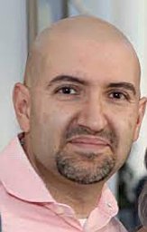 Nima Mosammaparast合作论文数Brigham and Women's Hospital, Harvard Medical School13
 Brittany A Townley合作论文数4Clement Oyeniran合作论文数4Tsai Miaw-Sheue合作论文数4Soll Jennifer M合作论文数4
 Tsao Ning合作论文数a Graduate Institute of Biochemistry and Molecular Biology;College of Medicine;National Taiwan University;Taipei;College of Medicine, National Taiwan University4
 John A. Tainer合作论文数Department of Molecular and Cellular Oncology, Division of Basic Science Research, The University of Texas MD Anderson Cancer Center;Department of Structural Biology, The University of Texas MD Anderson Cancer Center;Department of Integrated Diffraction Analysis Technologies Program, The University of California Lawrence Berkeley National Laboratory;Skaggs Institute for Chemical Biology, Scripps Research Institute4
 Albino Bacolla合作论文数Institute of Biosciences and Technology, The Texas A&M University System Health Science Center4Rebecca Rodell合作论文数3
 Hua Sun合作论文数Chinese Acad Sci, Inst Genet & Dev Biol, State Key Lab Plant Cell & Chromosome Engn, Beijing 100101, Peoples R China3Zhao Yu合作论文数3Alessandro Vindigni合作论文数3
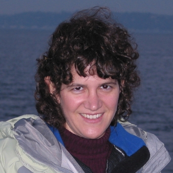 Karlene Cimprich合作论文数Cimprich Lab, Department of Chemical and Systems Biology Operations, Stanford University3Prachi Bagadia合作论文数2Miranda C Mudge合作论文数2Nicolas Schmidt合作论文数2Jesse Davidson合作论文数2Drew McDonald合作论文数2
 Chenlin Song合作论文数2Patrick M Lombardi合作论文数2
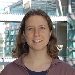 Magdalena P Crossley合作论文数Department of Chemical and Systems Biology, Stanford University School of Medicine2Vivek Durai合作论文数2
 Tanveer Ahmad合作论文数CNRS 5309, Grenoble Alpes Univ2
 Sridhar Nonavinkere Srivatsan合作论文数Div Oncol, Washington Univ2Reilly A Sample合作论文数2
 Alexandre Casanova合作论文数UMR5309, Grenoble Alpes Univ2
 Elvin J Lauron合作论文数2Luke Buerer合作论文数2
 Valentina Lukinovic合作论文数UMR5309, Grenoble Alpes Univ2Timur Rusanov合作论文数Temple Univ, Lewis Katz Sch Med, Dept Med Genet & Mol Biochem, Fels Inst Canc Res, Philadelphia, PA 19140 USA2
 William Fairbrother合作论文数Department of Molecular Biology, Cell Biology, and Biochemistry, Brown University2
 Qiuling Wang合作论文数Department of Molecular Microbiology, Washington University School of Medicine2Jackson Jessica合作论文数2
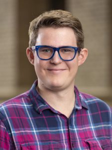 Alex Holehouse合作论文数Department of Biochemistry and Molecular Biophysics, School of Medicine, Washington University2
 Derek Theisen合作论文数Schreiber Lab, Department of Pathology & Immunology, Washington University in St. Louis2
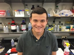 Briseño Carlos合作论文数Dept Pathol & Immunol, Washington Univ2
 Herbert W. Virgin合作论文数Department of Pathology & Immunology, School of Medicine, Washington University2
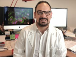 Marco Gargaro合作论文数Department of Experimental Medicine, University of Perugia2Clark Nathaniel E合作论文数2
 Pritesh Desai合作论文数Division of Infectious Diseases, John T. Milliken Department of Medicine, School of Medicine, Washington University in St. Louis2Majid Mona C合作论文数2
 William Gillanders合作论文数2
 Wayne M. Yokoyama合作论文数Roy and Diana Vagelos Division of Biology & Biomedical Sciences, School of Medicine, Washington University in St. Louis;Division of Rheumatology, Department of Medicine, School of Medicine, Washington University in St. Louis;Howard Hughes Medical Institute2
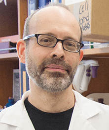 Michael S. Diamond合作论文数Division of Infectious Diseases, John T. Milliken Department of Medicine, School of Medicine, Washington University in St. Louis2
 Kenneth Murphy合作论文数Division of Immunobiology, Department of Pathology & Immunology, School of Medicine, Washington University in St. Louis;Howard Hughes Medical Institute2David F. Wozniak合作论文数2
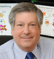 Robert Schreiber合作论文数Department of Pathology & Immunology, School of Medicine, Washington University;Center for Human Immunology and Immunotherapy Programs, Washington University;Siteman Comprehensive Cancer Center, Washington University2
 Wandy L. Beatty合作论文数Department of Molecular Microbiology, School of Medicine, Washington University in St. Louis2
 David Sibley合作论文数Department of Molecular Microbiology, School of Medicine, Washington University in St. Louis2




