Chen, Claire1; Shih, Juliann1; Sutton, Jenna1; Cabugao, Paul2; Romero, Arthur1 Author Information
Figure S1. Super-enhancers near the KLF5 gene are focally amplified in cancer cell lines;Figure S2. The regulatory potential of the KLF5/KLF12 intervening region;Figure S3. Expression level of KLF12, DIS3 and PIBF1 after repression of KLF5 superenhancers;Figure S4. Genomic analysis of KLF5 binding sites in BICR31 cells;Figure S5. Target genes of KLF5 in head and neck squamous cell carcinomas;Figure S6. The effect of KLF5 silencing on H3K27ac profile;Figure S7. Mutation hotspots in KLF5;Figure S8. Mutation profile in FBXW7 in colorectal cancers; Figure S9. Ectopic expression of KLF5 mutants in HEK293T cells; Figure S10. Ectopic expression of KLF5 E419Q in HCC95 cells
Table S7 contains microbe screening results.
Table S1 contains cohort description, Master Patient Table and MutSigCV results.
Supplementary Methods. Supplementary Figure 1. Somatic alterations in PPARG and RXRA are hallmarks of luminal bladder cancer. Supplementary Figure 2. PPARG pathway is activated by overexpression of RXRA S427F/S427Y, but not other mutant alleles in bladder cancer cells. Supplementary Figure 3. Representation of the effects of ligand-dependent modulation on the PPARG interactome. Supplementary Figure 4. Downregulation of FABP4 protein by treatment of PPARG-activated bladder cancer cell lines by inverse-agonist T0070907. Supplementary Figure 5. Genome engineering scheme for generating reporter cell line. Supplementary Figure 6. Basal expression of FABP4 is reduced by PPARG inverse agonists, but not antagonists. Supplementary Figure 7. Lipid metabolism genes are inhibited by PPARG inverse-agonists. Supplementary Figure 8. PPARG inverse-agonists inhibit proliferation of PPARG activated bladder cancer cell lines in clonogenic assays. Supplementary Figure 9. PPARG inverse-agonists, but not antagonists, inhibit proliferation of PPARG-activated bladder cancer cell lines. Supplementary Figure 10. Somatic alterations in RXRA and PPARG.
Table S2 contains BAP1 analysis results, as well as detailed lists of YY1 and IRF8 target genes.
Table S6 contains results from the analysis of DNA methylation in SETD2 mutated and BAP1 inactivated samples.
Aneuploidies—whole-chromosome or whole-arm imbalances—are the most prevalent alteration in cancer genomes 1 , 2 . However, it is still debated whether their prevalence is due to selection or ease of generation as passenger events 1 , 2 . Here we developed a method, BISCUT, that identifies loci subject to fitness advantages or disadvantages by interrogating length distributions of telomere- or centromere-bounded copy-number events. These loci were significantly enriched for known cancer driver genes, including genes not detected through analysis of focal copy-number events, and were often lineage specific. BISCUT identified the helicase-encoding gene WRN as a haploinsufficient tumour-suppressor gene on chromosome 8p, which is supported by several lines of evidence. We also formally quantified the role of selection and mechanical biases in driving aneuploidy, finding that rates of arm-level copy-number alterations are most highly correlated with their effects on cellular fitness 1 , 2 . These results provide insight into the driving forces behind aneuploidy and its contribution to tumorigenesis.
Table S3 contains the karyotypes of 16 genome-wide LOH MPM cases from the BWH cohort.
INTRODUCTION: Congenital bullous emphysema (CBE) is a rare disorder that affects an estimated 1/25,000 births, and is most commonly found in male children with bronchial and cardiovascular malformations.We present a case of CBE with atypical phenotype and multiple complications eventually requiring lobectomy. CASE PRESENTATION:A 52 year old female with a two pack year smoking history presented with cough and dyspnea.Respiratory biofire showed H. influenza infection, while imaging revealed large, bibasilar bullae with near complete effacement of the regional lung parenchyma and mosaic attenuation.The patient improved with standard treatment for an acute exacerbation of chronic obstructive pulmonary disease and only required 3L of supplemental oxygen on discharge; however, the size of the bullae and extent of the emphysema was inconsistent with her smoking history.Notably, the patient had a negative serum alpha antitrypsin, alpha-1 antitrypsin S allele, Z allele, SERPINA1 gene sequencing, antinuclear nuclear antitrypsin, antineutrophilic cytoplasmic antibody, autoimmune superpanel, histoplasma and aspergillus antigen.The patient denied any complications at birth, occupational exposure, or records of previous imaging.Unfortunately, the patient would present again multiple times for hydro-and pneumothoracies over the next two years.Ultimately, the patient underwent bullectomy with resection of the right middle and lower lobe.The procedure was complicated by right upper lobe collapse and tension pneumothorax, requiring bilateral chest tube placement.Despite heavy pulmonary trauma, the patient was discharged on only Stiolto (LAMA/LABA) and 3L of supplemental oxygen.Pathology only showed normal lung tissue with chronic inflammatory changes.The patient has not suffered another pneumothorax to this date.DISCUSSION: It is hypothesized that hypoplastic cartilaginous folds develop abnormal lung architecture that predispose children to recurrent pulmonary infections, eventually growing into hyperinflated bullae that compress surrounding lobes, compromise vascular flow, and can even cause mediastinal shift to the point of herniation into the contralateral hemithorax.Bullae are typically asymmetric, apical, paraseptal, and progressive, although our female patient had bibasilar, paraseptal bullae.CONCLUSIONS: CBE is a rare disorder that can be complicated with recurrent pneumothoracies.Lobectomy may prove preventative against pneumothorax, and bridge the patient to lung transplant evaluation.
The role of PPM1D mutations in de novo gliomagenesis has not been systematically explored. Here we analyze whole genome sequences of 170 pediatric high-grade gliomas and find that truncating mutations in PPM1D that increase the stability of its phosphatase are clonal driver events in 11% of Diffuse Midline Gliomas (DMGs) and are enriched in primary pontine tumors. Through the development of DMG mouse models, we show that PPM1D mutations potentiate gliomagenesis and that PPM1D phosphatase activity is required for in vivo oncogenesis. Finally, we apply integrative phosphoproteomic and functional genomics assays and find that oncogenic effects of PPM1D truncation converge on regulators of cell cycle, DNA damage response, and p53 pathways, revealing therapeutic vulnerabilities including MDM2 inhibition.
Aneuploidy, which we define as whole chromosome or chromosome arm copy number imbalance, is a near-universal characteristic of human cancers. Cancer subtypes are often characterized by tumor specific patterns of chromosome arm copy number alterations; for example, squamous cell carcinomas (SCCs) from different tissues of origin (including lung, head and neck, esophagus, and bladder) have a pattern of chromosome 3p loss and chromosome 3q gain. Although these alterations are frequent, they are not well understood due to difficulty in modeling specific aneuploidy alterations in the matching cell type. However, recent advances in genome engineering allow generation of large chromosomal alterations. We used the CRISPR-Cas9 system to delete one copy of chromosome 3p in human immortalized lung epithelial cells most similar to upper airway basal cells. Deletion of chromosome 3p was validated by whole genome sequencing and karyotyping. Consistent with patient data, expression of 3p genes was decreased upon deletion, as well as increased expression of interferon response genes. Phenotypic characterization revealed that cells with chromosome 3p deletion initially proliferated more slowly than their siblings. These chromosome 3p deleted cells had increased G1 arrest but did not undergo increased apoptosis or cell death. Interestingly, after several passages in culture, the proliferation defect was rescued in chromosome 3p deleted cells. Genome sequencing and karyotype analyses found that evidence of chromosome 3 duplication, transitioning the cell to a state of chromosome 3q gain. We isolated sibling cells with chromosome 3p deletion or chromosome 3q gain and demonstrated that duplication of chromosome 3 could indeed rescue proliferation rates. With our cellular model of chromosome arm-level aneuploidy, we uncovered a selection mechanism that allowed aneuploidy tolerance in vitro, and a possible explanation for joint alteration of both arms of chromosome 3. In conclusion, our genome engineering approach to model chromosome arm-level deletions provides a robust model that will address a gap in our understanding of aneuploidy in cancer. Citation Format: Alison Marie Taylor, Sejal Jain, Juliann Shih, Andrew D. Cherniack, Rameen Beroukhim, Matthew Meyerson. Functional models of chromosome arm aneuploidies in lung squamous cell carcinoma [abstract]. In: Proceedings of the American Association for Cancer Research Annual Meeting 2021; 2021 Apr 10-15 and May 17-21. Philadelphia (PA): AACR; Cancer Res 2021;81(13_Suppl):Abstract nr 2135.
Aneuploidies, defined as whole-arm or whole-chromosome imbalances, are the most prevalent alteration in cancer genomes. However, the extent to which they are enriched due to selection is unclear, against the alternative hypothesis that they are passenger events that are simply highly prone to occur. We developed a novel method, BrISCUT, that identifies loci under selective advantage or disadvantage due to arm-level copy-number alterations by interrogating length distributions of events that are bounded at either the telomere or centromere. These loci were significantly enriched for known cancer driver genes, including genes not detected through analysis of focal copy-number events, and were often lineage-specific. We also formally quantified the role of selection and mechanistic biases in driving aneuploidy, finding that rates of arm-level SCNAs are most highly correlated with selective pressures. These results provide insight into the causes of aneuploidies and their contributions to tumorigenesis.
 Andrew Cherniack合作论文数Meyerson Laboratory Dana-Farber Cancer Institute;Broad Institute of MIT34
 Matthew L. Meyerson合作论文数Harvard Medical School;Center for Cancer Genomics, Dana-Farber Cancer Institute25
 Rameen Beroukhim合作论文数Division of Medical Sciences, Harvard Medical School;Dana-Farber Cancer Institute19
 Rehan Akbani合作论文数Department of Bioinformatics and Computational Biology, Graduate School of Biomedical Sciences, University of Texas MD Anderson Cancer Center17
 Gordon Robertson合作论文数15
 Ludmila Danilova合作论文数Sidney Kimmel Comprehensive Cancer Center, Johns Hopkins University;School of Medicine, Johns Hopkins University13
 Bowlby Reanne合作论文数BC Cancer Agency13
 Katie Hoadley合作论文数Department of Genetics, School of Medicine, University of North Carolina at Chapel Hill13Ina Felau合作论文数12Tara M Lichtenberg合作论文数12
 Ashton Berger合作论文数Canc Program, Broad Inst MIT & Harvard12
 Peter W. Laird合作论文数Department of Epigenetics, Van Andel Institute12
 Newton Yulia合作论文数12Jean C. Zenklusen合作论文数University of Texas;Fox Chase Cancer Center;Department Ã12Kristen Leraas合作论文数11Esther Drill合作论文数11
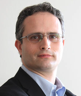 Gad A. Getz合作论文数Harvard Medical School;Department of Pathology, Massachusetts General Hospital;Broad Institute of Harvard and MIT11
 Heiman David合作论文数Broad Institute of MIT and Harvard, Cambridge, Massachusetts 02142, USA.11Lixia Diao合作论文数MD Anderson Cancer Center, The University of Texas11
 Lauren Averett Byers合作论文数Department of Thoracic-Head and Neck Medical Oncology, Division of Cancer Medicine, The University of Texas MD Anderson Cancer Center11
 Ewan A. Gibb合作论文数Department of Microbiology, University of Manitoba11
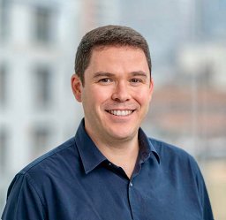 Francisco Sanchez-Vega合作论文数Johns Hopkins University11Michael S. Noble合作论文数11
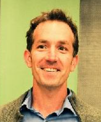 Joshua M Stuart合作论文数Baskin School of Engineering, University of California Santa Cruz;Genomics Institute, University of California Santa Cruz11Karen Mungall合作论文数Wellcome Trust Genome Campus, The Wellcome Trust Sanger Institute11Lisa Iype合作论文数10Yiling Lu合作论文数10Gordon Saksena合作论文数10Denise Brooks合作论文数10
 Leslie Cope合作论文数Department of Otolaryngology-Head and Neck Surgery, School of Medicine, Johns Hopkins University10
 Nikolaus Schultz合作论文数Department of Biostatistics and Epidemiology, Memorial Sloan Kettering Cancer Center;Center for Molecular Oncology, Memorial Sloan Kettering Cancer Center10
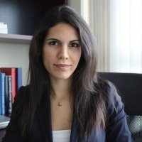 Kasaian Katayoon合作论文数Canada's Michael Smith Genome Sciences Centre, British Columbia Cancer Agency10Bowen Jay合作论文数10
 Jaegil Kim合作论文数Broad Institute of MIT and Harvard, Cambridge, Massachusetts.10
 Armenia Joshua合作论文数CRO, National Cancer Institute10Cho Juok合作论文数10
 Michael S. Lawrence合作论文数Harvard Medical School;Lawrence Laboratory, Mass General Cancer Center, Massachusetts General Hospital10
 Gordon Mills合作论文数School of Medicine, Oregon Health Sciences University10
 Marc Ladanyi合作论文数Memorial Sloan Kettering Cancer Center10
 Chris Sander合作论文数Dana–Farber Cancer Institute;Cell Biology Department, Harvard Medical School;Division of Medical Sciences, Harvard University10
 Carolyn M. Hutter合作论文数Office of Strategic Coordination, Division of Program Coordination, Planning, and Strategic Initiatives, Office of the Director, National Institutes of Health10
 Nina Thiessen合作论文数10
 Todd Auman合作论文数National Institute of Environmental Health Sciences;Research Triangle Park10
 Steven E Schumacher合作论文数Cancer Program, The Broad Institute of MIT and Harvard10
 Ronglai Shen合作论文数Department of Epidemiology and Biostatistics, Sloan-Kettering Cancer Center10
 Gavin Ha合作论文数10
 Chip Stewart合作论文数Broad Inst10
 Vesteinn Thorsson合作论文数Department of Physics;University of Illinois at Urbana-Champaign;Department of Physics, University of Illinois at Urbana-Champaign10Pavana Anur合作论文数Oregon Health & Science University10




