BACKGROUND:Intraoperative nerve monitoring (IONM) is widely used in endocrine surgery, yet substantial heterogeneity exists in its documentation, interpretation, and reporting. This limits comparability across studies and hampers the evaluation of clinical outcomes. METHODS:A consensus process was conducted by the European Society of Endocrine Surgeons (ESES), combining a systematic literature review, analysis of Eurocrine® registry data, a survey among ESES members, and a structured Delphi consensus process. RESULTS:Considerable variability was identified in monitoring techniques, signal interpretation, and outcome reporting. Key areas requiring standardization include the definition of loss of signal, documentation of neuromonitoring parameters, reporting of technical settings, and objective assessment of vocal cord function. Based on these findings, a set of consensus statements was developed to define minimum requirements for IONM research and reporting. CONCLUSION:This consensus statement provides a structured framework to improve standardization, data quality, and comparability in IONM research. Adoption of the recommendations may facilitate more robust evaluation of clinical outcomes and support future evidence generation in endocrine surgery.
OBJECTIVES:To examine (1) the relationship between anthropometric measurements and recurrent laryngeal nerve (RLN) diameter; (2) whether thin RLNs have different baseline electromyographic (EMG) characteristics; (3) if thin or branched morphology is associated with increased risk of EMG adverse events, loss of signal (LOS) or vocal cord paresis/paralysis (VCP). METHODS:In this prospective study, anthropometric data were collected, including weight, height, body mass index (BMI), neck circumference, shoulder diameter, and circumference of the right middle finger (RMF) or right ring finger (RRF). RESULTS:We enrolled 216 patients who underwent surgery from 2016 to 2020 with 307 RLNs at risk. Thin nerves were defined as those ≤ 1.5 mm in diameter. The mean RLN diameter was 2 mm; the RLN was thinner in females and in patients with BMI < 20 kg/m2, body weight < 120 kg, height < 175 cm, neck circumference < 40 cm, shoulder diameter < 50 cm, and RMF or RRF < 7.6 cm (all p < 0.05). No statistically significant differences were detected in baseline EMG characteristics between thin and thick RLNs bilaterally (except for right RLN R2 latency). We had zero cases of LOS or VCP. In all branched nerves, motor fibers resided in the anterior branch. CONCLUSION:Multiple anthropometric factors were associated with RLN diameter. Most of the studied EMG signal characteristics did not statistically differ by RLN diameter. With zero events of LOS or VCP, our study is not powered to determine if thin diameter or branching are independent risk factors for RLN injury in a high-volume endocrine surgery practice utilizing IONM.
OBJECTIVE:Little is known about thyroid cancer survivors' risk of chronic conditions. We, therefore, investigated the prevalence of drugs used for chronic conditions among thyroid cancer patients using population-wide register data. METHODS:We linked data from the Cancer Registry of Norway to the Norwegian Prescription Database and other databases for a study population of 3.52 million individuals, including 3486 individuals with thyroid cancer diagnosed during 2005-2019. Prevalence ratios (PRs) with 95% CIs of reimbursed prescribed drugs in thyroid cancer patients up to 15 years after thyroid cancer diagnosis were estimated by log-binomial regression, with the cancer-free population as reference. RESULTS:Individuals (both males and females) with thyroid cancer had higher use of drugs for several chronic conditions in the years after diagnosis; eg, 5 years after thyroid cancer diagnosis, there was elevated use of drugs for hypoparathyroidism (PRmales = 35.4, 95% CI, 25.2-49.7; PRfemales = 42.8, 95% CI, 34.2-53.6), hypertension (PRfemales = 1.20, 95% CI, 1.12-1.28), anxiety and tension (PRmales = 4.01, 95% CI, 1.80-8.92; PRfemales = 2.01, 95% CI, 1.15-3.52), gastric acid disorders (PRmales = 1.52, 95% CI, 1.22-1.91; PRfemales = 1.45, 95% CI, 1.27-1.66), and pain (PRmales = 1.48, 95% CI, 1.11-1.97; PRfemales = 1.24, 95% CI, 1.08-1.42) as compared with the cancer-free population. In addition, males with thyroid cancer had long-term elevated use of drugs for depression (eg, year 10+, PRmales = 1.66, 95% CI, 1.06-2.59). Individuals with thyroid cancer also had higher use of drugs for several conditions prior to the thyroid cancer diagnosis, eg, hypertension, gastric acid disorders, and pain. CONCLUSIONS:Individuals diagnosed with thyroid cancer had elevated long-term use of drugs for several chronic conditions, as compared with the cancer-free population.
Abstract Background Techniques for autofluorescence have been introduced to visualize the parathyroid glands during surgery and to reduce hypoparathyroidism after thyroidectomy. Methods This parallel multicentre RCT investigated the use of Fluobeam® LX to visualize the parathyroid glands by autofluorescence during total thyroidectomy compared with no use. There was no restriction on the indication for surgery. Patients were randomized 1 : 1 and were blinded to the group allocation. The hypothesis was that autofluorescence enables identification and protection of the parathyroid glands during thyroidectomy. The primary endpoint was the rate of low parathyroid hormone (PTH) levels the day after surgery. Results Some 535 patients were randomized, and 486 patients received an intervention according to the study protocol, 246 in the Fluobeam® LX group and 240 in the control group. Some 64 patients (26.0 per cent) in the Fluobeam® LX group and 77 (32.1 per cent) in the control group had low levels of PTH after thyroidectomy (P = 0.141; relative risk (RR) 0.81, 95 per cent c.i. 0.61 to 1.07). Subanalysis of 174 patients undergoing central lymph node clearance showed that 15 of 82 (18 per cent) in the Fluobeam® LX group and 31 of 92 (33 per cent) in the control group had low levels of PTH on postoperative day 1 (P = 0.021; RR 0.54, 0.31 to 0.93). More parathyroid glands were identified during operation in patients who had surgery with Fluobeam® LX, and fewer parathyroid glands in the surgical specimen on definitive histopathology. No specific harm related to the use of Fluobeam® LX was reported. Conclusion The use of autofluorescence during thyroidectomy did not reduce the rate of low PTH levels on postoperative day 1 in the whole group of patients. It did, however, reduce the rate in a subgroup of patients. Registration number: NCT04509011 (http://www.clinicaltrials.gov).
Thyroid lobectomy is sufficient for differentiated
Children are more likely to experience recurrent laryngeal nerve (RLN) injury during thyroid surgery. Intraoperative nerve monitoring (IONM) may assist in nerve identification and surgical decision making. A literature review of pediatric IONM was performed and used to inform a monitoring technique guide and expert opinion statements. Pediatric IONM is achieved using a variety of methods. When age-appropriate endotracheal tubes with integrated surface electrodes are not available, an alternative method should be used. Patient age and surgeon experience with laryngoscopy influence technique selection; four techniques are described in detail. Surgeons must be familiar with the nuances of monitoring technique and interpretation; opinion statements address optimizing this technology in children. Adult IONM guidelines may offer strategies for surgical decision making in children. In some cases, delay of second-sided surgery may reduce bilateral RLN injury risk.
86 Objectives: To assess the diagnostic performance of integrated parathyroid scintigraphy and cervical ultrasound in a large cohort of patients with primary hyperparathyroidism (pHPT) imaged and operated at a single tertiary institution. Methods: The retrospective study included all parathyroid scintigraphies conducted at a single tertiary institution in Norway between January 2006 and December 2018. Exclusion criteria were: Secondary or tertiary HPT, repeat imaging, deviations from the standard imaging protocol. In 2006, patients underwent planar subtraction scintigraphy with [99mTc]pertechnetate. Since 2007, imaging was performed as 3-phase scintigraphy with [99mTc]MIBI with initial dynamic planar imaging, SPECT (since 2009: SPECT/CT on a Siemens Symbia T6; Siemens Healthineers, Hoffman Estates/IL) 30 min p.i. (Fig. panels A-C) and a 10-min planar image 3 h p.i. (Panel D). After the completion of scintigraphy, high-resolution ultrasound was conducted by the nuclear medicine specialist on a Hitachi EUB 5500 or Hitachi Preirus (Hitachi Medical Corp., Akihabara, Japan) (Panel E). All images were digitally archived in the department’s PACS. Parathyroidectomy was performed at the same institution. The diagnosis of a parathyroid adenoma was regarded as true positive if it was confirmed by pathology and follow-up revealed the resolution of HPT. All data were recorded in a custom-developed relational database. Statistical analysis was performed with R. The study was approved as a quality control study with no regulatory requirement for patient consent. Results: 1319 patients with pHPT underwent their first parathyroid scintigraphy and ultrasound between January 2006 and December 2018. 1001 (76 %) were female. Age was 62 years (interquartile range (IQR) 54-73). Imaging was positive in 1038 patients (79 %) and negative in 281 (21 %). Of the 1038 patients with positive imaging, 810 (78 %) underwent operation, of the imaging-negative patients 110 (39 %; p < 0.001; Fisher’s exact test). Mean volume of the 737 correctly identified parathyroid adenomas was 0.3 ml (IQR 0.15; 0.67). 72 % were positive on SPECT(/CT) and 63 % on the late planar images. Using intra- and postoperative outcomes as a reference standard in the 920 operated patients, lesion-based sensitivity for the detection of a parathyroid adenoma was 89 %. The false-positive rate among lesions that were diagnosed as parathyroid glands on imaging was 9 %. Conclusions: Multimodal imaging of patients with pHPT with [99mTc]sestamibi and ultrasound in the hands of the same operator is both sensitive and specific, achieving a lesion-based sensitivity of 89 % with a false-positive rate of 9 %.
Background: The recurrent laryngeal nerve (RLN) can be injured during thyroid surgery, which can negatively affect a patient's quality of life. The impact of intraoperative anatomic variations of the RLN on nerve injury remains unclear. Objectives of this study were to (1) better understand the detailed surgical anatomic variability of the RLN with a worldwide perspective; (2) establish potential correlates between intraoperative RLN anatomy and electrophysiologic responses; and (3) use the information to minimize complications and assure accurate and safe intraoperative neuromonitoring (IONM). Methods: A large international registry database study with prospectively collected data was conducted through the International Neural Monitoring Study Group (INMSG) evaluating 1000 RLNs at risk during thyroid surgery using a specially designed online data repository. Monitored thyroid surgeries following standardized IONM guidelines were included. Cases with bulky lymphadenopathy, IONM failure, and failed RLN visualization were excluded. Systematic evaluation of the surgical anatomy of the RLN was performed using the International RLN Anatomic Classification System. In cases of loss of signal (LOS), the mechanism of neural injury was identified, and functional evaluation of the vocal cord was performed. Results: A total of 1000 nerves at risk (NARs) were evaluated from 574 patients undergoing thyroid surgery at 17 centers from 12 countries and 5 continents. A higher than expected percentage of nerves followed an abnormal intraoperative trajectory (23%). LOS was identified in 3.5% of NARs, with 34% of LOS nerves following an abnormal intraoperative trajectory. LOS was more likely in cases of abnormal nerve trajectory, fixed splayed or entrapped nerves (including at the ligament of Berry), extensive neural dissection, cases of cancer invasion, or when lateral lymph node dissection was needed. Traction injury was found to be the most common form of RLN injury and to be less recoverable than previous reports. Conclusions: Multicenter international studies enrolling diverse patient populations can help reshape our understanding of surgical anatomy during thyroid surgery. There can be significant variability in the anatomic and intraoperative characteristics of the RLN, which can impact the risk of neural injury.
AbstractObjectiveIf bilateral thyroid surgery is planned and staged thyroidectomy considered in case of loss of neuromonitoring signal (LOS), a waiting time of 20 minutes is suggested for evaluation of early nerve recovery. This recommendation is based on clinical observations and has not been thoroughly validated experimentally.MethodsSixteen pigs were randomly studied, and electromyogram (EMG) was continuously recorded during traction injury until an amplitude decrease of 70% from baseline (BL) (16 nerves) or LOS (16 nerves), and further during 40‐minute recovery time. At the end of the experiments, vocal cord twitch was evaluated by video‐laryngoscopy.ResultsIn the 70% group, 8 of 16 nerves recovered to or above an amplitude of 50% of baseline after 20 minutes and finally one more after 40 minutes. In the LOS group, only one nerve showed recovery after 20 minutes and one more after 40 minutes. Video‐laryngoscopy revealed good or strong vocal cord twitches, in 10 of 14 nerves in the 70% group and in only 2 of 14 nerves in the LOS group.ConclusionsThe overall intraoperative recovery was low after LOS. Even after 70% amplitude depression, only half of the nerves showed recovery to amplitudes ≥50% of BL. Nerve recovery is dynamic, and a waiting time of 20 minutes seems appropriate for the identification of early nerve recovery before decisions are taken to continue or terminate surgery. The final EMG amplitude was not always well correlated with estimated vocal cord twitch, evaluated by video‐laryngoscopy. This observation needs further investigation.
Purpose of review Thyroid cancer is the most common endocrine cancer in adults with rising incidence. Challenges in imaging thyroid cancer are twofold: distinguishing thyroid cancer from benign thyroid nodules, which occur in 50% of the population over 50 years; and correct staging of thyroid cancer to facilitate appropriate radical surgery in a single session. The clinical management of thyroid cancer patients has been covered in detail by the 2015 guidelines of the American Thyroid Association (ATA). The purpose of this review is to state the principles underlying optimal multimodal imaging of thyroid cancer and aid clinicians in avoiding important pitfalls. Recent findings Recent additions to the literature include assessment of ultrasound-based scoring systems to improve selection of nodules for fine needle biopsy (FNB) and the evaluation of new radioactive tracers for imaging thyroid cancer. Summary The mainstay of diagnosing thyroid cancer is thyroid ultrasound with ultrasound-guided FNB. Contrast-enhanced computed tomography and PET with [ 18 F]-fluorodeoxyglucose (FDG) and MRI are reserved for advanced and/or recurrent cases of differentiated thyroid cancer and anaplastic thyroid cancer, while [ 18 F]FDOPA and [ 68 Ga]DOTATOC are the preferred tracers for medullary thyroid cancer.
Clinical ThyroidologyVol. 31, No. 6 Thyroid CancerFree AccessPreoperative PET/CT Helps Decide the Extent of Surgery for Medullary Thyroid Cancer When Basal Calcitonin Is ≥1000 pg/mlMartin Biermann and Katrin BrauckhoffMartin BiermannNuclear Medicine/PET-Center, Department of Radiology, Haukeland University Hospital, Bergen, NorwayDepartment of Clinical Medicine, University of Bergen, Bergen, NorwaySearch for more papers by this author and Katrin BrauckhoffEndocrine Surgery, Haukeland University Hospital, Bergen, NorwaSearch for more papers by this authorPublished Online:7 Jun 2019https://doi.org/10.1089/ct.2019;31.240-243AboutSectionsPDF/EPUB Permissions & CitationsPermissionsDownload CitationsTrack CitationsAdd to favorites Back To Publication ShareShare onFacebookTwitterLinked InRedditEmail Review of: Brammen L, Niederle MB, Riss P, Scheuba C, Selberherr A, Karanikas G, Bodner G, Koperek O, Niederle B 2018 Medullary thyroid carcinoma: do ultrasonography and F-DOPA-PET-CT influence the initial surgical strategy? Ann Surg Oncol 25:3919–3927. PMID: 30306375.SUMMARYBackgroundMedullary thyroid cancer (MTC) is a neuroendocrine tumor originating from the parafollicular C cells, which produce the proteohormone calcitonin. MTC accounts for about 2 to 3% of all thyroid neoplasms. The only curative approach to date is radical surgery (1). At the time of diagnosis, one third of MTC patients show lymph node or distant metastases. Exact preoperative staging may help in selecting a stage-adapted surgical strategy. Neck ultrasound is the primary imaging method used to assess local extent and lymphatic spread (1). As a neuroendocrine tumor, MTC is able to take up and decarboxylate dopamine. Positron-emission tomography (PET) with fluorine-18 labeled dihydroxyphenylalanine (F-DOPA) is the preferred functional imaging method for detecting recurrent or persistent MTC after initial surgery in patients with a basal calcitonin level >150 pg/ml (1). Two recent studies have assessed the value of F-DOPA-PET before initial surgery (2,3)one-third of medullary thyroid carcinoma (MTC.MethodsThe study at the University of Vienna in Austria extends a previously published series of 32 patients with newly diagnosed MTC (3) and included 50 patients who fulfilled the biochemical criteria of MTC after routine calcitonin screening.Under the screening policy, serum calcitonin was determined in all patients with thyroid nodules regardless of serum thyroid function. A basal calcitonin of ≥8 pg/ml for males or ≥6 pg/ml for females in combination with a calcium-stimulated calcitonin level of > 100 pg/ml were considered highly suspicious for MTC and were an indication for surgery.A standardized neck ultrasound of the cervical and supraventricular regions was performed by one radiologist who was aware of the biochemical diagnosis on a General Electric LOGIQ E9 ultrasound machine with a 9-MHz probe.PET scanning was performed 60 minutes after the intravenous administration of F-DOPA (3 MBq/kg of body weight) on a Siemens Biograph TruePoint 64 hybrid scanner in combination with contrast-enhanced computed tomography (CECT). Lymph nodes were considered suspicious for malignancy when they had visually higher DOPA uptake than the surrounding background activity or when they had a round shape and showed contrast enhancement (3).All patients underwent total thyroidectomy with bilateral central neck dissection (level VI) and bilateral lateral lymphadenectomy (level II–V) sparing vessels, muscles, and nerves. In patients with positive F-DOPA PET/CT imaging in the mediastinum but negative imaging for distant metastases, mediastinal lymph node dissection was conducted. In patients with distant metastases, a less radical lateral lymph node dissection was performed.Histopathologic studies, including immunohistochemistry, were performed for all macroscopically suspect lesions by a single pathologist who was aware of the elevated basal calcitonin levels.ResultsThe mean age of the 50 patients was 57 years (range, 9–79), and 26 of the patients were female. Basal serum calcitonin was 202 pg/ml (range, 82–1074). Seventeen patients were UICC stage pT1a (tumor diameter ≤10 mm), 15 pT1b (11–20 mm), nine pT2 (21–40 mm), and nine pT3 (>40 mm, or with minimal extrathyroidal extension). Eight MTCs were multifocal, and eight were hereditary. Twenty-one patients had lymph node metastases (18 central, 16 lateral, and 3 mediastinal). A mean (±SD) of 74±7 lymph nodes were removed per patient, and 6.7±2 of these were positive. Three patients had distant metastases. Seven patients suffered temporary hypoparathyroidism. Temporary recurrent laryngeal-nerve paralysis occurred in 4 of 100 nerves at risk. There were no permanent complications. Biochemical cure was achieved in 34 of 50 patients (68%). Five patients died of progressive MTC after a mean follow-up of 60±31 months.Neck ultrasound was true positive for detection of the primary tumor in 45 of 50 patients and F-DOPA PET/CT in 43 of 50, with a sensitivity of 90% and 86%, respectively.Lymph node metastases were detected on ultrasound in 9 of 21 patients and F-DOPA–PET/CT in 12 of 21 patients, yielding a sensitivity of 43% and 57%, respectively. For diagnosing central lymph node metastases, ultrasound was true positive in only 1 of 18 patients and F-DOPA PET/CT in 5 of 18 patients, with a sensitivity of 6% and 28%, respectively. In the lateral neck, neck ultrasound was true positive in 9 of 16 patients and false positive in 1 (sensitivity, 56%; specificity, 97%), while F-DOPA–PET/CT was true positive in 12 of 16 patients with no false positives (sensitivity, 75%; specificity, 100%). In a subanalysis of 11 patients with 1 to 10 lymph node metastases, the sensitivity of ultrasound was 9% and of F-DOPA PET/CT 27%.F-DOPA-PET/CT was true positive in three patients with mediastinal lymph node metastases. None of the patients with mediastinal lymph node metastases had distant metastases, and basal calcitonin values were above 12,000 pg/ml. Three patients had distant metastases to the liver only (n = 2) or to the lungs, bone, and liver (n = 1), all with basal calcitonin values above 1900 pg/ml.ConclusionsIn newly diagnosed MTC, neither ultrasound nor F-DOPA PET/CT is sensitive for the presence and location of lymph node metastases in the central and lateral neck. Preoperative F-DOPA–PET/CT is most useful for the diagnosis of mediastinal lymph node metastasis and distant metastases in patients with a basal calcitonin ≥500–1000 pg/ml.COMMENTARYThe only curative approach to treating MTC is surgery (1). The current guidelines of the American Thyroid Association recommend total thyroidectomy and dissection of the lymph nodes of the central compartment (level VI). Dissection of the ipsilateral and contralateral lateral compartments (levels II–V) is recommended dependent on preoperative imaging and/or preoperative serum calcitonin (1).As shown in a series of 715 patients operated on at a single institution, the number of metastatic lymph nodes is a major prognostic indicator of MTC (4). In patients with ≤10 metastatic lymph nodes, the risk of distant metastases (especially in the lung) is less than 4%, and surgical cure is possible (4). It is therefore appealing to attempt to improve preoperative imaging for the identification of cervical and upper mediastinal lymph node metastases so that the extent of surgery can be better adapted to the risk profile of the patient. However, in the subgroup of patients with ≤10 lymph node metastases in this study, sensitivity of both ultrasound and F-DOPA–PET/CT were disappointing. The authors concluded that in patients in whom only a few or microscopic lymph node metastases are expected, F-DOPA–PET/CT was of no practical diagnostic value.The chief value of F-DOPA–PET/CT was for the diagnosis of mediastinal lymph node metastases and for distant metastases. In the former situation, mediastinal lymph node dissection was performed, while in the latter condition, radicality of the lateral neck dissection was reduced. This affected 6 of 50 patients in the series, all of whom had a basal calcitonin level in excess of 1000 pg/ml.The limited sensitivity of ultrasound for the detection of central lymph node metastases has been described in many studies of thyroid cancer of follicular cell origin and agrees with our own experience (5). However, contrast-enhanced CT is highly useful, particularly when it is combined with ultrasound performed by the same operator. Regrettably, the authors did not include a separate analysis of the performance of CECT alone. It is unclear how many of the six patients with mediastinal or distant metastases could have been correctly diagnosed based on CECT alone. Of note, in the initial publication from the same institution, all patients with a basal calcitonin level >500 pg/ml (14 of 32 patients) underwent magnetic resonance imaging (MRI) of the liver (3), which presumably would have identified all three patients harboring distant metastases in this series.F-DOPA is the tracer of choice for detecting recurrent MTC after initial radical surgery. When available, it is recommended in patients with the basal calcitonin above 150 pg/ml (1). Based on the data from the current series, it is too early to conclude the extent to which F-DOPA PET/CT should be included in the preoperative workup of patients with suspected MTC. However, improving preoperative workup, rather than spending the same resources for diagnosing recurrences, is laudable. Unfortunately, the availability of F-DOPA is still very limited in most countries, including our own. We have adopted a similarly aggressive imaging strategy for the preoperative workup of thyroid cancer of follicular-cell origin, including preoperative PET/CT using 18F- fluorodeoxyglucose (18F-FDG). This approach modifies our intended surgical approach in about 20% of our patients not only with recurrent thyroid cancer (5), but also in our newly diagnosed patients.The present study has the following limitations: (i) The cohort included patients detected through serum calcitonin screening and thus a high proportion of patients with early-stage disease. (ii) All patients except those with distant metastases underwent modified radical bilateral neck dissection. This approach is more radical than that recommended by the current ATA guidelines. While this provides histopathology as a gold standard for evaluating the performance of the diagnostic methods, it makes it difficult to show the potential therapeutic benefit of more extended diagnostic imaging. (iii) Only patients with positive mediastinal F-DOPA–PET/CT underwent mediastinal lymph node detection. The sensitivity of F-DOPA–PET of 100% is most probably overestimated since the rate of false negative examinations cannot be ascertained.In conclusion, F-DOPA–PET/CT allows the timely diagnosis of mediastinal lymph node metastases and distant metastases in the preoperative workup of patients with MTC, while the sensitivity for detecting lymph node metastases in the central and lateral neck is limited.References1. Wells SA, Asa SL, Dralle H, Elisei R, Evans DB, Gagel RF, Lee N, Machens A, Moley JF, Pacini F, et al. 2015 Revised American Thyroid Association guidelines for the management of medullary thyroid carcinoma. Thyroid 25:567–610. Link, Google Scholar2. Brammen L, Niederle MB, Riss P, Scheuba C, Selberherr A, Karanikas G, Bodner G, Koperek O, Niederle B 2018 Medullary thyroid carcinoma: do ultrasonography and F-DOPA-PET-CT Influence the initial surgical strategy? Ann Surg Oncol 25:3919–3927. Crossref, Medline, Google Scholar3. Rasul S, Hartenbach S, Rebhan K, Göllner A, Karanikas G, Mayerhoefer M, Mazal P, Hacker M, Hartenbach M 2018 [18F]DOPA PET/ceCT in diagnosis and staging of primary medullary thyroid carcinoma prior to surgery. Eur J Nucl Med Mol Imaging 45:2159–2169. Crossref, Medline, Google Scholar4. Machens A, Dralle H 2013 Prognostic impact of N staging in 715 medullary thyroid cancer patients: proposal for a revised staging system. Ann Surg 257:323–329. Crossref, Google Scholar5. Biermann M, Kråkenes J, Brauckhoff K, Haugland HK, Heinecke A, Akslen LA, Varhaug JE, Brauckhoff M 2015 Post-PET ultrasound improves specificity of 18F-FDG-PET for recurrent differentiated thyroid cancer while maintaining sensitivity. Acta Radiol 56:1350–1360. Crossref, Google ScholarFiguresReferencesRelatedDetailsCited byPET/CT with [68Ga]DOTANOC Is More Sensitive Than [18F]FDG for Restaging of Metastatic Medullary Thyroid Cancer Martin Biermann10 September 2019 | Clinical Thyroidology, Vol. 31, No. 9 Volume 31Issue 6Jun 2019 InformationCopyright 2019 American Thyroid Association, Inc.To cite this article:Martin Biermann and Katrin Brauckhoff.Preoperative PET/CT Helps Decide the Extent of Surgery for Medullary Thyroid Cancer When Basal Calcitonin Is ≥1000 pg/ml.Clinical Thyroidology.Jun 2019.240-243.http://doi.org/10.1089/ct.2019;31.240-243Published in Volume: 31 Issue 6: June 7, 2019PDF download
BACKGROUND:Preoperative predictors for the need of prophylactic lymph node dissection in the lateral neck have been studied in patients with medullary thyroid carcinoma (MTC). OBJECTIVES:To evaluate the ability of serum calcitonin to predict the extent of surgery needed in the lateral neck. METHODS:This retrospective population-based cohort study includes data from 94 of 139 patients with MTC surgically treated in Norway from 2003 to 2016. Patients were identified in the 4 regional centers treating MTC and by the Cancer Registry of Norway, and grouped according to calcitonin levels. In 58 patients without distant metastases or disease progression to the next tumor level (NPNL), data were compared in prognostic groups (N0-NPNL), (N1a-NPNL), and (N1b-NPNL). RESULTS:At calcitonin levels ≤500, 501-1,000, and >1,000 pmol/L, metastatic lymph nodes in the lateral neck were found in 16, 50, and 71% of the patients, respectively. In the prognostic groups, 19% of N0-NPNL patients had calcitonin >500 pmol/L and 17% of N1b-NPNL patients had calcitonin ≤500 pmol/L. In multivariate analysis, factors predicting biochemical cure and calcitonin level ≤500 pmol/L were no metastatic lymph nodes in the lateral neck (p = 0.030) and tumor diameter ≤20 mm (p < 0.001), respectively. Factors related to metastatic lymph nodes in the lateral neck were extrathyroidal extension (p = 0.007) and no biochemical cure (p = 0.028). CONCLUSIONS:Basal calcitonin cannot predict the need for prophylactic lateral lymph node dissection in patients with MTC. Further prospective, randomized studies are warranted.
ObjectiveGradual impairment of nerve conduction is expected to be tightly associated with simultaneous gradual loss of vocal cord contractility, related to the fact that injured axons are connected to a defined number of muscle cells. In clinical studies, there is a time gap between observed adverse electromyographic (EMG) changes and examination of vocal cord function. This study evaluates the impact of intraoperative EMG changes on synchronous vocal cord contractility by simultaneous use of continuous intraoperative neuromonitoring (C‐IONM) and accelerometry for registration of actual vocal cord function at a given change of EMG amplitude.MethodsEMG was obtained following vagus nerve stimulation by use of C‐IONM. A vocal cord accelerometer probe that could be attached to the vocal cords was developed based on a LIS3DH ultra low‐power high performance three axis linear accelerometer (STMicroelectronics, Geneva, Switzerland). Accelerometer data were registered continuously together with EMG data during traction injury of the recurrent laryngeal nerve (RLN) until an amplitude depression ≤100 μV.ResultsSix RLN from four immature domestic pigs were studied. Vocal cord contractility assessed by vocal cord accelerometry decreased in parallel with EMG amplitude, with significant correlations ranging from 0.707 to 0.968.ConclusionDecrease of EMG amplitude during traction injury to the RLN injury is closely associated with a parallel drop in vocal cord contractility.Level of EvidenceNA Laryngoscope, 130:1090–1096, 2020
The purpose of this publication was to inform surgeons as to the modern state-of-the-art evidence-based guidelines for management of the recurrent laryngeal nerve invaded by malignancy through blending the domains of 1) surgical intraoperative information, 2) preoperative glottic function, and 3) intraoperative real-time electrophysiologic information. These guidelines generated by the International Neural Monitoring Study Group (INMSG) are envisioned to assist the clinical decision-making process involved in recurrent laryngeal nerve management during thyroid surgery by incorporating the important information domains of not only gross surgical findings but also intraoperative recurrent laryngeal nerve functional status and preoperative laryngoscopy findings. These guidelines are presented mainly through algorithmic workflow diagrams for convenience and the ease of application. These guidelines are published in conjunction with the INMSG Guidelines Part I: Staging Bilateral Thyroid Surgery With Monitoring Loss of Signal. Level of Evidence: 5 Laryngoscope, 128:S18-S27, 2018
Background: Recurrent laryngeal nerve (RLN) injury during surgery may reveal differences in electromyographic (EMG) changes after sustained compression or traction. Methods: In 20 pigs with the NIM-FLEX EMG-endotracheal tube, EMG was recorded at baseline, during sustained RLN compression, or traction until 70% amplitude decrease and during 30 minutes of recovery. Results: Seventy percent amplitude decrease from baseline was reached after 110 +/- 98 seconds (compression group) and 2034 +/- 2108 seconds (traction group). Traction induced a pronounced latency increase, peaking at 122 +/- 8% in contrast to compression with 106 +/- 5% (P <.001). The EMG amplitude recovery to >= 50% of baseline failed in 7 nerves after compression and 8 nerves after traction. Conclusion: Compression caused a fast decrease of EMG amplitude with minor effects on latency. In contrast, RLN traction showed early and significant latency increase preceding a delayed amplitude decrease. Recovery rate of the EMG signals were similar in both groups.
Clinical ThyroidologyVol. 30, No. 3 SummariesFree AccessMost "Recurrences" of Thyroid Cancer Represent Persistent Rather Than Recurrent DiseaseMartin Biermann and Katrin BrauckhoffMartin BiermannSearch for more papers by this author and Katrin BrauckhoffDepartment of Endocrine Surgery, Haukeland University Hospital, Bergen/NorwaySearch for more papers by this authorPublished Online:1 Mar 2018https://doi.org/10.1089/ct.2018;30.108-111AboutSectionsPDF/EPUB Permissions & CitationsPermissionsDownload CitationsTrack CitationsAdd to favorites Back To Publication ShareShare onFacebookTwitterLinked InRedditEmail SUMMARYBackgroundUp to 20% of patients with papillary thyroid carcinoma (PTC) experience recurrent disease at some point during follow-up (1,2). The following prognostic factors are associated with an increased risk of recurrence: lymph node metastases, histologic subtype, tumor size, extrathyroidal extension, male sex, and age over 45 years at the time of diagnosis (3). However, it is often unclear whether "recurrent" disease truly represents a relapse after achieving disease-free status after initial curative therapy or persistent disease after incomplete therapy. The present study retrospectively examined a series of 69 patients who underwent reoperation for PTC between 2000 and 2016 at a single tertiary institution.MethodsThe study included all patients who underwent reoperation for PTC between 2000 and 2016 after previous total thyroidectomy, either through a 1-stage or a 2-stage procedure.Based on the 2016 American Thyroid Association guidelines, the following criteria were assumed for disease-free status after initial ablative therapy: (i) no clinical evidence of tumor, (ii) no evidence of tumor on radioactive iodine imaging and/or cervical ultrasound, (iii) unstimulated serum thyroglobulin (Tg) <0.2 ng/ml, or stimulated Tg <1 ng/ml in the absence of interfering antibodies (Ab) (4).The study defined disease as a true recurrence if a patient had had an undetectable Tg in the absence of TgAb and a negative neck ultrasound examination within 1 year after the previous operation. Persistent disease was defined as positive Tg, an abnormal neck ultrasound, or persisting increased levels of TgAb. Time of recurrence was measured from the date of the most recent neck operation. The study had been approved by the regional ethics committee.ResultsBetween 2000 and 2016, of 3370 thyroid operations and neck dissections performed, 92 procedures in 69 patients were reoperations after previous thyroid cancer surgery; 19 patients had more than one reoperation. Median time to reoperation was 24 months.When evaluating the initial operation, 46% of patients had undergone some form of neck dissection in conjunction with their initial thyroidectomy; 78% of initial operations were performed at referring institutions, and the remainder were performed at the same tertiary center. Mean (±SD) patient age was 43±15 years at initial surgery, 70% were female. Tumor size was 27±16 mm. Multifocal disease was present in 51%, extracapsular invasion in 35%, and lymphovascular invasion in 30%. When classified according to the ATA risk stratification guidelines, 15% were in the high-risk, 57% in the intermediate-risk, and 16% in the low risk group, while 13% could not be classified. Radioiodine therapy was administered in 77% of the patients after initial thyroidectomy.Of the 92 reoperations, 71 (77%) were classified as persisting disease. Only in 3 cases were both cervical ultrasound and Tg and TgAb negative within the first year after initial therapy. These were counted as true recurrences. In 18 patients, either ultrasound or Tg/TgAb was negative, but the others were missing this data and were counted as "unable to classify".Median follow-up time was 24 months; 19 patients were lost to follow-up while 6 patients died, all from complications of metastatic PTC.ConclusionsMore than three quarters of patients who underwent reoperation after initial total thyroidectomy for papillary thyroid carcinoma suffered from persistent disease rather than true recurrence after an initial disease-free interval.ANALYSIS AND COMMENTARYRepeat surgery and reevaluating patients for potential recurrence are major activities at every tertiary thyroid cancer center. However, it is not always clear whether "recurrent" disease represents true recurrence after an initial disease-free interval or just persistent disease.In the present study, at least 77% of all reoperations were due to persisting disease. Only 3 of 90 cases were true recurrences in patients in whom cervical ultrasound and Tg/TgAb had both been negative in the first year after initial therapy. The remaining cases could not be classified because of missing data.The high proportion of persisting disease in patients who underwent treatment for PTC suggests that initial therapy had not been effective in eradicating disease in these patients. Thus, the present study addresses a central problem in the surgical management of differentiated thyroid cancer. However, the present study has two limitations: (i) The number of primary operations for thyroid cancer at the institution is not given, so no estimate of the frequency of reoperations in the cohort is possible. (ii) The study presents no data as to why initial therapy was incomplete.Incomplete primary therapy could be due to deficiencies in preoperative staging (e.g., an overlooked lymph node metastasis on preoperative ultrasound) or surgical therapy (such as incomplete resection, accidental incision through the tumor, or inadequate lymph node dissection within a given cervical compartment).Prompted by the present study, we have reanalyzed our published series of 51 patients restaged with positron-emission tomography (PET) with 18F-fluorodeoxyglucose (FDG) and cervical ultrasound at our institution (5). Of the 51 patients, 33 had disease that was detectable on multimodal imaging. When the 12 patients with distant metastatic disease were excluded, 21 patients (19 papillary thyroid carcinoma, 2 follicular thyroid carcinoma) were diagnosed with recurrent/persisting differentiated thyroid carcinoma amenable to reoperation in the neck. Of the 21 patients, at least 13 had persistent disease. Eight cases of persistent disease can be attributed to incomplete presurgical staging, while five cases were due to incomplete resection. We include a case from our own institution in which preoperative staging with ultrasound (Figure 1) overlooked a lymph node metastasis in right level IV (Figure 2), necessitating reoperation.Figure 1.# Preoperative ultrasound in a 41-year-old man demonstrated multiple cystic lymph node metastases in the right lateral neck (levels II–IV) due to a 10-mm papillary thyroid carcinoma in the upper pole of the right thyroid lobe. Cytology of lymph node 1 (upper right panel) was positive. The patient underwent total thyroidectomy and systematic lymph node dissection of the central and the right lateral neck. Histologic examination showed that 15 of 42 lymph nodes were positive. Despite ablation with 3 GBq of iodine-131, Tg was persistently elevated to 5 ng/ml, indicating persistent disease.Figure 2.# Restaging with FDG–PET/CT 5 years after initial therapy revealed focal uptake in an enhancing lymph node (right arrows) behind the right clavicle, which also could be demonstrated on ultrasonography. Histology was positive on reoperation confirming the diagnosis. The patient has since been in complete remission.Since 2015, we have shifted our clinical focus toward improving preoperative staging before initial surgery rather than restaging recurrences in the same patients later. After being diagnosed with thyroid cancer on fine-needle cytology, the patient is referred to repeat ultrasonography at our imaging center. We carefully look for signs of extrathyroidal extension, multifocal disease and lymph node metastases. Suspicious lymph nodes (one per major compartment) undergo ultrasound-guided fine-needle cytology with measurement of Tg (or calcitonin in case of patients with positive serum-calcitonin screening) in the washout. In cases of advanced thyroid cancer, we routinely perform 18F-FDG-PET with a diagnostic computed tomography of neck and mediastinum, most often with intravenous contrast, to accurately delineate the precise extent of disease. Whenever FDG-PET/CT reveals new findings, we repeat the ultrasound, if necessary with fine-needle cytology (5). By this we hope to increase the proportion of patients achieving a disease-free state after initial surgery. More extensive imaging occupies one imaging specialist between 0.5 and 3 hours, while repeat surgery will mean even more time spent by an entire surgical team. We aim to publish the results of our new approach for pretherapeutic staging of DTC shortly.In conclusion, most "recurrent" disease following initial ablative therapy for papillary thyroid cancer represents persistent disease due to incomplete preoperative staging and/or incomplete surgery (6). This should induce us to redouble our efforts to achieve radical treatment in the first round of therapy.References1 Mazzaferri EL, Jhiang SM 1994 Long-term impact of initial surgical and medical therapy on papillary and follicular thyroid cancer. Am J Med 97:418–428. Crossref, Medline, Google Scholar2 Grogan RH, Kaplan SP, Cao H, Weiss RE, Degroot LJ, Simon CA, Embia OMA, Angelos P, Kaplan EL, Schechter RB 2013 A study of recurrence and death from papillary thyroid cancer with 27 years of median follow-up. Surgery 154:1436–1447. Epub 2013 Sep 26. Crossref, Medline, Google Scholar3 Ito Y, Kudo T, Kobayashi K, Miya A, Ichihara K, Miyauchi A 2012 Prognostic factors for recurrence of papillary thyroid carcinoma in the lymph nodes, lung, and bone: analysis of 5,768 patients with average 10-year follow-up. World J Surg 36:1274–1278. Crossref, Medline, Google Scholar4 Haugen BR, Alexander EK, Bible KC, Doherty GM, Mandel SJ, Nikiforov YE, Pacini F, Randolph GW, Sawka AM, Schlumberger M, et al. 2016 2015 American Thyroid Association management guidelines for adult patients with thyroid nodules and differentiated thyroid cancer: the American Thyroid Association Guidelines Task Force on Thyroid Nodules and Differentiated Thyroid Cancer. Thyroid 26:1–133. Link, Google Scholar5 Biermann M, Kråkenes J, Brauckhoff K, Haugland HK, Heinecke A, Akslen LA, Varhaug JE, Brauckhoff M 2015 Post-PET ultrasound improves specificity of 18F-FDG-PET for recurrent differentiated thyroid cancer while maintaining sensitivity. Acta Radiol 56:1350–1360. Epub 2015 Mar 13. Crossref, Medline, Google Scholar6 Bates MF, Lamas MR, Randle RW, Long KL, Pitt SC, Schneider DF, Sippel RS 2018 Back so soon? Is early recurrence of papillary thyroid cancer really just persistent disease? Surgery 163:118–123. Crossref, Medline, Google ScholarFiguresReferencesRelatedDetailsCited byDiagnostic Hybrid PET/CT and PET/MR with 18F-FDG Perform Similarly in Recurrent Differentiated Thyroid Cancer Gunnar Moen and Martin Biermann13 January 2021 | Clinical Thyroidology, Vol. 33, No. 1Persistent Disease is 3.5-Fold More Common Than Recurrent Disease After Initial Therapy for Differentiated Thyroid Cancer Jerome M Hershman11 October 2018 | Clinical Thyroidology, Vol. 30, No. 10 Volume 30Issue 3Mar 2018 InformationCopyright 2018 American Thyroid Association, Inc.To cite this article:Martin Biermann and Katrin Brauckhoff.Most "Recurrences" of Thyroid Cancer Represent Persistent Rather Than Recurrent Disease.Clinical Thyroidology.Mar 2018.108-111.http://doi.org/10.1089/ct.2018;30.108-111Published in Volume: 30 Issue 3: March 1, 2018PDF download
Background: Medullary thyroid carcinoma (MTC) is rare. Nationwide population-based studies are important to evaluate its clinical course. Objectives: To describe all patients with MTC in Norway during 1994-2016 and compare time-related trends in diagnostics and surgical treatment, including prognostic factors for biochemical cure and disease-specific survival (DSS). Methods: This retrospective population-based cohort study includes data for 228 out of 237 patients (96%) with MTC; 201 patients were surgically treated. Patients were identified in the 4 regional centers treating MTC and by the Cancer Registry of Norway. Data were collected from patients' files. Trends were compared over 2 study periods. Results: MTC accounted for 4.2% of thyroid carcinomas. During the study periods, the incidence increased from 0.18 to 0.25: 100,000 per year, preoperative diagnostics improved with increased use of calcitonin, ultrasound, and fine-needle cytology (p = 0.010, p < 0,001, and p = 0.001), patients were diagnosed at an earlier tumor stage (p = 0.004), and more patients were cured (p = 0.002). Via multivariate analysis of patients with metastatic lymph nodes, independent prognostic factors for cure were: a low ratio of metastatic and total number of dissected lymph nodes (p = 0.021) and no extrathyroidal extension (p = 0.030). Independent prognostic factors for DSS were: no distant metastasis, a younger age, and a low ratio of metastatic and dissected lymph nodes (p = 0.005, p = 0.020, p = 0.022). Conclusions: Preoperative diagnostics have improved over time with increased therapeutic control. A low ratio of metastatic and dissected lymph nodes predicts better outcomes in patients with metastatic lymph nodes.
This publication offers modern, state-of-the-art International Neural Monitoring Study Group (INMSG) guidelines based on a detailed review of the recent monitoring literature. The guidelines outline evidence-based definitions of adverse electrophysiologic events, especially loss of signal, and their incorporation in surgical strategy. These recommendations are designed to reduce technique variations, enhance the quality of neural monitoring, and assist surgeons in the clinical decision-making process involved in surgical management of recurrent laryngeal nerve. The guidelines are published in conjunction with the INMSG Guidelines Part II, Optimal Recurrent Laryngeal Nerve Management for Invasive Thyroid Cancer-Incorporation of Surgical, Laryngeal, and Neural Electrophysiologic Data. Laryngoscope, 128:S1-S17, 2018.
Background Positron emission tomography (PET) with fluor-18-deoxy-glucose (FDG) is widely used for diagnosing recurrent or metastatic disease in patients with differentiated thyroid cancer (DTC). Purpose To assess the diagnostic accuracy of FDG-PET for DTC in patients after ablative therapy. Material and Methods A systematic search was conducted in Medline/PubMed, EMBASE, Cochrane Library, Web of Science, and Open Grey looking for all English-language original articles on the performance of FDG-PET in series of at least 20 patients with DTC having undergone ablative therapy including total thyroidectomy. Diagnostic performance measures were pooled using Reitsma’s bivariate model. Results Thirty-four publications between 1996 and 2014 met the inclusion criteria. Pooled sensitivity and specificity were 79.4% (95% confidence interval [CI], 73.9–84.1) and 79.4% (95% CI, 71.2–85.4), respectively, with an area under the curve of 0.858. Conclusion F18-FDG-PET is a useful method for detecting recurrent DTC in patients having undergone ablative therapy.
 Henning Dralle合作论文数15Michael Brauckhoff合作论文数13
 Martin Biermann合作论文数Department of Nuclear Medicine, University Hospital Münster11Marcin Barczynski合作论文数8Phuong Nguyen Thanh合作论文数8Kerstin Lorenz合作论文数8
 Oliver Gimm合作论文数Division of Surgery, Orthopedics and Oncology, Department of Biomedical and Clinical Sciences, Linköping University7
 Gregory W. Randolph合作论文数Department of Otolaryngology Head and Neck Surgery, Harvard Medical School, Harvard University;Division of Thyroid and Parathyroid Endocrine Surgical Service, Massachusetts Eye and Ear Infirmary;Massachusetts General Hospital7Schneider Rick合作论文数6
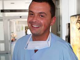 Gianlorenzo Dionigi合作论文数Dipartimento di Patologia Umana dell'adulto e dell'età evolutiva Gaetano Barresi, University of Messina6
 Turid Aas合作论文数6
 Neil Tolley合作论文数Department of Surgery and Cancer, Imperial College5Kamani Dipti合作论文数5
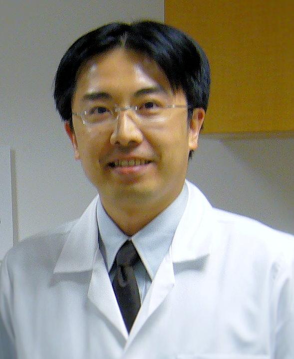 Che-Wei Wu (吳哲維)合作论文数Department of Otolaryngology, Kaohsiung Medical University5Feng-Yu Chiang合作论文数5
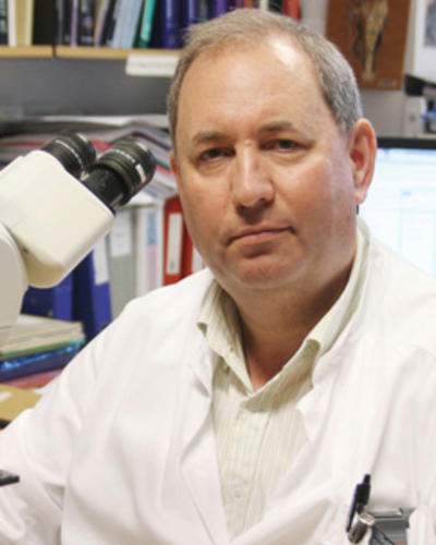 Lars Andreas Akslen合作论文数Department of Clinical Medicine, Faculty of Medicine, University of Bergen;Centre for Cancer Biomarkers, University of Bergen4Jan Erik Varhaug合作论文数4
 Paul Husby合作论文数Methods: Three groups were studied: the control group (CT group;n 10), the;Methods: Three groups were studied: the control group (CT group; n 10), the4Whitney Liddy合作论文数3Bjøro Trine合作论文数3
 Sam Van Slycke合作论文数3Øyvind S Svendsen合作论文数3Bo Wang (王波)合作论文数3Lodve Stangeland合作论文数3
 Jonathan Serpell合作论文数3Eva Sigstad合作论文数3Emad Kandil合作论文数Department of Surgery, Tulane Medical Center3Dana M. Hartl合作论文数3
 Ellen Schlichting合作论文数3John-Helge Heimdal合作论文数3
 Jennifer J. Shin合作论文数Department of Otolaryngology Head and Neck Surgery, Harvard Medical School, Harvard University;Department of Surgery, Brigham and Women's Hospital3Krystyna Kotanska Grøholt合作论文数Department of Pathology, Oslo University Hospital3Joseph Scharpf合作论文数3Else Opsahl合作论文数3Carsten Sekulla合作论文数3
 Erivelto Volpi合作论文数2Okenwa C Okose合作论文数2Peter E. Goretzki合作论文数2Natalia Kyriazidis合作论文数2Nadia Innaro合作论文数2
 John M Chaplin合作论文数2Anthony Y. Cheung合作论文数2
 Martin Almquist合作论文数2Ayaka J Iwata合作论文数2Amy Chen合作论文数2Amr H Abdelhamid Ahmed合作论文数2
 Brendan C. Stack合作论文数Dept Otolaryngol Head & Neck Surg, Univ Arkansas Med Sci2Singer Michael C合作论文数Department of Otolaryngology-Head and Neck Surgery, Georgia Health Sciences University2
 Gianluca Donatini合作论文数Department of General and Endocrine Surgery, CHR Huriez2




