目的 分析颅内压监测在治疗重症颅脑损伤中的价值.方法 选择2016年3月~2018年3月于我院就诊的重症颅脑损伤患者,共60例,按入院编号随机分为两组,每组30例.对照组和观察组分别给予常规、颅内压监测治疗,对比两组患者的具体治疗情况.结果 观察组取得了较高的总有效率,且各项临床指标明显优于对照组,具有统计学意义(P<0.05).结论 在治疗重症颅脑损伤时,通过颅内压监测能够取得理想的治疗效果,值得临床推广与应用.
目的 探讨采用单孔等量盐水置换引流术治疗慢性硬膜下血肿(CSDH)的治疗效果及临床体会.方法 选取2013年12月~2017年10月我院收治的CSDH患者76例作为研究对象,46例采用单孔等量盐水置换冲洗引流术,30例患者采用标准钻孔置管冲洗引流术.统计两组患者住院时间,手术并发症(气颅、颅内感染),术后复发情况,以及治疗效果.结果 置换组颅内积气发生率明显降低、术后痊愈率明显提高.结论 单孔等量盐水置换引流术治疗慢性硬膜下血肿保留了原术式的简单、易行、创伤小的特点,又降低了张力性气颅的危险性,提高了有效性及安全性.
目的 探讨亚低温治疗对降低重症高血压脑出血患者术后颅内压的作用.方法 对我院2013年12月-2016年12月接收的58例重症高血压脑出血患者资料加以探究,随机分成2组,每组29例;对照组行常规治疗,实验组行亚低温治疗,比对2组术后颅内压治疗前后NIHSS与ADL评分变化情况.结果 实验组术后1h、2h、24h及72h颅内压水平均显著低于对照组(P<0.05);实验组治疗后NIHSS评分(10.36±3.18)分显著低于对照组(17.42±3.61)分,ADL评分(85.69±7.42)分显著高于对照组(79.30±5.11) (P<0.05).结论 重症HICH患者行亚低温治疗可取得良好效果,有效降低术后颅内压,并改善其神经功能及生活能力,可被临床推广、应用.
假性缺氧性脑肿胀(PHBS)由van Roost等[1]2003年首次提出,是神经外科术后少见、病情进展迅速,甚至致命性的并发症. 现将我院1 例慢性硬膜下血肿术后出现PHBS 报告如下. 1 病例 男,39岁. 因"间断性头痛10 d,加重伴意识障碍2 d"于2015年6月14日入院. 患者入院前10 d因不慎摔伤头部,当时未到医院诊治,近10 d内感间断性头痛,双颞为主,口服去痛片缓解. 近2d内患者头痛症状进行性加重,精神不振,意识模糊. 既往体健,否认家族性遗传病史. 查体:昏睡,刺痛睁眼,问话不答,刺痛定位,GCS评分8分,颈软无抵抗,双侧瞳孔等大等圆,直径3.0 mm,对光反射灵敏,四肢肌力Ⅴ级,肌张力正常,双侧病理征未引出. 头颅CT平扫示双侧额颞顶颅骨内板下稍高密度影,考虑慢性硬膜下血肿(图1A).
急性高血压脑出血是脑血管病中病死率和致残率都很高的一种疾患.西方国家脑出血占全部脑卒中的8%~15%,而我国则高达21%~48%[1].急性高血压脑出血是以中老年患病为主的疾患,正在严重威胁着人们的健康.因此,必须引起重视,不断研究对其防治有效的措施.笔者应用醒脑静注射液联合盐酸钠美芬注射液治疗急性高血压脑出血,旨在观察二者联用的早期疗效,并探讨其机制.
Objective To evaluate the new approach of minimal invasive treatment of ventricular hemorrhage.Methods Neuroendoscopy was applied to treat 20 cases of ventricular hemorrhage. Twenty-two patients who were treated by ventricular drainage were taken as control. Results In the neuroendoscope treatment group, nearly compete evacuation of intraventricular hematoma was achieved in 16 cases, partial evacuation in 4 cases. Intracranial infection after surgery happened in 1 case, there was no secondary hemorrhage after surgery in all cases. All patients were followed up for two months. According to GOS, the outcome was excellent in 5 cases, good in 8 cases, fare in 3 cases, poor in 2 cases,death in 2 cases. In external ventricular drainage group, only 2 cases achieved partial evacuation of intraventricular hematoma and no change in 20 cases. Secondary hemorrhage after surgery in 1 case and intracranial infection in 5 cases.All patients were followed up for two months. According to GOS, the result was excellent in 2 case, good in 5 cases, fare in8 cases, poor in 5 cases, death in 2 cases. More patients in the neuroendoscope treatment group showed good recovery after two months(P 0.05). The difference between the two groups in mortality rate and secondary hemorrhage after surgery rate was not statistically significant(P 0.05). Conclusion Neuroendoscopic neurosurgery for ventricular hemorrhage is a good surgical treatment approach because it is characterized by visualized manipulation, effective hemorrhage evacuation and excellent postoperative outcomes.
Objective To explore the prognostic factors in surgical practices for hypertensive basal ganglia hemorrhage(HBGH).Methods The clinical data of 80 cases of hypertensive basal ganglia hemorrhage were analyzed retrospectively.The correlation of clinical features and the prognosis was analyzed.The prognostic outcomes were assessed according to GOS scale after 6-month follow-up.GOS scores 3-5 were defined good outcomes,and 1-2 scores were defined as poor outcomes.Results Among 80 cases,43 cases achieved good outcomes and 37 cases got poor outcomes.The results of univariate analysis showed that there were 10 variables related to the prognostic outcomes,including preoperative GCS score,volume of the intracerebral hematoma,intraventricular hematoma,brain hernia,surgical ways,operative hemorrhage,residual hematoma,pneumonia,hemorrhage of stress ulcer and electrolyte disturbance.Significant factors selected by univariate analysis were further evaluated by logistic regression analysis with forward stepwise selection procedures.Only 2 predictors showed statistical significance in the multivariate analysis,including brain hernia(P=0.002,OR=9.000,95%CI 2.269-35.701),preoperative GCS scores(P=0.023,OR=1.335,95%CI 1.040-1.715).Conclusion The related factors of operative prognosis could be helpful for doctors to make a correct decision for treating patients with HBGH.
<正>持续意识障碍患者气管切开能够通畅气道、改善氧合、控制肺部感染,经皮扩张钳气管切开术(percutaneous dilational tracheostomy,PDT)已被报道安全便捷,我院神经外科经过109例意识障碍患者PDT实践,总结经验如下。一、资料和方法1.一般资料:2009年1月至2011年10月神经外科意识障
一、资料与方法 1.一般资料:2008年10月至2011年8月收治高血压基底节区脑出血80例.常规开颅手术35例,男23例,女12例,年龄37~79岁,血肿量30~147 ml;显微镜辅助小骨窗手术26例,男19例,女7例,年龄40~84岁,血肿量30~100 ml;神经内镜辅助小骨窗手术19例,男9例,女10例,年龄43 ~76岁,血肿量34~110ml.
目的介绍冠状切口双额去骨瓣减压术治疗双额叶脑挫裂伤的临床体会。方法回顾性分析42例双额叶脑挫裂伤并接受双额去骨瓣减压术病例,总结资料临床特点、手术时机、手术特点及效果。结果 42例中3例急诊手术,39例入院时不具备手术指征,病情逐渐加重而中转手术治疗。术后并发枕部迟发硬膜外血肿3例(7.1%),硬膜下积液6例(14.3%),皮瓣下积液12例(28.6%),切口局部延迟愈合7例(16.7%)、局部脑脊液漏5例(11.9%),颅内感染3例(7.1%)。随访6个月,按GOS评分评定预后,恢复良好24例,中残6例,重残3例,植物生存3例,死亡6例。结论双额叶脑挫裂伤患者在保守治疗过程中有很大比例需中转急诊开颅手术,应严密观察病情进展,把握合理手术时机,冠状切口双额去骨瓣减压术在该病的治疗中仍能发挥着重要作用。
It's important for a doctor to master medical English in the teaching hospital.It can helps him to work well in many fields such as in clinic works,education works and science research works.In order to draw up a effective plan about how to improve the doctors' medical English level,some strategies were carried out from five aspects in this paper such as building the medical English department,encouraging doctors to use English in daily clinic works and in their spare time,making a regular studying plan for each clinic department,education of teachers and application of internet resources.
目的通过重症脑血管病患者经皮扩张钳气管切开术(PDT)与常规气管切开(OT)两种术式比较,分析PDT在重症脑血管病患者的应用价值及注意事项。方法回顾分析北京潞河医院同期气管切开资料齐全的66例重症脑血管病例,按气管切开方式分为PDT组(n=49)和OT组(n=17),比较两组的切口长度、操作时间、参与人员、术中出血、生命体征波动及其他并发症的发生率。结果 PDT组切口长度及操作时间明显短于OT组,差异有统计学意义[(1.7±0.2)cmvs.(3.4±0.7)cm,P<0.001;(4.4±3.4)minvs.(19.6±3.0)min,P<0.001],PDT单人完成8例,OT均由两人完成,术中出血、心率、血压波动及并发症总例数两组比较无统计学差异(44.9%vs.64.7%,P=0.159;38.8%vs.29.4%,P=0.489;40.8%vs.41.2%,P=0.979)。结论 PDT具有切口小、操作快捷、可单人完成优势,熟练操作技巧后可在重症脑血管病患者中应用,术中应准备吸引器、气管插管包及常规气管切开器械,积极防控出血、缺氧及气道损伤。
Objective To compare the advantages and disadvantages of conventional craniotomy,microscope-assisted minicraniotomy and endoscope-assisted minicraniotomy for hypertensive basal ganglia hemorrhage(HBGH).Methods From October 2008 to September 2011,80 consecutive patients with HBGH were collected,including 35 cases of conventional craniotomy,26 cases of microscope-assisted minicraniotomy,and 19 cases of endoscope-assisted minicraniotomy.The clinical characteristics and results including age,gender,preoperative GCS score,time from ictus to operation,volumes and locations of intracerebral hematoma,operation time,residual hematoma and GOS score six months after operation were analyzed by SPSS17.0.In each group GOS score was evaluated in 6 months after operation,patients with 3-5 scores were defined as a column with good outcome,and 1-2 scores as a column with poor outcome.Results There was no significant difference in age,gender,preoperative GCS score,time from ictus to operation,volumes and locations of the intracerebral hematoma among three groups(P 0.05),but there was significant difference in the operation time,blood loss and residual hematoma volume(P0.05).Those data between the microscope-assisted group and endoscope-assisted group were not statistically different(P0.05).Compared with conventional craniotomy group,those data in the other two groups were significantly different(P0.01).Patients in microscope-assisted and endoscope-assisted groups had better outcome than those in conventional craniotomy group(P0.01),but the prognosis was not statistically different between microscope-assisted and endoscope-assisted groups(P0.05).Conclusion Compared with conventional craniotomy,both microscope-assisted and endoscope-assisted minicraniotomy are better in the treatment of HBGH due to short operation time,less blood loss and residual hematoma,and better patients'outcome.
Objective:To probe into the skills and time for decompression operation and predictors associated with outcomes of decompression by standard large craniectmy to treat large ischemic cerebral infarction.Methods:Reviewing and analyzing 9 patients with large ischemic cerebral infarction who underwent decompression by standard large craniectomy.Summarizing the predictors including patients′ age,time and score of GCS before the operation.The patients were followed 3 and 6 months after the operation,to evaluate the prognosis according the score of the MRS and barthel index(BI).Results:The average time before the operation is(59.67±15.63) hours.The ischemic areas of 3 cases locate on the territory supplied by the right middle cerebral artery,those of 3 cases located on that by the left middle cerebral artery,and those of 3 cases locate on that by the left middle cerebral artery and anterior cerebral artery.The average score of GCS for patients at admission is 11.78±2.69,that before the operation is 8.22±2.07.Two cases who account for 22.2% of the nine patients died after the operation,7 cases were survival.Three months after the operation,4 cases got 4 scores,3 cases got 3 scores according to the MRS assessment.4 cases got 55 scores,2 cases got 65 scores,1 case got 75 scores according to the BI.Six months after the operation,according to the MRS assessment 1 cases got 4 scores,5 cases got 3 scores,1 cases got 2 scores.According to the BI,3 cases got 70 scores,2 cases got 75 scores,1 case got 65 scores,1 case got 85 scores.Conclusion:The decompression by standard craniectomy is an effective way to treat the large ischemic cerebral infarction.
<正>三叉神经显微血管减压术是治疗三叉神经痛最有效的方法。但术中岩静脉的损伤可导致小脑、脑干出血或梗死等严重并发症,因此,减少岩静脉的损伤是减少手术并发症的关键。选择2007年1月
Objective To systematically study the neural structures in the carotid bifurcate area for providing anatomical parameters for operative procedures.Methods Ten adult head specimen(20 sides) were studied.The course,relationship and spatial distribution of the cervical plexus cutaneous branch,cerebral nerves and sympathetic nerves in the carotid bifurcation area were observed and measured.Results The distance from the intersection of auricular nerve and sternocleidomastoid anterior edge to the mastoidle was 27.5±5.4 mm.The distance from the intersection of the transverse cervical nerve and sternocleidomastoid anterior edge to the mastoidle was 54.0±6.3 mm.The distance from the bifurcation of the hypoglossal nerve and the superior root of the ansa cervicalis to the lowest depression of the carotid bifurcation area was 8.2±5.0 mm.The distance from the bifurcation of superior laryngeal nerve to the lowest depression of the carotid bifurcation area was 8.1±4.2 mm.Conclusions Neuroanatomical study of the carotid bifurcate area provides the reference for guiding operation,protecting the nerves and reducing surgical complications,thus possessing good theoretical and practical meaning.
OBJECTIVE To investigate the characteristics of pathogens causing pulmonary infection in patients with craniocerebral injury and the drug resistance.METHODS The clinical data of 257 patients with pulmonary infection in patients with craniocerebral injury were retrospectively analyzed.RESULTS A total of 257 strains of pathogens were isolated,and the Gram-positive bacteria(51 strains),Gram-negative bacteria(199 strains),and fungi(7 strains) took proportions of 19.84%,77.43% and 2.73%,respectively.The main pathogens were Klebsiella pneumoniae,Pseudomonas aeruginosa,Staphylococcus aureus,and Escherichia coli,accounting for 25.29%,15.95%,15.56% and 14.79%,respectively.The pathogens showed high drug resistance.CONCLUSION Gram-negative bacteria are the main pathogens that cause pulmonary infection in patients with craniocerebral injury and show high drug resistance which should be paid more attention.
Objective:To evaluate the curative effects and advantages of stereotactic operation on hypertensive basal ganglia hemorrhage. Methods:86 cases were taken stereotactic aspiration;80 cases were taken traditional operation to eliminate hematoma;to compare the two methods and to find which operation method is the better.Results:The cases adopted with the stereotactic aspiration had a lower mortality and a better prognosis than the cases adopted traditional operations.Conclusion:Stereotactic operation is an effective way to treat hypertensive basal ganglia hemorrhage with advantages of accurate location,minimal invasiveness and low mortality and it had a better curative effect.
Objective To explore the techniques of surgical treatment for hypertensive intracerebral hematoma via minicraniotomy approach assisted by neuroendoscopy. Method The clinical data of 24patients treated surgically by external neuroendoscopic lumina operating via minicraniotomy approach for hypertensive intracerebral hematomas were retrospectively reviewed. Results The mean time of the procedure was 65 minutes. Complete evacuation of the intracerebral hematoma was achieved in 19 patients,and partial evacuation in 5. No patients recurred intracerebral hematoma. The period of follow - up for all of the patients was 1 to 10 months. According to Glasgow Outcome Scale, the results were excellent in 3patients, good in 7, fare in 11, dead in 3. Conclusions The procedure of minicraniotomy approach assisted by neuroendoscopy for treatment of hypertensive intracerebral hematoma has some advantages such as miniinvasion, time - saving, good exposure, and complete hemostasis. It is worth to be spread in clinical work.
Objective:To compare the three operation methods of hypertensive intracerebral hemorrhage.Methods:From May 2005 to May 2009,132 patients with hypertensive intracerebral hemorrhage were randomly divided into 3 groups(44 cases for each),received conventional skull operation(conventional group),microscopic skull operation (microscopic group) and stereotaxic miniinvasive skull operation(stereotaxic group) respectively.The clinical effects were compared.Results:For the short term efficacy,there were significant differences among the three groups(x~2=16.178,P=0.039 9<0.05),stereotaxic group was superio to conventional one,and the difference between stereotaxic group and microscopic group was no statistically significant(x~2=1.527,P=0.821 8),For the prostecdtive efficacy,stereotaxic group was superio to the other two groups(x~2was 23.990 and 10.048 respectively,P was 0.000 1 and 0.039 6 respectively).Conclusion:Because of the improvement of short term efficacy and prostecdtive efficacy,stereotaxic miniinvasive skull operation is worthy of wide clinical use in the treatment of patients with hypertensive intracerebral hemorrhage.
 HongBing Zhang (张洪兵)合作论文数25
 Gang Wang (王刚)合作论文数The pathological department of Medical Faculty of Qinghai University19XinBing Lv (吕新兵)合作论文数14Jianhua Lv (吕建华)合作论文数13
 Chao Sun (孙超)合作论文数北京协和医学院北京医院11Lianhu Yin (尹连虎)合作论文数Beijing Luhe Hospital, Capital Medical University10ShiQi Zheng (郑仕奇)合作论文数9WANG Chang-jiang (王长江)合作论文数7JianXin Liu (刘建鑫)合作论文数4Wen-Jun Cao (曹文军)合作论文数Department of Electrical & Computer Engineering, National University of Singapore 10 Kent Ridge Crescent, Singapore 119260, Singapore4hongyan han (韩宏彦)合作论文数3ZHANG Hong-bing (张红兵)合作论文数3LingWei Cui (崔玲伟)合作论文数3YanLong Hu (胡艳龙)合作论文数3
 Weidong Zhu (朱蔚东)合作论文数Dongzhimen Hospital, Beijing University of Chinese Medicine3Qi Fan (樊琪)合作论文数3
 Weidong Sun (孙卫东)合作论文数Dongzhimen Hospital, Beijing University of Chinese Medicine3zhigang tian (田志钢)合作论文数2yue liu (刘玥)合作论文数2DingJun Ma (马定军)合作论文数2YaXiong Sun (孙亚雄)合作论文数2TengYu Li (李腾宇)合作论文数2WenJun Cao (曹文军)合作论文数2Chao Wang (王超)合作论文数2Mo Yun (运陌)合作论文数2Shengli Liu (刘胜利)合作论文数2SiXun Zhang (张思迅)合作论文数1WeiQing Wan (万伟庆)合作论文数1JianHua Cai (蔡建华)合作论文数1WeiDong Zhang (张伟东)合作论文数1Fang Han (韩芳)合作论文数1Jun Yang (杨俊)合作论文数1
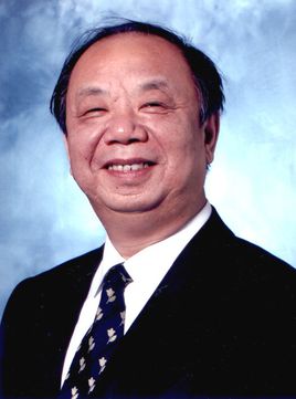 Huancong Zuo (左焕琮)合作论文数School of Medicine, Tsinghua University;Tsinghua University Yuquan Hospital1
 Weiqing Wan (万伟庆)合作论文数Beijing Tiantan Hospital, Capital Medical Univerity1
 Qing Huang (黄庆)合作论文数,, Test Center of Affiliated Hospital of, Youjiang Ethnomedicine1
 Hongshuan Liu (刘红栓)合作论文数Dongzhimen Hospital, Beijing University of Chinese Medicine1
 Yue Yuan (袁越)合作论文数China-Japan Friendship Hospital1Chunmei Zhang (张春梅)合作论文数1
 Hao Wang (王浩)合作论文数阜外心血管病医院影像学1
 Yakui Zhang (张亚奎)合作论文数Beijing Luhe Hospital, Capital Medical University1Xu-Yi Zong (宗绪毅)合作论文数1
 RuChen Peng (彭如臣)合作论文数Beijing Luhe Hospital, Capital Medical University1




