To evaluate the utility of microRNAs (miRNAs) integrated with current clinical risk models as predictive models for hepatocellular carcinoma (HCC) recurrence after liver transplantation (LT). This retrospective proof-of-concept study included 20 patients with HCC who underwent LT between 2007 and 2021 (n = 10 recurrent, n = 10 5-year recurrence-free). MiRNA profiling was performed on formalin-fixed, paraffin-embedded (FFPE) HCC explant tissue at the time of transplantation and clinical data were collected. The predictive value of miRNA expression for HCC recurrence was evaluated in a hybrid data- and hypothesis-driven approach and combined with clinical risk models (Milan, UCSF, Metroticket 2.0 and AFP). Kaplan-Meier analysis was performed to analyze recurrence-free survival (RFS). We identified a 3-miRNA signature - miR-3692-5p, miR-424, and miR-718 - that revealed discriminatory capacity between recurrence and non-recurrence. Adding this signature to clinical models increased the area under the receiver operating characteristic curve (AUC) for modeling HCC recurrence from 0.5 to 0.7 to 0.94-0.96. The combined models were used to categorize patients as high- or low-risk, with patients in the high-risk group having a shorter estimated median RFS (17.0 months vs. 38.5 months, p < 0.05). Integrating tissue-derived molecular miRNA signatures with existing clinical risk models may enhance the prediction of HCC recurrence following LT. Incorporating molecular approaches into current protocols could refine post-transplant risk stratification and surveillance guidance.
Background and Aims ReMELD-Na and MELD 3.0 are newly introduced prognostic scores for liver graft allocation, but their ability to predict the outcome after transjugular intrahepatic portosystemic shunt (TIPS) for refractory ascites in Western populations remains uncertain. This study compared the prognostic performance of ReMELD-Na and MELD 3.0 with FIPS, MELD-Na, MELD. Methods In this multicenter retrospective study, 1,621 cirrhotic patients undergoing TIPS for refractory ascites at eight German centers (January 2004-June 2024) were analyzed. The outcome was the event of either death or LTx within 90 days (primary) and one year (secondary) after TIPS. Prognostic performance was evaluated using receiver operator characteristic (ROC) and area under the curve (AUC), including sex-stratified analyses, and compared using DeLong’s test. High-risk groups (above the 85th/75th for the 90- day and one-year endpoint, respectively) were compared to non-high-risk groups using Kaplan-Meier analysis, scatter plots and descriptive score-vs-score spline smooth analysis. Results All scores showed limited predictive performance, with ROC-AUC values of 0.635 to 0.675 for the 90-day and 0.644 to 0.672 for the one-year outcome. Female patients demonstrated higher ROC-AUC values, reaching 0.714 for FIPS at 90 days. ReMELD-Na showed significantly lower ROC-AUC values than FIPS, MELD 3.0, and MELD-Na. Beyond this, MELD 3.0´s ROC-AUC values were comparable to those of the other scores. All models identified high-risk groups with increased event rates of death and LTx. Conclusions After TIPS for refractory ascites, all scores exhibited limited prognostic performance, but adequately distinguished high- and low-risk patients. MELD 3.0 performed comparably to established models, while ReMELD-Na was inferior to FIPS, MELD 3.0, and MELD-Na. Higher ROC-AUCs in women indicate sex-specific differences and the need for sex-sensitive prognostic tools. IMPACT AND IMPLICATIONS Accurate prediction of post-TIPS outcomes is essential to optimize management strategies for patients with cirrhosis and refractory ascites. In this large multicenter study, MELD 3.0 demonstrated prognostic performance comparable to established models, whereas ReMELD-Na - recently implemented for liver allocation in the Eurotransplant region - showed inferior predictive performance, raising concerns about its applicability in this setting. These results are particularly relevant as existing models may inadequately capture post-TIPS risk, especially in male patients. Collectively, the findings advocate for a cautious application of ReMELD-Na in clinical decision-making and emphasize the need to develop sex-sensitive, multidimensional prognostic tools to improve patient selection and surveillance.
BACKGROUND & AIMS:ReMELD-Na and MELD 3.0 are newly introduced prognostic scores for liver graft allocation, but their ability to predict outcomes after transjugular intrahepatic portosystemic shunt (TIPS) for refractory ascites in Western populations remains uncertain. This study compared the prognostic performance of ReMELD-Na and MELD 3.0 with FIPS, MELD-Na, and MELD. METHODS:In this multicenter retrospective study, 1,621 patients with cirrhosis undergoing TIPS for refractory ascites at eight German centers (January 2004-June 2024) were analyzed. Outcomes were the composite of death or liver transplantation (LTx) within 90 days (primary endpoint) and one year (secondary endpoint) after TIPS. Prognostic performance was evaluated using the area under the receiver-operating characteristic curve (AUROC), including sex-stratified analyses, and compared using DeLong's test. High-risk groups (above the 85th percentile for the 90-day endpoint and the 75th percentile for the 1-year endpoint) were compared with non-high-risk groups using Kaplan-Meier analysis, scatter plots, and descriptive score-vs.-score spline smoothing. RESULTS:All scores showed limited predictive performance, with AUROC values ranging from 0.635 to 0.675 for the 90-day outcome and from 0.644 to 0.672 for the 1-year outcome. Female patients demonstrated higher AUROC values, reaching 0.714 for FIPS at 90 days. ReMELD-Na showed significantly lower AUROC values than FIPS, MELD 3.0, and MELD-Na. In contrast, MELD 3.0 demonstrated AUROC values comparable to those of the other scores. All models identified high-risk groups with increased rates of death and LTx. CONCLUSIONS:After TIPS for refractory ascites, all scores exhibited limited prognostic performance, but adequately distinguished high- and low-risk patients. MELD 3.0 performed comparably to established models, while ReMELD-Na was inferior to FIPS, MELD 3.0, and MELD-Na. Higher AUROC values in women suggest sex-specific differences and highlight the need for sex-sensitive prognostic tools. IMPACT AND IMPLICATIONS:Accurate prediction of post-TIPS outcomes is essential to optimize management strategies for patients with cirrhosis and refractory ascites. In this large multicenter study, MELD 3.0 demonstrated prognostic performance comparable to established models, whereas ReMELD-Na - recently implemented for liver allocation in the Eurotransplant region - showed inferior predictive performance, raising concerns about its applicability in this setting. These results are particularly relevant as existing models may inadequately capture post-TIPS risk, especially in male patients. Collectively, the findings advocate for a cautious application of ReMELD-Na in clinical decision-making and emphasize the need to develop sex-sensitive, multidimensional prognostic tools to improve patient selection and surveillance.
Abstract Large language models (LLMs) like GPT have been proposed to support complex clinical decision-making. This study evaluated the performance of GPT-based LLM in analyzing clinical, radiological, and laboratory data from patients with hepatocellular carcinoma (HCC) to assess liver function, assign BCLC stage, and recommend treatment. Data from 106 HCC patients (82% male, median age 65 [22–86]) were compiled into anonymized integrated reports. Four GPT-versions (4, o1, o3, 5.4) were prompted—using both short and long instructions—to calculate MELD, ALBI, and Child–Pugh scores, assign BCLC stage, and generate treatment recommendations based on current guidelines. Outputs were compared to expert consensus and tumor board decisions. Errors were categorized by type and source. Time and cost analyses compared GPT to clinical staff. All GPT versions achieved high accuracy (> 85%) in liver function assessment, with MELD calculation being the most error-prone. BCLC staging accuracy ranged from 46.2% (version 4) to 84.0% (o3), with misclassification of radiological reports as the main error source. Reasoning-optimized models (o1, o3) performed best for treatment recommendations, achieving an overall accuracy (correct suggestions and acceptable alternatives) of up to 90.6%. In 9–14% of cases, GPT suggestions were retrospectively more guideline-concordant than tumor board decisions. GPT processing was significantly faster and reduced costs by approximately 300- to 1300-fold compared to clinical staff. GPT-based LLMs show potential as decision-support tools for liver function assessment, BCLC staging, and treatment guidance in HCC. Particularly with reasoning-optimized models and detailed prompting, LLMs may serve as valuable adjuncts in multidisciplinary HCC workflows. However, a non-negligible error rate requires expert oversight and further model refinement.
Purpose Before selective internal radiation therapy (SIRT), 99mTc macroaggregated albumin (MAA) particles are injected from the same catheter position(s) as a surrogate for later resin sphere distribution and to enable predictive dosimetry. Deviations in tumor segmentation affect predicted tumor and normal-liver absorbed doses and therefore prescribed activity. This study investigates the magnitude of interobserver variability in tumor segmentation for inexperienced and experienced observers when using biphasic contrast-enhanced CT as part of the 99mTc-MAA SPECT/CT imaging protocol and how it impacts the resulting tumor and normal-liver doses. Methods One inexperienced observer (performing two segmentations eight weeks apart) and one experienced observer used MIM SurePlan LiverY90 software to create tumor regions of interest (ROIs) from the SPECT/CT data of 19 patients. These three sets of ROIs were compared to the original clinical ROIs that had been defined similarly by another experienced physician together with a physicist. The Dice Similarity Coefficient (DSC) was calculated as a measure of tumor ROI overlap. Additionally, the resulting tumor and normal-liver dose differences using the partition model (assumed homogeneity within tumor and normal-liver compartments, doses represented by their mean values) between the three retrospectively determined sets of ROIs and the clinical ROIs were calculated and compared between inexperienced and experienced observers. Results DSC values between 0.73 and 0.75 were observed, indicating moderate to good agreement of segmentations, for inexperienced and experienced observers alike. Tumor dose differences were -3±13%, 2±10%, 2±10%; normal-liver dose differences were 9±16%, -9±14%, -4±14%, showing no relevant differences between inexperienced and experienced observers. Conclusion Significant interindividual variability in tumor segmentation and thus non-negligible deviations in tumor and normal-liver doses exist even with high-quality SPECT/CT imaging; however, for inexperienced and experienced observers alike. Hence, inexperienced observers with limited training can perform acceptably well. The magnitude of this variability should be considered when choosing injected activity based on predictive dosimetry, nevertheless.
BACKGROUND:Pain recurrence following regular radiofrequency ablation (RFA) in osteoid osteoma (OO) remains incompletely understood, yet impacts quality of life. PURPOSE:To detect predictive imaging parameters in OO with symptomatic recurrence. MATERIAL AND METHODS:This retrospective, monocentric study included 122 OO patients [median age 19 (16; 27) years, male 78%] January 2010 to May 2024, which underwent T1-weighted dynamic contrast-enhanced magnetic resonance imaging (DCE-MRI) (3T) before CT-guided RFA. Pain recurrence was defined as visual analogue scale increase >4/10 occurring >2 weeks post-intervention (22 (18%) patients). Nidus was categorized intra-/extracortical. DCE-MRI relative/maximum (relative) enhancement (M(R)E) (%), time to peak (TTP) (s), wash-in/out rate (s-1), brevity of enhancement (s) were compared by Wilcoxon rank-sum test. Hazard ratios (HR), multivariate cox proportional hazard regression, receiver operating characteristics and Bayesian were performed. RESULTS:Extracortical nidus occurred more frequently in patients with recurrence (14%) compared to intracortical nidus (5%, p < 0.001). Patients with recurrence had lower MRE [median, 327.2 (IQR, 227.5-393.6)% vs 386.1 (261, 511.6)%; p = 0.038], higher TTP [47.1 (34.5-59.5) vs 32.3 (24.7-46.7); p = 0.002], lower wash-in rate [38.9 (0-107.5) vs. 83.7(30.5-159.7); p = 0.028]. ME [HR 0.2 (95% CI: 0.06, 0.04); p < 0.001], MRE [HR 0.3 (0.1,1); p = 0.042], TTP [HR 6 (1.4, 25.4); p = 0.016], wash-in rate [HR 0.1(0.02, 0.8); p = 0.03] were predictors of recurrence. Extracortical nidus [HR 6.5 (2.1, 19.7); p < 0.001] was predictive. In extracortical nidus-subgroup recurrence probability ≥55% occurred with TTP >40 s, MRE <300%. CONCLUSION:Imaging-based risk stratification enables personalized treatment approaches for osteoid osteoma patients, who may benefit from alternative ablation protocols such as extended ablation time, higher temperatures.
BACKGROUND/OBJECTIVES:To develop a decision framework integrating computed tomography (CT) radiomics and clinical factors to guide the selection of transarterial chemoembolization (TACE) technique for optimizing treatment response in non-resectable hepatocellular carcinoma (HCC). METHODS:A retrospective analysis was performed on 151 patients [33 conventional TACE (cTACE), 69 drug-eluting bead TACE (DEB-TACE), 49 degradable starch microsphere TACE (DSM-TACE)] who underwent TACE for HCC at a single tertiary center. Pre-TACE contrast-enhanced CT images were used to extract radiomic features of the TACE-treated liver tumor volume. Patient clinical and laboratory data were combined with radiomics-derived predictors in an elastic net regularized logistic regression model to identify independent factors associated with early response at 4-6 weeks post-TACE. Predicted response probabilities under each TACE technique were compared with the actual techniques performed. RESULTS:Elastic net modeling identified three independent predictors of response: radiomic feature "Contrast" (OR = 5.80), BCLC stage B (OR = 0.92), and viral hepatitis etiology (OR = 0.74). Interaction models indicated that the relative benefit of each TACE technique depended on the identified patient-specific predictors. Model-based recommendations differed from the actual treatment selected in 66.2% of cases, suggesting potential for improved patient-technique matching. CONCLUSIONS:Integrating CT radiomics with clinical variables may help identify the optimal TACE technique for individual HCC patients. This approach holds promise for a more personalized therapy selection and improved response rates beyond standard clinical decision-making.
Abstract Objective This study aimed to evaluate medical students’ perception of a new radiology teaching format for abdominal diagnostics. The format transitioned traditional lectures and seminars to a case- and competency-based course that incorporates technology-enhanced individual case-work, small group discussions, and concise lectures. Materials and methods 235 students (23.5 ± 2.6 years, 72.3% female, 93.3% response rate, November 2023–June 2024) completed a questionnaire before (12 items) and after (20 items) the course, assessing perceived importance of course content, competency gains in abdominal imaging, enjoyment of learning, interest in a radiology career, and pedagogical perception of the teaching concept. Responses were recorded on a 1–10 scale (no agreement to strong agreement) or dichotomously (yes/no). The new course format was compared with a cohort of students who had previously (May 2022–June 2023) attended traditional lectures (n = 169) and/or seminars (n = 234). Results Students strongly agreed before the course that radiology content in abdominal diagnostics is important, and they found the content highly relevant and applicable to their work as doctors following the course. Significant improvement was observed in perceived competency in modality selection and description and interpretation of common pathologies, with the strongest effect for CT and MRI data. The new format was rated more motivating and significantly better in pedagogical and content quality than traditional lectures and seminars, although it did not influence students’ interest in pursuing a radiology career. Conclusion From the students’ perspective, case- and competency-based teaching enhances skill acquisition, learning success, and enjoyment in radiology. Clinical relevance statement From a student perspective, case- and competency-based teaching in radiology may enhance imaging competency, contributing to the development of more skilled healthcare providers. Key Points Case- and competency-based teaching concepts may improve students’ learning. Students reported improved perceived competency in decision-making and image interpretation with the new teaching method. Case- and competency-based teaching was perceived as more engaging, motivating, and pedagogically superior to traditional lectures. Graphical Abstract
Contrast agents (CAs) are essential in biomedical imaging to aid in the diagnosis and therapy monitoring of disease. However, they are typically restricted to one imaging modality and have fixed properties such as size, shape, toxicity profile, or photophysical characteristics, which hampers a comprehensive view of biological processes. Herein, rationally designed dye assemblies are introduced as a unique CA platform for simultaneous multimodal and multiscale biomedical imaging. To this end, a series of amphiphilic aza-BODIPY dyes are synthesized with varying hydrophobic domains ( C 1 , C 8 , C 12, and C 16 ) that self-assemble in aqueous media into nanostructures of tunable size (50 nm–1 µm) and photophysical properties. While C 1 exhibits oblique-type exciton coupling and negligible emission, C 8 - C 16 bearing longer alkyl chains undergo J -type aggregation with NIR absorption and emission and excellent photoacoustic properties. Given these advantageous features, aza-BODIPY specific, semi-quantitative fluorescence reflectance and photoacoustic imaging both in vitro and in vivo are established. Additionally, in vitro cell viability as well as murine in vivo biodistribution analysis with ex vivo validation showed excellent biocompatibility and a size-dependent biodistribution of nanostructures to different organ beds. These results broaden the scope of aqueous self-assembly to multimodal imaging and highlight its great potential for rationalizing numerous biomedical questions.
This retrospective study aims to develop a deep learning-based approach to whole-body CT segmentation out of standard PSMA-PET-CT to assess body composition in metastatic castration resistant prostate cancer (mCRPC) patients prior to [177Lu]Lu-PSMA radioligand therapy (RLT). Our goal is to go beyond standard PSMA-PET-based pretherapeutic assessment and identify additional body composition metrics out of the CT-component, with potential prognostic value. We used a deep learning segmentation model to perform fully automated segmentation of different tissue compartments, including visceral- (VAT), subcutaneous- (SAT), intra/intermuscular- adipose tissue (IMAT) from [68 Ga]Ga-PSMA-PET-CT scans of n = 86 prostate cancer patients before RLT. The proportions of different adipose tissue compartments to total adipose tissue (TAT) assessed on a 3D CT-volume of the abdomen or on a 2D single slice basis (centered at third lumbal vertebra (L3)) were compared for their prognostic value. First, univariate and multivariate Cox proportional hazards regression analyses were performed. Subsequently, the subjects were dichotomized at the median tissue composition, and these subgroups were evaluated by Kaplan–Meier analysis with the log-rank test. The automated segmentation model was useful for delineating different adipose tissue compartments and skeletal muscle across different patient anatomies. Analyses revealed significant correlations between lower SAT and higher IMAT ratios and poorer therapeutic outcomes in Cox regression analysis (SAT/TAT: p = 0.038; IMAT/TAT: p < 0.001) in the 3D model. In the single slice approach only IMAT/SAT was significantly associated with survival in Cox regression analysis (p < 0.001; SAT/TAT: p > 0.05). IMAT ratio remained an independent predictor of survival in multivariate analysis when including PSMA-PET and blood-based prognostic factors. In this proof-of-principle study the implementation of a deep learning-based whole-body analysis provides a robust and detailed CT-based assessment of body composition in mCRPC patients undergoing RLT. Potential prognostic parameters have to be corroborated in larger prospective datasets.
PURPOSE:To evaluate the pathological response after degradable starch microspheres transarterial chemoembolization (DSM-TACE) conducted to bridge or downstage hepatocellular carcinoma (HCC) patients to liver transplantation. MATERIALS AND METHODS:A multi-center retrospective study was conducted on consecutive patients with HCC who underwent liver transplantation after receiving DSM-TACE as a stand-alone therapy for bridging or downstaging between January 1, 2010, and December 31, 2022. Pathological response was evaluated histologically by board certified surgical pathologists. Tumor necrosis was estimated histologically as a percentage area of the total tumor area. The criteria for estimation of pathological response were established as follows: (1) Complete pathological response: 100% tumor necrosis and absence of any viable tumor cells; (2) Significant pathological response: 50-99% tumor necrosis in cross section; (3) Mild pathological response: 1-49% tumor necrosis in cross section and (4) No pathological response: no tumor necrosis present. RESULTS:Twenty-two patients (16 men; median age, 65 years) with 73 HCCs (median largest diameter: 2.7 cm) were bridged or downstaged with DSM-TACE and subsequently underwent liver transplantation. Histopathological examination of the explanted livers showed complete pathological response in 27 % of patients, significant pathological response in 32 %, mild pathological response in 27 % and no pathological response in 14 %. CONCLUSION:The present study provides important clinical evidence supporting the efficacy of DSM-TACE in inducing tumor necrosis, despite its short embolization time.
Objective: The benign nature of perimesencephalic subarachnoid hemorrhage (pmSAH) can be challenged by the occurrence of complications. Given the limited prognostic value of established clinical parameters for the development of complications in patients with pmSAH, this study evaluates the potential of volumetric hemorrhage quantification for risk assessment and the evaluation of the clinical outcome. Material and Methods: In this retrospective single-center study, we analyzed all consecutive patients diagnosed with pmSAH between 2010 and 2023 at a tertiary care academic medical center in Germany. The volumetric quantification of the hemorrhage in cm3 was performed using non-contrast CT imaging. The occurrence of clinical complications, including hydrocephalus, vasospasm, and delayed cerebral ischemia (DCI), were assessed. Clinical outcomes were determined by the Glasgow Outcome Scale (GOS) at discharge. Multivariable logistic regression models were used to assess the correlation between quantified hemorrhage volumes and the occurrence of complications and clinical outcomes (GOS) controlled for other variables such as age, sex, cardiovascular risk factors, clinical symptoms, and the modified Fisher scale. Results: A total of 82 patients (58.5% male, 54.8 ± 12.1 years) were enrolled. The median World Federation of Neurosurgical Societies (WFNS) score for all patients at admission was 1.0 (IQR 1.0–1.0). During the clinical course, hydrocephalus occurred in 29%, vasospasm in 14.6%, and DCI in 8.5% of all patients. Hemorrhage volume quantification was found to be the strongest independent predictor for hydrocephalus (OR 1.28; 95% CI 1.02–1.61; p = 0.032) and vasospasm (OR 1.25; 95% CI 1.07–1.46; p = 0.007) and showed a high predictive accuracy in ROC analyses (AUC = 0.77 and 0.76, respectively). Conversely, neither clinical parameters nor the modified Fisher scale were associated with these complications. A higher hemorrhage volume was also significantly correlated with a worse functional outcome (GOS; β = –0.07, CI: −0.12–−0.02, p = 0.021). Conclusions: In patients with pmSAH, the volumetric quantification of hemorrhage may be an adequate prognostic parameter regarding the occurrence of hydrocephalus and vasospasm. In addition, the quantitative assessment of hemorrhage volumes was strongly associated with clinical outcomes in these patients. Despite the generally benign nature of pmSAH, this imaging biomarker could improve individualized clinical management strategies and inform about the risk for the occurrence of complications.
PURPOSE:Time-lapse MRI allows for the dynamic tracking of single iron-labeled cells. However, the time required for spatial encoding creates a temporal blur and, therefore, a limited ability to resolve moving cells. To study fast moving cells, such as rolling immune cells along the endothelium during inflammatory processes, advanced accelerated acquisition techniques are required. METHODS:Balanced SSFP (bSSFP) imaging is applied to phantom and in vivo murine brain time-lapse MRI measurements at 9.4 T. Its detection capability of moving iron-labeled cells is compared with conventional gradient echo imaging (GRE) for 2D Cartesian sampling and evaluated for fully sampled and accelerated reconstructions with compressed sensing for 3D interleaved radial sampling in bSSFP. RESULTS:Both phantom and in vivo time-lapse MRI measurements show that single cells can be followed dynamically using bSSFP. High temporal resolution of less than 2 min reduces geometric distortion. The velocity detection limit increases to 0.8 mm/min in vitro and previously hidden fast-moving cells are recovered. Interleaved 3D radial sampling enables 3D cell tracking and simultaneous imaging at varying acceleration factors. Fivefold acceleration with compressed sensing optimizes cell visibility, image quality, and temporal resolution. CONCLUSION:bSSFP time-lapse MRI improves single-cell tracking by enhancing temporal resolution. In vitro, the velocity detection limit is increased fourfold compared to conventional GRE. Interleaved 3D radial bSSFP offers whole-brain coverage at isotropic spatial resolution and retrospective reconstruction of both fully sampled and high temporal resolution images.
 Moritz Wildgruber合作论文数Department for Radiology, Ludwig-Maximilians-Universität in Munich73
 Walter Heindel合作论文数Department of Radiology at the University Hospital Muenster44
 Anne Helfen合作论文数32
 Philipp Schindler合作论文数30
 Michael Kohler合作论文数23
 Mirjam Gerwing合作论文数23
 Cornelius Faber合作论文数Department of Experimental Physics, University of Würzburg22
 P Schindler合作论文数15M Köhler合作论文数15
 Eisenblätter Michel合作论文数King's College London, University of Münster13Hauke Heinzow合作论文数13Max Seidensticker合作论文数12Andreas Pascher合作论文数10Wachsmuth Lydia合作论文数10Jing Claussen合作论文数10
 Kambiz Rahbar合作论文数Young Researchers Club, Islamic Azad University of Mashhad10Carsten Höltke合作论文数10
 Vanessa F. Schmidt合作论文数9Schäfers Michael合作论文数9
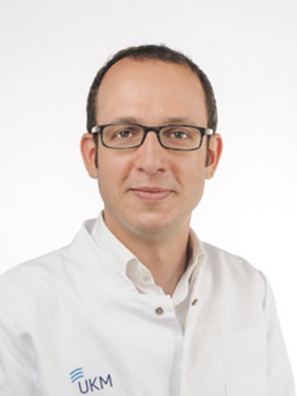 Mehmet Haluk Morgül合作论文数UKM Uniklinikum Muenster;Department of General and Visceral Surgery, University of Münster9M Eisenblätter合作论文数8Jonel Trebicka合作论文数8Georg Gosheger合作论文数8Hartmut Schmidt合作论文数8
 Enrica Wilken合作论文数8
 Kristian Nikolaus Schneider合作论文数8
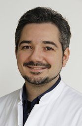 Angelos Karlas合作论文数DZHK (German Centre for Cardiovascular Research),, Munich Heart Alliance8
 Christian Wilms合作论文数8
 Dominik Bettinger合作论文数8
 Lars Stegger合作论文数8
 Walter A. Wohlgemuth合作论文数8
 Shadi Katou合作论文数7
 Wolfgang Roll合作论文数7Geyer Christiane合作论文数7
 Jens Ricke合作论文数Klinik Und Poliklinik Für Radiologie, LMU Klinikum;Medizinischen Fakultät Der LMU München7
 Uwe Karst合作论文数Institute of Inorganic and Analytical Chemistry, University of Munster7
 Vasilis Ntziachristos合作论文数School of Medicine and Health, Technical University of Munich;Institute of Biological and Medical Imaging, Helmholtz Zentrum Münche7W. Roll合作论文数6Richard Brill合作论文数6Sturm Lukas合作论文数6
 Thomas Vogl合作论文数5
 Emily Hoffmann合作论文数5Paul Stracke合作论文数5Felix Piecha合作论文数5
 Leon Louis Seifert合作论文数5
 Bastian Maus合作论文数5
 Michael Praktiknjo合作论文数5
 Peter Sporns合作论文数Universitätsspital Basel5Evers Georg合作论文数5




