Early detection and activity assessment of posterior cerebral artery (PCA) involvement in moyamoya disease (MMD) are challenging. We aimed to assess the cross-sectional association between PCA involvement and PCA wall enhancement on vessel wall MRI (DANTE T1-SPACE). We analyzed consecutive patients with MMD who underwent both non-contrast-enhanced and contrast-enhanced DANTE T1-SPACE. Positive wall enhancement was defined as location-appropriate arterial wall enhancement with an enhancement ratio ≥ 1.5. PCA involvement was graded 0–2 on MRA obtained during the same period. Of 368 hemispheres from 184 patients, 56 (15.2
BACKGROUND:Neovascularization contributes to plaque vulnerability in carotid artery stenosis. We aimed to assess the association between adventitial enhancement, which reflects neovascularization, on contrast-enhanced magnetic resonance imaging (MRI), and carotid plaque volume and clinical outcomes. METHODS:We retrospectively analyzed 72 patients with 75 carotid plaques who underwent preoperative contrast-enhanced MRI using a delay alternating with nutation for tailored excitation-prepared T1-weighted turbo spin echo (DANTE T1-SPACE) sequence at Kyoto University Hospital (April 2019-March 2025). Adventitial enhancement was visually graded across the entire lesion on postcontrast DANTE T1-SPACE as category 0 (no enhancement), category 1 (<50% of outer wall circumference), or category 2 (≥50%). Plaque volume was measured from precontrast DANTE T1-SPACE. Clinical outcomes included symptomatic events and ipsilateral cerebral ischemic lesions. Intraplaque hemorrhage (IPH) presence and volume were also evaluated. RESULTS:Of the 75 plaques, 9 were category 0, 40 category 1, and 26 category 2. The proportion of either symptomatic plaques or plaques with cerebral ischemic lesions increased across categories: 11% (n = 1), 50% (n = 20), and 62% (n = 16) (P = 0.018). Mean plaque volumes were 0.59, 0.67, and 0.88 mL, respectively (P = 0.004). No significant trend was seen in IPH frequency or volume. Among plaques without IxPH, volume still increased across categories: 0.27, 0.60, and 0.92 mL (P = 0.005). CONCLUSIONS:Whole-lesion adventitial enhancement on DANTE T1-SPACE MRI may serve as a noninvasive imaging biomarker of plaque burden and vulnerability for risk stratification in carotid artery stenosis.
BackgroundConventional MRI sequences are insufficient for the detailed depiction of intracranial atherosclerotic disease (ICAD) plaques. The aim of this study was to investigate the association between ischemic events and intracranial atherosclerotic plaque characteristics using a high-resolution T1-weighted black-blood MRI technique (DANTE T1-SPACE) in the anterior circulation in the Japanese population.MethodsPatients with a total of 108 lesions causing ≥40% stenosis on the C1–5 segments of the intracranial internal carotid artery (ICA) or M1 segment of the middle cerebral artery (MCA) were included. Hyperintense plaques (HIPs) were defined as plaques with a spot of signal intensity (SI) higher than 1.5-fold SI of the ipsilateral temporal muscle on DANTE T1-SPACE. The vessel wall lesions were divided into symptomatic and asymptomatic groups. The lesions in the symptomatic group were classified as artery-to-artery embolism, hemodynamic infarction, cardiac embolism, undetermined and transient ischemic attack (TIA).ResultsAmong the 108 plaques, 19 were symptomatic and 89 were asymptomatic. The percentage of HIPs in the symptomatic group was significantly higher than in the asymptomatic group (57.9% vs. 24.7%, p = 0.01). In the symptomatic group, the proportion of HIPs in the A-to-A embolism subgroup was higher than in the other subgroups.ConclusionsDANTE T1-SPACE may aid in the identification of intracranial plaques with imaging characteristics suggestive of increased stroke risk, particularly hyperintensity potentially reflecting intraplaque hemorrhage.
In mild traumatic brain injury, imaging biomarkers are needed to support clinical management. In four antecedent publications, we used two new (sodium and fingerprinting) and two established (spectroscopy and diffusion) MR techniques in a longitudinally followed patient cohort. Here we report final results and combine all data to determine which marker(s) from the four modalities offer the greatest utility for detecting injury and predicting outcomes. We also leverage the independent specificities offered by each modality to explore injury mechanisms. The longitudinal spectroscopy data were analysed to complete a full data set of proton (spectroscopy, fingerprinting, diffusion) and sodium MRI, acquired alongside symptomatic, cognitive, and functional assessments in 27 patients at 1, 3, and 12 months following injury. Twenty-three matched controls were scanned once. Testing for associations between nine MR markers and three outcome measures was standardized across the entire data set, and performed using Spearman correlations and logistic regression. Previously elevated white matter choline and creatine from spectroscopy (markers of membrane turnover and cellular energetics, respectively) normalized to control levels by 3 months, at rates which correlated with the rate of symptom recovery. Sodium and spectroscopy showed findings coinciding in pattern and timepoint, but there were no associations between them, suggesting independent origin. Choline and creatine met the greatest number of biomarker properties, followed by water T1 from fingerprinting (marker of the cellular microenvironment). We identified independent, dynamic, metabolic and ionic changes, with choline and creatine from spectroscopy fulfilling the most criteria for a clinical biomarker.
Observation of the clearance of gadolinium-based contrast agents (GBCA) in patients with cerebral infarction and Moyamoya disease (MMD) using vessel-wall MRI (VW-MRI). This prospective study included 11 patients with recent cerebrovascular disorder and 6 patients with MMD who were scheduled for VW-MRI. All participants underwent whole brain delay alternating with nutation for tailored excitation-prepared T1-weighted variable flip angle turbo spin echo (DANTE T1-SPACE) imaging before, immediately after, and 1–6 h after gadolinium injection. Additionally, a phantom experiment was conducted to assess the signal intensity according to T1 values using DANTE T1-SPACE and 3D real inversion recovery sequences. All five large infarction cases (≥ 5 cm) had delayed meningeal enhancement on the ipsilateral side, while none of the remaining six small infarction cases (< 5 cm) exhibited delayed meningeal enhancement on MRI. More than half of the entire cases had delayed white matter enhancement adjacent to the infarction. No MMD cases showed delayed enhancement outside postoperative regions. In the phantom experiment, DANTE T1-SPACE (TR = 700–1300 ms) demonstrated stable signal intensity across a T1 range of approximately 500–1000 ms. This T1 range corresponds to signal intensity ≈ 200–600 typically observed in regions showing delayed enhancement in clinical stroke and MMD cases. Delayed meningeal enhancement observed in patients with large cerebral infarctions likely reflects alterations in brain clearance pathways. The absence of such enhancement in MMD supports that these enhancements are not attributable to collateral circulation. Phantom validation confirmed that the imaging parameters used in the DANTE T1-SPACE sequence were sufficient to detect subtle contrast enhancement in the clinically relevant T1 range.
PURPOSE:The pathological hallmarks of Alzheimer's disease (AD), amyloid, tau, and associated neurodegeneration, are present in the cortical gray matter (GM) years before symptom onset, and at significantly greater levels in carriers of the apolipoprotein E4 (APOE4) allele. Their respective biomarkers, A/T/N, have been found to correlate with aspects of brain biochemistry, measured with magnetic resonance spectroscopy (MRS), indicating a potential for MRS to augment the A/T/N framework for staging and prediction of AD. Unfortunately, the relationships between MRS and A/T/N biomarkers are unclear, largely due to a lack of studies examining them in the context of the spatial and temporal model of T/N progression. Advanced MRS acquisition and post-processing approaches have enabled us to address this knowledge gap and test the hypotheses, that glutamate-plus-glutamine (Glx) and N-acetyl-aspartate (NAA), metabolites reflecting synaptic and neuronal health, respectively, measured from regions on the Braak stage continuum, correlate with: (i) cerebrospinal fluid (CSF) p-tau181 level (T), and (ii) hippocampal volume or cortical thickness of parietal lobe GM (N). We hypothesized that these correlations will be moderated by Braak stage and APOE4 genotype. METHODS:We conducted a retrospective imaging study of 34 cognitively unimpaired elderly individuals who received APOE4 genotyping and lumbar puncture from pre-existing prospective studies at the NYU Grossman School of Medicine between October 2014 and January 2019. Subjects returned for their imaging exam between April 2018 and February 2020. Metabolites were measured from the left hippocampus (Braak II) using a single-voxel semi-adiabatic localization by adiabatic selective refocusing sequence; and from the bilateral posterior cingulate cortex (PCC; Braak IV), bilateral precuneus (Braak V), and bilateral precentral gyrus (Braak VI) using a multi-voxel echo-planar spectroscopic imaging sequence. Pearson and Spearman correlations were used to examine the relationships between absolute levels of choline, creatine, myo-inositol, Glx, and NAA and CSF p-tau181, and between these metabolites and hippocampal volume or parietal cortical thicknesses. Covariates included age, sex, years of education, Fazekas score, and months between CSF collection and MRI exam. RESULTS:There was a direct correlation between hippocampal Glx and CSF p-tau181 in APOE4 carriers (Pearson's r = 0.76, p = 0.02), but not after adjusting for covariates. In the entire cohort, there was a direct correlation between hippocampal NAA and hippocampal volume (Spearman's r = 0.55, p = 0.001), even after adjusting for age and Fazekas score (Spearman's r = 0.48, p = 0.006). This relationship was observed only in APOE4 carriers (Pearson's r = 0.66, p = 0.017), and was also retained after adjustment (Pearson's r = 0.76, p = 0.008; metabolite-by-carrier interaction p = 0.03). There were no findings in the PCC, nor in the negative control (late Braak stage) regions of the precuneus and precentral gyrus. CONCLUSIONS:Our findings are in line with the spatially- and temporally-resolved Braak staging model of pathological severity in which the hippocampus is affected earlier than the PCC. The correlations, between MRS markers of synaptic and neuronal health and, respectively, T and N pathology, were found exclusively within APOE4 carriers, suggesting a connection with AD pathological change, rather than with normal aging. We therefore conclude that MRS has the potential to augment early A/T/N staging, with the hippocampus serving as a more sensitive MRS target compared to the PCC.
Purpose:To evaluate the use of delay alternating with nutation for tailored excitation-prepared T1-weighted turbo spin echo (DANTE T1-SPACE) imaging for diagnosing optic neuritis and to analyze its correlation with clinical findings before and after treatment. Patients and Methods:Patients diagnosed with optic neuritis or non-arteritic anterior ischemic optic neuropathy (NA-AION) were evaluated at the Ophthalmology Department of Kyoto University Hospital. All patients underwent magnetic resonance (MR) studies before treatment initiation and ophthalmic examinations before and after treatment. Three ophthalmologists independently reviewed the MR scans for abnormalities. The magnetic resonance imaging (MRI) assessments included post-contrast DANTE T1-SPACE, post-contrast volumetric interpolated breath-hold examination (VIBE), and short T1 inversion recovery (STIR) scans. The presence of abnormalities in each sequence was determined. Results:Of 36 eyes from 30 patients, 21 eyes from 17 patients were diagnosed with optic neuritis, and 15 eyes from 13 patients were diagnosed with NA-AION. DANTE T1-SPACE sequences showed better sensitivity for detecting optic neuritis than STIR sequences (100% vs 67%, p = 0.009). VIBE images did not confirm enhancement of lesions in some cases with optic neuritis. No differences were observed among the sequences for NA-AION. Lesion length evaluated by DANTE T1-SPACE sequences was associated with circumpapillary retinal nerve fiber layer thickness at the initial visit, eye pain, and the time interval from symptom onset to MRI scan. Conclusion:Contrast-enhanced DANTE T1-SPACE was better than other sequences of MRI for diagnosing optic neuritis.
OBJECTIVE:Certain patients must undergo frequent postoperative digital subtraction angiography (DSA) after flow diversion (FD) therapy. No imaging modality with an efficacy comparable to that of DSA has been established. This study was conducted to determine the efficacy of contrast-enhanced delay alternating with nutation for tailored excitation (DANTE) T1-sampling perfection with application-optimized contrasts by using different flip angle evolution (SPACE), a high-resolution vessel wall magnetic resonance imaging technique for evaluating the occlusion status of intracranial aneurysms after FD treatment, with DSA serving as the reference standard. METHODS:This retrospective study included 23 patients with 26 aneurysms who had undergone FD treatment between April 2016 and May 2022. Contrast-enhanced DANTE T1-SPACE and DSA were performed as postoperative follow-up imaging studies at 45 time points, both in the same period. The agreement rates for aneurysm occlusion status in the 45 imaging studies were examined. RESULTS:Contrast-enhanced DANTE T1-SPACE had a sensitivity and specificity of 96.3% (26/27) and 83.3% (15/18), respectively, for detecting aneurysm remnants. Overall, 91.1% (41/45) of findings detected on contrast-enhanced DANTE T1-SPACE were consistent with those on DSA. The findings detected on contrast-enhanced DANTE T1-SPACE were completely consistent with those of intraaneurysmal residual blood flow identified on DSA or high-resolution cone-beam computed tomography images in 74.1% (20/27) of the examinations that showed incomplete occlusion on DSA. Furthermore, parent artery status after FD treatment on contrast-enhanced DANTE T1-SPACE was consistent with that observed on DSA in 97.8% (44/45) of examinations. CONCLUSIONS:Contrast-enhanced DANTE T1-SPACE is a very useful option in the follow-up of aneurysms after FD treatment.
PURPOSE:CEST MRI has been used to probe changes in cardiac metabolism via assessment of CEST contrast from Cr. However, B1 variation across the myocardium leads to spatially variable Cr CEST contrast in healthy myocardium. METHODS:We developed a spatial-spectral (SPSP) saturation pulsed CEST protocol to compensate for B1 variation. Flip angle maps were used to individually tailor SPSP pulses comprised of a train of one-dimensional spatially selective subpulses selective along the principal B1 gradient dimension. Complete Z-spectra in the hearts of (n = 10) healthy individuals were acquired using conventional Gaussian saturation and SPSP schemes and supported by phantom studies. RESULTS:In simulations, the use of SPSP pulses reduced the average SD of the effective saturation B1 values within the myocardium (n = 10) from 0.12 ± 0.02 μT to 0.05 ± 0.01 μT (p < 0.01) and reduced the average SD of Cr CEST contrast in vivo from 10.0 ± 4.3% to 6.1 ± 3.5% (p < 0.05). Results from the hearts of human subjects showed a significant reduction of CEST contrast distribution at 2 ppm, as well as amplitude, when using SPSP saturation. Corresponding phantom experiments revealed PCr-specific contrast generation at body temperature when SPSP saturation was used but combined PCr and Cr contrast generation when Gaussian saturation was used. CONCLUSION:The use of SPSP saturation pulsed CEST resulted in PCr-specific contrast generation and enabled ratiometric mapping of PCr to total Cr CEST contrast in the human heart at 3T.
To investigate the relationship of followings for patients with moyamoya disease (MMD): arterial wall enhancement on vessel wall MRI (VW-MRI), cross-sectional area (CSA), time-of-flight MR angiography (MRA), age, locations from intracranial internal carotid artery (ICA) to proximal middle cerebral artery (MCA), disease progression, and transient ischemic attack (TIA). Patients who underwent VW-MRI between October 2018 and December 2020 were enrolled in this retrospective study. We measured arterial wall enhancement (enhancement ratio, ER) and CSA at five sections of ICA and MCA. Also, we scored MRA findings. Multiple linear regression (MLR) analysis was performed to explore the associations between ER, age, MRA score, CSA, history of TIA, and surgical revascularization. We investigated 102 sides of 51 patients with MMD (35 women, 16 men, mean age 31 years ± 18 [standard deviation]). ER for MRA score 2 (signal discontinuity) was higher than ER for other scores in sections D (end of ICA) and E (proximal MCA) on MLR analysis. ER in section E was significantly higher in patients for MRA score 2 with TIA history than without. ER significantly increased as CSA increased in section E, which suggests ER becomes less in decreased CSA due to negative remodeling. Arterial wall enhancement in MMD varies by age, location, and disease progression. Arterial wall enhancement may be stronger in the progressive stage of MMD. Arterial wall enhancement increases with history of TIA at proximal MCA, which may indicate the progression of the disease. Arterial wall enhancement in moyamoya disease varies by age, location of arteries, and disease progression, and arterial wall enhancement may be used as an imaging biomarker of moyamoya disease.
The newly developed “Impulse” head gradient (200 mT/m Gmax, 900 T/m/s) on the NexGen 7T scanner has been specifically designed to allow for high-resolution single-shot EPI with minimal distortions and blurring by minimizing echo spacing. In conjunction with the increased SNR and reduced g-factor noise afforded by the 96 channel receive array coil, mesoscale fMRI at 0.45mm and 0.6mm isotropic resolutions can be robustly achieved in human subjects.
To increase granularity in human neuroimaging science, we designed and built a next-generation 7 Tesla magnetic resonance imaging scanner to reach ultra-high resolution by implementing several advances in hardware. To improve spatial encoding and increase the image signal-to-noise ratio, we developed a head-only asymmetric gradient coil (200 mT m-1, 900 T m-1s-1) with an additional third layer of windings. We integrated a 128-channel receiver system with 64- and 96-channel receiver coil arrays to boost signal in the cerebral cortex while reducing g-factor noise to enable higher accelerations. A 16-channel transmit system reduced power deposition and improved image uniformity. The scanner routinely performs functional imaging studies at 0.35-0.45 mm isotropic spatial resolution to reveal cortical layer functional activity, achieves high angular resolution in diffusion imaging and reduces acquisition time for both functional and structural imaging.
1 H-MRS has the potential to provide biomarkers for mild traumatic brain injury (mTBI), a diagnosis in which damage is often imaging-occult, but a lack of replicability and reproducibility studies hampers clinical translation. Here, we tested the replicability of previous MRSI results in mTBI with a different patient cohort. Five out of seven hypotheses were consistent with previous work, with key findings of globally diffuse white matter (WM) injury and limited gray matter injury. However, we report differences in choline and creatine, not N -acetyl-aspartate. Correlations were found between metabolite levels in WM and both symptomatology and neuropsychological testing.
Rationale and objectives: Macromolecules (MMs) affect the precision and accuracy of neurochemical quantification in magnetic resonance spectroscopy. A measured MM basis is increasingly used in LCModel analysis combined with a spline baseline, whose stiffness is controlled by a parameter named DKNTMN. The effects of measured MM basis and DKNTMN were investigated.Materials and methods: Twenty-six healthy subjects were prospectively enrolled and scanned twice using a short echo-time Stimulated Echo Acquisition Mode (STEAM) at 7-T. Using LCModel, analyses were conducted using the simulated MM basis (MMsim) with DKNTMN 0.15 and an MM basis measured inhouse (MMmeas) with DKNTMN of 0.15, 0.30, 0.60 and 1.00. Crame & PRIME;r-Rao lower bound (CRLB) and the concentrations of gamma-aminobutyric acid (GABA), glutamate and excitatory-inhibitory ratio (EIR), in addition to MMs were statistically analyzed. Measurement stability was evaluated using coefficient of variation (CV).Results: CRLBs of GABA were significantly lower when using MMsim than MMmeas; those of glutamate were 2-3. GABA concentrations were significantly higher in the analysis using MMsim than MMmeas where concentrations were significantly higher with DKNTMN of 0.15 or 0.30 than 0.60 or 1.00. Difference in glutamate concentration was not significant. EIRs showed the same difference as in GABA depending on the DKNTMN values. CVs between test-retest scans were relatively stable for glutamate but became larger as DKNTMN increased for GABA and EIR.Conclusion: Neurochemical quantification depends on the parameters of the basis sets used for fitting. Analysis using MMmeas with DKNTMN of 0.30 conformed best to previous studies and is recommended.
PURPOSE:Proton magnetic resonance spectroscopy (1H MRS) offers biomarkers of metabolic damage after mild traumatic brain injury (mTBI), but a lack of replicability studies hampers clinical translation. In a conceptual replication study design, the results reported in four previous publications were used as the hypotheses (H1-H7), specifically: abnormalities in patients are diffuse (H1), confined to white matter (WM) (H2), comprise low N-acetyl-aspartate (NAA) levels and normal choline (Cho), creatine (Cr) and myo-inositol (mI) (H3), and correlate with clinical outcome (H4); additionally, a lack of findings in regional subcortical WM (H5) and deep gray matter (GM) structures (H6), except for higher mI in patients' putamen (H7). METHODS:26 mTBI patients (20 female, age 36.5 ± 12.5 [mean ± standard deviation] years), within two months from injury and 21 age-, sex-, and education-matched healthy controls were scanned at 3 Tesla with 3D echo-planar spectroscopic imaging. To test H1-H3, global analysis using linear regression was used to obtain metabolite levels of GM and WM in each brain lobe. For H4, patients were stratified into non-recovered and recovered subgroups using the Glasgow Outcome Scale Extended. To test H5-H7, regional analysis using spectral averaging estimated metabolite levels in four GM and six WM structures segmented from T1-weighted MRI. The Mann-Whitney U test and weighted least squares analysis of covariance were used to examine mean group differences in metabolite levels between all patients and all controls (H1-H3, H5-H7), and between recovered and non-recovered patients and their respectively matched controls (H4). Replicability was defined as the support or failure to support the null hypotheses in accordance with the content of H1-H7, and was further evaluated using percent differences, coefficients of variation, and effect size (Cohen's d). RESULTS:Patients' occipital lobe WM Cho and Cr levels were 6.0% and 4.6% higher than controls', respectively (Cho, d = 0.37, p = 0.04; Cr, d = 0.63, p = 0.03). The same findings, i.e., higher patients' occipital lobe WM Cho and Cr (both p = 0.01), but with larger percent differences (Cho, 8.6%; Cr, 6.3%) and effect sizes (Cho, d = 0.52; Cr, d = 0.88) were found in the comparison of non-recovered patients to their matched controls. For the lobar WM Cho and Cr comparisons without statistical significance (frontal, parietal, temporal), unidirectional effect sizes were observed (Cho, d = 0.07 - 0.37; Cr, d = 0.27 - 0.63). No differences were found in any metabolite in any lobe in the comparison between recovered patients and their matched controls. In the regional analyses, no differences in metabolite levels were found in any GM or WM region, but all WM regions (posterior, frontal, corona radiata, and the genu, body, and splenium of the corpus callosum) exhibited unidirectional effect sizes for Cho and Cr (Cho, d = 0.03 - 0.34; Cr, d = 0.16 - 0.51). CONCLUSIONS:We replicated findings of diffuse WM injury, which correlated with clinical outcome (supporting H1-H2, H4). These findings, however, were among the glial markers Cho and Cr, not the neuronal marker NAA (not supporting H3). No differences were found in regional GM and WM metabolite levels (supporting H5-H6), nor in putaminal mI (not supporting H7). Unidirectional effect sizes of higher patients' Cho and Cr within all WM analyses suggest widespread injury, and are in line with the conclusion from the previous publications, i.e., that detection of WM injury may be more dependent upon sensitivity of the 1H MRS technique than on the selection of specific regions. The findings lend further support to the corollary that clinic-ready 1H MRS biomarkers for mTBI may best be achieved by using high signal-to-noise-ratio single-voxels placed anywhere within WM. The biochemical signature of the injury, however, may differ and therefore absolute levels, rather than ratios may be preferred. Future replication efforts should further test the generalizability of these findings.
Whether brain temperature noninvasively extracted by magnetic resonance imaging has a role in identifying brain changes in the later phases of mild to moderate traumatic brain injury (TBI) is not known. This prospective study aimed to evaluate if TBI patients in subacute and chronic phases had altered brain temperature measured by whole-brain magnetic resonance spectroscopic imaging (WB-MRSI) and if the measurable brain temperature had any relationship with cognitive function scores. WB-MRSI was performed on eight TBI patients and fifteen age- and sex-matched control subjects. Brain temperature (T) was extracted from the brain's major metabolites and compared between the two groups. The T of the patients was tested for correlation with cognitive function test scores. The results showed significantly lower brain temperature in the TBI patients (p < 0.05). Brain temperature derived from N-acetylaspartate (TNAA) strongly correlated with the 2 s paced auditory serial addition test (PASAT-2s) score (p < 0.05). The observation of lower brain temperature in TBI patients may be due to decreased metabolic activity resulting from glucose and oxygen depletion. The correlation of brain temperature with PASAT-2s may imply that noninvasive brain temperature may become a noninvasive index reflecting cognitive performance.
Major depressive disorder (MDD) afflicts up to 20% of the total population. MRS findings are different among studies. One of the reasons is differences in MRS sequences used. This study compared two sequences: sSPECIAL and STEAM, which consistently found significant and marginally significant reduction of taurine in the anterior cingulate cortex in MDD compared with healthy controls, although concentration of neurochemicals in sSPECIAL and STEAM was different. The difference was lost when concentration was normalized by total creatine due to its significant positive correlation to Taurine. Differences between the two sequences did not result in different neurochemical findings for MDD.
Hepatic encephalopathy (HE) is a common neurological manifestation of liver cirrhosis and is characterized by an increase of ammonia in the brain accompanied by a disrupted neurotransmitter balance, including the GABAergic and glutamatergic systems. The aim of this study is to investigate metabolic abnormalities in the cerebello-thalamo-cortical system of HE patients using GABA-edited MRS and links between metabolite levels, disease severity, critical flicker frequency (CFF), motor performance scores, and blood ammonia levels. GABA-edited MRS was performed in 35 participants (16 controls, 19 HE patients) on a clinical 3 T MRI system. MRS voxels were placed in the right cerebellum, left thalamus, and left motor cortex. Levels of GABA+ and of other metabolites of interest (glutamine, glutamate, myo-inositol, glutathione, total choline, total NAA, and total creatine) were assessed. Group differences in metabolite levels and associations with clinical metrics were tested. GABA+ levels were significantly increased in the cerebellum of patients with HE. GABA+ levels in the motor cortex were significantly decreased in HE patients, and correlated with the CFF (r = 0.73; p < .05) and motor performance scores (r = -0.65; p < .05). Well-established HE-typical metabolite patterns (increased glutamine, decreased myo-inositol and total choline) were confirmed in all three regions and were closely linked to clinical metrics. In summary, our findings provide further evidence for alterations in the GABAergic system in the cerebellum and motor cortex in HE. These changes were accompanied by characteristic patterns of osmolytes and oxidative stress markers in the cerebello-thalamo-cortical system. These metabolic disturbances are a likely contributor to HE motor symptoms in HE. In patients with hepatic encephalopathy, GABA+ levels in the cerebello-thalamo-cortical loop are significantly increased in the cerebellum and significantly decreased in the motor cortex. GABA+ levels in the motor cortex strongly correlate with critical flicker frequency (CFF) and motor performance score (pegboard test tPEG), but not blood ammonia levels (NH3).
 John Grinstead合作论文数12
 Yasutaka Fushimi合作论文数Graduate School of Medicine, Kyoto University10
 Shantanu Sinha合作论文数Department of Radiology, School of Medicine, University of California, San Diego8Sheriff Sulaiman合作论文数8
 Gerhard Laub合作论文数Dr. Laub Consulting, LLC.8
 David Saloner合作论文数Department of Radiology and Biomedical Imaging, School of Medicine, University of California, San Francisco8Ivan I. Kirov合作论文数Department of Neurology, Department of Medicine, New York University7
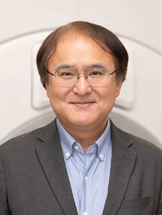 Tomohisa Okada (岡田知久)合作论文数Riken Center For Brain Science7Rosemary Peralta合作论文数6Anna M Chen合作论文数6Yuta Urushibata合作论文数6
 Gajdošík Martin合作论文数6
 Zhu Chengcheng合作论文数6Tadashi Isa合作论文数6
 Xiaoping P Hu合作论文数Department of Bioengineering, Marlan and Rosemary Bourns College of Engineering, University of California, Riverside6
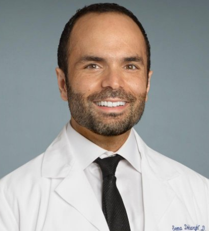 Seena Dehkharghani合作论文数Department of Neurology, Department of Medicine, New York University5
 Stephen P. Wall合作论文数Department of Emergency Medicine, Grossman School of Medicine, New York University5
 Huddleston Daniel E合作论文数Department of Neurology, Emory University;Center for Health Research Southeast, Kaiser Permanente;Center for Health Research Southeast, Emory University5
 Tamara Bushnik合作论文数5
 Yoshiki Arakawa合作论文数Cell Motility Laboratory, London Research Institute5
 Guillaume Madelin合作论文数Magnetic Resonance Center, University of Rennes 1 and Centre Eugène Marquis5
 Dongsuk Shin合作论文数the Advanced Integrated Systems Lab., Korea University5
 Henrik Haraldsson合作论文数Department of Theoretical Physics;Lund University;Department of Theoretical Physics, Lund University5Hideto Kuribayashi合作论文数5Alejandro Zarate合作论文数4Mickael Tordjman合作论文数Dept Radiol, AP HP4Jonathan M. Silver合作论文数Department of Psychiatry, Grossman School of Medicine, New York University4
 Teresa Gerhalter合作论文数4
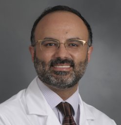 Faraji F合作论文数4Xiangchuan Chen (陈湘川)合作论文数University of Science and Technology of China Center for Brain Science4Masakazu Okawa合作论文数4James S. Babb合作论文数NYU Langone Medical Center4Peter Dietz合作论文数3Takakuni Maki合作论文数Department of Neurology, Kyoto University Graduate School of Medicine3Satoshi Nakajima合作论文数3
 Hideo Chihara合作论文数3
 Brian S. Im合作论文数Rusk Rehabilitation Education & Research, Grossman School of Medicine, New York University3
 David A Feinberg合作论文数University of California, Berkeley3
 Sarah Kefayati合作论文数Dept. of Phys. & Astron., Univ. of Western Ontario;c3
 Takeshi Funaki合作论文数3
 Papinutto Nico合作论文数Department of Neurology, University of California San Francisco3
 An Thanh Vu合作论文数3
 Jing Liu合作论文数1 Department of Radiology and Biomedical Imaging, University of California San Francisco;UCSF, UC Berkeley;1 Department of Radiology and Biomedical Imaging, University of California San Francisco3Nakamoto Yuji合作论文数3Okuchi Sachi合作论文数3
 Beckett Alexander合作论文数3
 Bing Tian (田冰)合作论文数3
 V. Reggie Edgerton合作论文数Department of Neurobiology, David Geffen School of Medicine, University of California, Los Angeles3
 Christopher Hess合作论文数Department of Radiology and Biomedical Imaging, School of Medicine, University of California, San Francisco3




