Background/Objectives: Carvedilol is an adrenergic blocker FDA-approved to improve outcomes in heart failure with reduced ejection fraction. Clinical trials examining whether carvedilol may be cardioprotective in the setting of cancer therapy-induced heart failure have generated mixed results that may depend on the cancer regimen, tumor, or comorbidities. Methods: To investigate the therapeutic potential of carvedilol to mitigate doxorubicin cardiotoxicity in cardiomyocytes, myocardial tissue, and in vivo, independent of confounding factors in clinical studies, we utilized disease-free cardiac slices and cardiomyocytes from mice, dogs, and human in vitro, and in wildtype mice injected with doxorubicin in vivo. We further evaluated the impact of carvedilol in dogs with cancer receiving doxorubicin. Results: In primary canine and murine cardiac slices, carvedilol treatment restored autophagy and prevented apoptosis from doxorubicin. Carvedilol restored mitochondrial energetics in human, canine, and murine models. In wildtype mice challenged with doxorubicin, carvedilol prevented declines in cardiac function and alterations in cardiac structure. In pet dogs with cancer and undergoing doxorubicin treatment, carvedilol was beneficial in preserving cardiac function and structure. Conclusions: Carvedilol activates cardioprotective autophagy, arrests doxorubicin-induced cell death, and improves energetics and cardiac structure and function across species.
Diffusion tensor magnetic resonance imaging of the heart is typically performed at millimetre-scale resolution, yielding only four to five voxels across the ventricular wall, limiting the measurement of local spatial variation in cardiomyocyte organization. Here we present a submillimetre in vivo cardiac diffusion tensor imaging method achieved during free breathing that facilitates voxel-level characterization of myocardial microstructure. We introduce a phenomapping framework that combines voxelwise diffusion magnitude and anisotropy with radial and circumferential gradients of cardiomyocyte helix angle to identify distinct microstructural environments. The approach was developed in healthy volunteers and applied to patients with severe aortic valve stenosis, who had preserved cardiac function and marked myocardial thickening. Comparisons to conventional resolution imaging, together with downsampling analyses and ex vivo and histological validation, show that these voxel-scale features are less optimally detected using standard techniques. Four data-driven microstructural classes, defined by combined diffusion properties and orientation gradients, were observed in both healthy and pressure-overloaded hearts. Despite substantial hypertrophy, pressure overload was associated with preserved cardiomyocyte spatial organization. This framework supports studies of myocardial microstructural remodelling in vivo.
AimPhysiological bone mineralization and ectopic vascular calcification share similarities in the composition of calcium phosphate minerals. Evidence suggests a connection between the underlying biological mechanisms driving the deposition of bone mineral and cardiovascular calcification. Therefore, understanding the chemistry and composition of bone mineral and vascular calcification may be important for the development of effective treatments and diagnostic tools for cardiovascular diseases, as pharmacological interventions for the treatment of one process might affect the other. The goal of this study was to identify and compare compositional features of calcium phosphates in bone and calcified vascular tissues using phosphorus-31 (31P) solid state cross-polarization magic angle spinning (CPMAS) magnetic resonance (MR) spectroscopy, a specialized technique that provides compositional information unattainable through conventional chemical analysis.MethodsSolid state MR spectra were acquired from biological specimens of human trabecular bone (n = 1), human vascular plaque (n = 1), human calcified aortic valves (n = 5), as well as calcified aortic tissues of apolipoprotein E-deficient mice (n = 1) fed a high cholesterol diet. Synthetic hydroxyapatite [Ca10(OH)2(PO4)6] and synthetic brushite (CaHPO4·2H2O) were used to model the solid state 31P MR spectra of the phosphate ion PO4–3 and hydrogen phosphate ion HPO4–2, respectively. Qualitative spectral features and quantitative metrics derived using Herzfeld-Berger analysis were assessed to characterize mineral composition and maturity.ResultsSolid state 31P MR spectra of all human specimens studied suggested a well-ordered crystal structure dominated by unprotonated phosphate (PO4–3), consistent with mature bone-like mineral. These specimens exhibited long CP time constants (700–900 µs) and modest chemical shift anisotropy. In contrast, the calcified mouse aorta spectrum showed pronounced sidebands, a short CP time constant (∼270 µs), and a more prominent HPO4–2 component—features indicative of immature, newly deposited mineral.Conclusion31P solid state MR spectroscopy reveals differences in the phosphate and hydrogen phosphate ion content among the calcified tissues studied. This technique could potentially be an important complement to basic studies of pathological calcification in atherosclerosis and related calcific disorders.
Background: Persistent scarring after myocardial infarction (MI) is a major driver of heart failure in mammals. However, unlike mammals, zebrafish exhibit a remarkable ability to resorb scar, which consists predominantly of collagen in both species. Aims: We aimed here to develop a toolbox of techniques to characterize the nature of collagen in infarcted mammalian and zebrafish hearts. Specifically, we aimed to characterize the early and late cross-links that form in collagen and determine whether differences in these cross-links exist in zebrafish and mammals. Methods: To characterize the initial oxidation and cross-linking of collagen we developed a novel fluorescent probe (TMR-O) that binds to lysine aldehydes on collagen and inform on the degree of collagen cross-linking. Additionally, we used high performance liquid chromatography (HPLC) to quantify degradation-resistant mature collagen cross-links, such as pyridinoline (Pyd), and its precursor, hydroxylysine. Results: TMR-O imaging revealed similar overall CCL levels in both zebrafish and mouse infarcts. However, the irreversible cross-link pyridinoline (Pyd) was abundant in mouse infarcts but completely absent in zebrafish (p<0.0001). High Pyd levels in mice were accompanied by a dynamic increase in lysine hydoxylation (>200%), which was not observed in zebrafish (<10%). Conclusions: While collagen in mammalian and zebrafish infarcts may appear histologically similar, marked biochemical differences exist. Mature degradation-resistant collagen cross-links, such as Pyd form, in murine but not zebrafish infarcts and likely explains why zebrafish can resorb myocardial scar. Targeting Pyd formation in mammals could pave the way for novel therapeutic strategies to promote scar resolution and improve cardiac function after MI.
Purpose:This study presents the biodistribution, clearance and dosimetry estimates of [64Cu]Fibrin Binding Probe #8 ([64Cu]FBP8) in healthy subjects. Procedures:This prospective study included 8 healthy subjects to evaluate biodistribution, safety and dosimetry estimates of [64Cu]FBP8, a fibrin-binding positron emission tomography (PET) probe. All subjects underwent up to 3 sessions of PET/Magnetic Resonance Imaging (PET/MRI) 0-2 hours, 4h and 24h post injection. Dosimetry estimates were obtained using OLINDA 2.2 software. Results:Subjects were injected with ~400 MBq of [64Cu]FBP8. Subjects did not experience adverse effects due to the injection of the probe. [64Cu]FBP8 PET images demonstrated fast blood clearance (half-life = 67 min) and renal excretion of the probe, showing low background signal across the body. The organs with the higher doses were: the urinary bladder (0.075 vs. 0.091 mGy/MBq for males and females, respectively); the kidneys (0.050 vs. 0.056 mGy/MBq respectively); and the liver (0.027 vs. 0.035 mGy/MBq respectively). The combined mean effective dose for males and females was 0.016 ± 0.0029 mSv/MBq, lower than the widely used [18F]fluorodeoxyglucose ([18F]FDG, 0.020mSv/MBq). Conclusions:This study demonstrates the following properties of the [64Cu]FBP8 probe: low dosimetry estimates; fast blood clearance and renal excretion; low background signal; and whole-body acquisition within 20 minutes in a single session. These properties provide the basis for [64Cu]FBP8 to be an excellent candidate for whole-body non-invasive imaging of fibrin, an important driver/feature in many cardiovascular, oncological and neurological conditions.
Current techniques to image the microstructure of the heart with diffusion tensor MRI (DTI) are highly under-resolved. We present a technique to improve the spatial resolution of cardiac DTI by almost 10-fold and leverage this to measure local gradients in cardiomyocyte alignment or helix angle (HA). We further introduce a phenomapping approach based on voxel-wise hierarchical clustering of these gradients to identify distinct microstructural microenvironments in the heart. Initial development was performed in healthy volunteers (n=8). Thereader, subjects with severe but well-compensated aortic stenosis (AS, n=10) were compared to age-matched controls (CTL, n=10). Radial HA gradient was significantly reduced in AS (8.0±0.8°/mm vs. 10.2±1.8°/mm, p=0.001) but the other HA gradients did not change significantly. Four distinct microstructural clusters could be idenJfied in both the CTL and AS subjects and did not differ significantly in their properties or distribution. Despite marked hypertrophy, our data suggest that the myocardium in well-compensated AS can maintain its microstructural coherence. The described phenomapping approach can be used to characterize microstructural plasticity and perturbation in any organ system and disease.
This study presents for the first time in humans the biodistribution, clearance and dosimetry estimates of [64Cu]Fibrin Binding Probe #8 ([64Cu]FBP8) in healthy subjects. [64Cu]FBP8-PET previously demonstrated its potential in two recent applications: thrombus imaging and pulmonary fibrosis. This prospective study included 8 healthy subjects to evaluate biodistribution, safety and dosimetry estimates of [64Cu]FBP8, a fibrin-binding positron emission tomography (PET) probe. All subjects underwent up to 3 sessions of PET/Magnetic Resonance Imaging (PET/MRI) 0–2 h, 4 h and 24 h post injection. Dosimetry estimates were obtained using OLINDA 2.2 software. Subjects were injected with 400 MBq of [64Cu]FBP8. Subjects did not experience adverse effects due to the injection of the probe. [64Cu]FBP8 PET images demonstrated fast blood clearance (half-life = 67 min) and renal excretion of the probe, showing low background signal across the body. The organs with the higher doses were: the urinary bladder (0.075 vs. 0.091 mGy/MBq for males and females, respectively); the kidneys (0.050 vs. 0.056 mGy/MBq respectively); and the liver (0.027 vs. 0.035 mGy/MBq respectively). The combined mean effective dose for males and females was 0.016 ± 0.0029 mSv/MBq, lower than the widely used [18F]fluorodeoxyglucose ([18F]FDG, 0.020mSv/MBq). This study demonstrates the following properties of the [64Cu]FBP8 probe: low dosimetry estimates; fast blood clearance and renal excretion; low background signal; and whole-body acquisition within 20 min in a single session. These properties provide the basis for [64Cu]FBP8 to be an excellent candidate for whole-body non-invasive imaging of fibrin, an important driver/feature in many cardiovascular, oncological and neurological conditions.
Thanks to recent developments in cardiovascular magnetic resonance (CMR), cardiac diffusion-weighted magnetic resonance is fast emerging in a range of clinical applications. Cardiac diffusion-weighted imaging (cDWI) and diffusion tensor imaging (cDTI) now enable investigators and clinicians to assess and quantify the tridimensional microstructure of the heart. Free-contrast DWI is uniquely sensitized to the presence and displacement of water molecules within the myocardial tissue, including the intracellular, extracellular, and intravascular spaces. CMR can determine changes in microstructure by quantifying: a) mean diffusivity (MD)—measuring the magnitude of diffusion; b) fractional anisotropy (FA)—specifying the directionality of diffusion; c) helix angle (HA) and transverse angle (TA)—indicating the orientation of the cardiomyocytes; d) absolute sheetlet angle (E2A) and E2A mobility—measuring the alignment and systolic-diastolic mobility of the sheetlets, respectively.This document provides recommendations for both clinical and research cDWI and cDTI, based on published evidence when available and expert consensus when not. It introduces the cardiac microstructure focusing on the cardiomyocytes and their role in cardiac physiology and pathophysiology. It highlights methods, observations, and recommendations in terminology, acquisition schemes, postprocessing pipelines, data analysis, and interpretation of the different biomarkers. Despite the ongoing challenges discussed in the document and the need for ongoing technical improvements, it is clear that cDTI is indeed feasible, can be accurately and reproducibly performed and, most importantly, can provide unique insights into myocardial pathophysiology.
Autophagy is a key biological process that has proven extremely difficult to detect noninvasively. To address this, an autophagy detecting nanoparticle (ADN) was recently developed, consisting of an iron oxide nanoparticle decorated with cathepsin-cleavable arginine-rich peptides bound to the near-infrared fluorochrome Cy5.5. Activation of the probe in autophagolysosomes results in the emission of Cy5.5 fluorescence and provides a measure of autophagosome flux. However, in the early autophagosome ADN fluorescence is silent due to fluorochrome stacking. Here, we introduce to ADN a second non-cleavable fluorophore that allows the probe to be tracked through all stages of autophagy. The nature of the secondary/tracking fluorophore has a profound effect on the activation of ADN and the emission of Cy5.5 fluorescence. The lead candidate, ADN2 (featuring AZDye546 as the secondary fluorophore) has the highest activation rate and change in Cy5.5 fluorescence. Absorbance and fluorescence spectrophotometry methods show that the negatively charged AZDye546 interacts with the positively charged polyarginine motifs of the Cy5.5-polyArg activatable fluorophore, resulting in enhanced baseline quenching of the Cy5.5 signal in the nanoprobe. Flow cytometry shows that the activation of ADN2 remains specific for autophagy and is strongly modulated by classical regulators of autophagy (starvation, bafilomycin) and genetic deletion of key autophagy proteins (ATG5, ATG7). ADN2 co-localized strongly with LC3-GFP positive autophagosomes and provided readouts of in vivo probe delivery and activation in the hearts of fed/starved mice. ADN2 enhances the ability to image autophagy without genetic transfection of cells/animals and underscores the possible effects for unanticipated interactions between fluorochromes and other moieties on the surface of decorated nanoparticles.
Patients with advanced gastric cancer (GCa) have limited treatment options, and alternative treatment approaches are necessary to improve their clinical outcomes. Because fibrin is abundant in gastric tumors but not in healthy tissues, we hypothesized that fibrin could be used as a high-concentration depot for a high-energy beta-emitting cytotoxic radiopharmaceutical delivered to tumor cells. We showed that fibrin is present in 64 to 75% of primary gastric tumors and 50 to 100% of metastatic gastric adenocarcinoma cores. First-in-human 64Cu-FBP8 fibrin-targeted positron emission tomography (PET) imaging in seven patients with gastric or gastroesophageal junction cancer showed high probe uptake in all target lesions with tumor-to-background (muscle) uptake ratios of 9.9 +/- 6.6 in primary (n = 7) and 11.2 +/- 6.6 in metastatic (n = 45) tumors. Using two mouse models of human GCa, one fibrin-high (SNU-16) and one fibrin-low (NCI-N87), we showed that PET imaging with a related fibrin-specific peptide, CM500, labeled with copper-64 (64Cu-CM500) specifically bound to and precisely quantified tumor fibrin in both models. We then labeled the fibrin-specific peptide CM600 with yttrium-90 and showed that 90Y-CM600 effectively decreased tumor growth in these mouse models. Mice carrying fibrin-high SNU-16 tumors experienced tumor growth inhibition and prolonged survival in response to either a single high dosage or fractionated lower dosage of 90Y-CM600, whereas mice carrying fibrin-low NCI-N87 tumors experienced prolonged survival in response to a fractionated lower dosage of 90Y-CM600. These results lay the foundation for a fibrin-targeted theranostic that may expand options for patients with advanced GCa.
NETosis, the process of neutrophil cell death due to the formation and release of neutrophil extracellular traps (NETs) has been implicated in the pathogenesis of heart disease. NETs are composed of immunogenic DNA fragments and citrullinated histones. The role of NETosis in the context of cardiac arrest and resuscitation (CA/R) is however unknown. We previously reported the development of a Dextran-Thiazole Orange (DTO) nanoprobe, with nanomolar affinity for nucleic acids, that exerts acute anti-inflammatory effects and reduces ischemia-reperfusion injury in mice. We aimed here to determine whether the DTO nanoprobe would also bind to the nucleic acid in NETS, whether DTO could attenuate the intensity of NETosis, and whether this would improve outcomes and survival after cardiac arrest. C57Bl6 mice were arrested for 8 minutes before resuscitation. DTO or unmodified dextran (vehicle control) was injected at the time of resuscitation, and again 4 hours later (n=38 mice). Survival and neurological function were scored daily. Time-domain resolved (lifetime) and conventional fluorescence imaging was performed at 4 hours after resuscitation in 6 additional mice injected with DTO. NETosis was evaluated by protein blot in the hearts of cardiac arrest mice injected with DTO or control. DTO significantly (p<0.05) improved 10-day survival (Fig. A), mean survival (from 4 to 7 days, Fig. B), and neurological function (Fig. C) following CA/R. Lifetime imaging (Fig. D), but not conventional fluorescence imaging (Fig. E) revealed the presence of DTO in the injured heart, liver and kidneys. In the heart, DTO signal was significantly increased in CA/R compared to sham mice injected with DTO or CA/R mice injected with control dextran (Fig. F). Citrullinated H3 and HMGB-1 (High mobility group box 1, a chromatin protein) were both upregulated in CA/R, and were significantly attenuated by DTO injection (Fig. G-I). We show for the first time that NETosis plays a key role in cardiac arrest and resuscitation. DTO is capable of detecting NETosis as well as attenuating it, likely via a reduction in HMGB-1 activity. The modulation of NETosis may provide a new avenue to improve survival after cardiac arrest.
Introduction: Cardiovascular calcification involving abnormal mineral depositions within the vessel walls and aortic valves underlies pathophysiologic mechanisms of cardiovascular diseases such as stroke and heart attack. To date, no effective therapeutic strategies are available to prevent or treat cardiovascular calcification, except for surgical interventions performed after formation of advanced calcification. Developing medical options requires a thorough understanding of mechanisms of the mineralization process, which are still insufficiently investigated. Goal and Method: The goal of the present study is to use a specialized solid state magnetic resonance spectroscopy (MRS) to characterize cardiovascular calcification. This MRS technique has been previously demonstrated to be effective in quantifying bone mineral and solid organic matrix, determining compositional features of minerals in solids, as well as characterizing the degree of bone mineral maturity, which is unattainable by existing non-destructive methods. The novelty of this study is the application of solid state MRS in combination with Herzfeld-Berger analysis to arterial calcification. A common model of cardiovascular calcification is apolipoprotein E-deficient (ApoE-/-) mice fed with a high cholesterol diet that develop substantial calcification in the aortic valves and carotid arteries. Hypothesis: We hypothesize that the specialized solid state MRS technique is capable of capturing differences in the mineralization of calcified human aortic valves and vascular calcification in the ApoE-/- knockout mouse. Results and Conclusions: Here we show the pattern of calcification in the human aortic valves resembles a dense, well-organized matrix characteristic of mature human trabecular bone and human calcified arterial plaque. In contrast, calcification in the mice resembles that of immature or embryonic bone, exhibiting a more disorganized pattern of mineralization that likely models early preclinical calcification of the aortic valve.
In mammalian hearts myocardial infarction produces a permanent collagen-rich scar. Conversely, in zebrafish a collagen-rich scar forms but is completely resorbed as the myocardium regenerates. The formation of cross-links in collagen hinders its degradation but cross-linking has not been well characterized in zebrafish hearts. Here, a library of fluorescent probes to quantify collagen oxidation, the first step in collagen cross-link (CCL) formation, was developed. Myocardial injury in mice or zebrafish resulted in similar dynamics of collagen oxidation in the myocardium in the first month after injury. However, during this time, mature CCLs such as pyridinoline and deoxypyridinoline developed in the murine infarcts but not in the zebrafish hearts. High levels of newly oxidized collagen were still seen in murine scars with mature CCLs. These data suggest that fibrogenesis remains dynamic, even in mature scars, and that the absence of mature CCLs in zebrafish hearts may facilitate their ability to regenerate.
We measured cardiac magnetostimulation thresholds in ten healthy pigs by discharging a 220-µF capacitor into a flat spiral coil placed close to the pigs’ torso. We used MR Dixon images to locate the porcine heart and determine the relative coil position to calculate the B-field in the heart (Biot-Savart). The average threshold for cardiac magnetostimulation during diastole was dB/dt≈1570±320 T/s at the center of the heart. This value is >10X greater than the IEC 60601-2-33 cardiac dB/dt limit for the effective stimulus duration of the magnetic stimulator system used in the experiments (0.45 ms).
Purpose: Modern high-amplitude gradient systems can be limited by the International Electrotechnical Commission 60601-2-33 cardiac stimulation (CS) limit, which was set in a conservative manner based on electrode experiments and E-field simulations in uniform ellipsoidal body models. Here, we show that coupled electromagnetic-electrophysiological modeling in detailed body and heart models can predict CS thresholds, suggesting that such modeling might lead to more detailed threshold estimates in humans. Specifically, we compare measured and predicted CS thresholds in eight pigs. Methods: We created individualized porcine bodymodels using MRI (Dixon for thewhole body, CINE for the heart) that replicate the anatomy and posture of the animals used in our previous experimental CS study. Wemodel the electric fields induced along cardiac Purkinje and ventricular muscle fibers and predict the electrophysiological response of these fibers, yielding CS threshold predictions in absolute units for each animal. Additionally, we assess the total modeling uncertainty through a variability analysis of the 25 main model parameters. Results: Predicted and experimental CS thresholds agreewithin 19% on average (normalized RMS error), which is smaller than the 27% modeling uncertainty. No significant difference was found between the modeling predictions and experiments (p < 0.05, paired t-test). Conclusion: Predicted thresholds matched the experimental data within the modeling uncertainty, supporting the model validity. We believe that our modeling approach can be applied to study CS thresholds in humans for various gradient coils, body shapes/postures, and waveforms, which is difficult to do experimentally.
HomeCirculation: Cardiovascular ImagingVol. 16, No. 10Metabolic Dysfunction in Aortic Stenosis: A Key Piece of the Pathophysiological Puzzle No AccessEditorialRequest AccessFull TextAboutView Full TextView PDFView EPUBSections ToolsAdd to favoritesDownload citationsTrack citationsPermissions ShareShare onFacebookTwitterLinked InMendeleyReddit Jump toNo AccessEditorialRequest AccessFull TextMetabolic Dysfunction in Aortic Stenosis: A Key Piece of the Pathophysiological Puzzle David E. Sosnovik and Sammy Elmariah David E. SosnovikDavid E. Sosnovik Correspondence to: David Sosnovik, MD, Martinos Center for Biomedical Imaging, Massachusetts General Hospital, 149 13th St, Charlestown MA 02129. Email E-mail Address: [email protected] https://orcid.org/0000-0002-6486-7434 Martinos Center for Biomedical Imaging (D.E.S.), Massachusetts General Hospital, Harvard Medical School, Boston. Cardiovascular Research Center and Cardiology Division (D.E.S.), Massachusetts General Hospital, Harvard Medical School, Boston. and Sammy ElmariahSammy Elmariah https://orcid.org/0000-0002-8013-8733 Division of Cardiology, Department of Medicine, University of California San Francisco (S.E.). Originally published17 Oct 2023https://doi.org/10.1161/CIRCIMAGING.123.015977Circulation: Cardiovascular Imaging. 2023;16This article is a commentary on the followingRole of Cardiac Energetics in Aortic Stenosis Disease Progression: Identifying the High-risk Metabolic PhenotypeFootnotesFor Sources of Funding and Disclosures, see page 790.The opinions expressed in this article are not necessarily those of the editors or of the American Heart Association.Correspondence to: David Sosnovik, MD, Martinos Center for Biomedical Imaging, Massachusetts General Hospital, 149 13th St, Charlestown MA 02129. Email dsosnovik@mgh.harvard.eduREFERENCES1. Dahl JS, Magne J, Pellikka PA, Donal E, Marwick TH. Assessment of subclinical left ventricular dysfunction in aortic stenosis.JACC Cardiovasc Imaging. 2019; 12:163–171. doi: 10.1016/j.jcmg.2018.08.040CrossrefMedlineGoogle Scholar2. Peterzan MA, Clarke WT, Lygate CA, Lake HA, Lau JYC, Miller JJ, Johnson E, Rayner JJ, Hundertmark MJ, Sayeed R, et al. Cardiac energetics in patients with aortic stenosis and preserved versus reduced ejection fraction.Circulation. 2020; 141:1971–1985. doi: 10.1161/CIRCULATIONAHA.119.043450LinkGoogle Scholar3. Dweck MR, Joshi S, Murigu T, Alpendurada F, Jabbour A, Melina G, Banya W, Gulati A, Roussin I, Raza S, et al. Midwall fibrosis is an independent predictor of mortality in patients with aortic stenosis.J Am Coll Cardiol. 2011; 58:1271–1279. doi: 10.1016/j.jacc.2011.03.064CrossrefMedlineGoogle Scholar4. Elmariah S, Farrell LA, Furman D, Lindman BR, Shi X, Morningstar JE, Rhee EP, Gerszten RE. Association of acylcarnitines with left ventricular remodeling in patients with severe aortic stenosis undergoing transcatheter aortic valve replacement.JAMA Cardiol. 2018; 3:242–246. doi: 10.1001/jamacardio.2017.4873CrossrefMedlineGoogle Scholar5. Popma JJ, Deeb GM, Yakubov SJ, Mumtaz M, Gada H, O'Hair D, Bajwa T, Heiser JC, Merhi W, Kleiman NS, et al; Evolut Low Risk Trial Investigators. Transcatheter aortic-valve replacement with a self-expanding valve in low-risk patients.N Engl J Med. 2019; 380:1706–1715. doi: 10.1056/NEJMoa1816885CrossrefMedlineGoogle Scholar6. Monga S, Valkovič L, Myerson SG, Neubauer S, Mahmod M, Rider OJ. Role of cardiac energetics in aortic stenosis disease progression: identifying the high-risk metabolic phenotype.Circ Cardiovasc Imaging. 2023; 16:e014863. doi: 10.1161/CIRCIMAGING.122.014863LinkGoogle Scholar7. Panagia M, Chen HH, Croteau D, Iris Chen YC, Ran C, Luptak I, Josephson L, Colucci WS, Sosnovik DE. Multiplexed optical imaging of energy substrates reveals that left ventricular hypertrophy is associated with brown adipose tissue activation.Circ Cardiovasc Imaging. 2018; 11:e007007. doi: 10.1161/CIRCIMAGING.117.007007LinkGoogle Scholar8. Perry AS, Zhao S, Murthy V, Gupta DK, Fearon WF, Kim JB, Kapadia S, Kumbhani DJ, Gillam L, Whisenant B, et al. Metabolic signatures of cardiac dysfunction, multimorbidity, and post-transcatheter aortic valve implantation death.J Am Heart Assoc. 2023; 12:e029542. doi: 10.1161/JAHA.123.029542LinkGoogle Scholar9. Stein EJ, Fearon WF, Elmariah S, Kim JB, Kapadia S, Kumbhani DJ, Gillam L, Whisenant B, Quader N, Zajarias A, et al. Left ventricular hypertrophy and biomarkers of cardiac damage and stress in aortic stenosis.J Am Heart Assoc. 2022; 11:e023466. doi: 10.1161/JAHA.121.023466LinkGoogle Scholar10. Lai L, Leone TC, Keller MP, Martin OJ, Broman AT, Nigro J, Kapoor K, Koves TR, Stevens R, Ilkayeva OR, et al. Energy metabolic reprogramming in the hypertrophied and early stage failing heart: a multisystems approach.Circ Heart Fail. 2014; 7:1022–1031. doi: 10.1161/CIRCHEARTFAILURE.114.001469LinkGoogle Scholar11. Otto CM, Nishimura RA, Bonow RO, Carabello BA, Erwin JP, Gentile F, Jneid H, Krieger EV, Mack M, McLeod C, et al. 2020 ACC/AHA Guideline for the management of patients with valvular heart disease: a report of the American College of Cardiology/American Heart Association Joint Committee on Clinical Practice Guidelines.Circulation. 2021; 143:e72–e227. doi: 10.1161/CIR.0000000000000923LinkGoogle Scholar Previous Back to top Next FiguresReferencesRelatedDetailsCited By Sengupta P and Chandrashekhar Y (2024) Advancing Myocardial Tissue Analysis Using Echocardiography, JACC: Cardiovascular Imaging, 10.1016/j.jcmg.2024.01.002, 17:2, (228-231), Online publication date: 1-Feb-2024. Related articlesRole of Cardiac Energetics in Aortic Stenosis Disease Progression: Identifying the High-risk Metabolic PhenotypeShveta Monga, et al. Circulation: Cardiovascular Imaging. 2023;16 October 2023Vol 16, Issue 10 Advertisement Article InformationMetrics © 2023 American Heart Association, Inc.https://doi.org/10.1161/CIRCIMAGING.123.015977PMID: 37847765 Originally publishedOctober 17, 2023 KeywordsEditorialsaortic valve stenosishypertrophymetabolismmyocardiumPDF download Advertisement SubjectsMagnetic Resonance Imaging (MRI)MetabolismMyocardial BiologyPathophysiologyTranslational Studies
 Choukri Mekkaoui合作论文数Yale University49
 Ralph Weissleder合作论文数Center for Systems Biology, Massachusetts General Hospital;Harvard Medical School48
 Matthias Nahrendorf合作论文数Center for Systems Biology, Massachusetts General Hospital;Harvard Medical School, Harvard University40
 Howard Chen合作论文数School of Medicine, Tufts University;Tufts Medical Center, Graduate School of Biomedical Sciences, Genetics, Molecular and Cellular Biology, Tufts University40
 Lee Josephson合作论文数Harvard Medical School, Massachusetts General Hospital;Dana-Farber / Harvard Cancer Center;MedChemImaging LLC26Timothy G. Reese合作论文数25
 Marcel Jackowski合作论文数Nooroot Inc25
 Peter Caravan合作论文数Center for Interdisciplinary Innovation in Imaging, Massachusetts General Hospital;Institute for Innovation in Imaging, Massachusetts General Hospital;Harvard Medical School21
 Christopher Nguyen合作论文数Cedars-Sinai Medical Center, Biomedical Imaging Research Institute;Department of Bioengineering, University of California Los Angeles;Biomedical Imaging Research Institute, University of California Los Angeles20
 Guangping Dai合作论文数20
 Elena Aikawa合作论文数Center for Interdisciplinary Cardiovascular Sciences, Brigham and Women’s Hospital;Department of Medicine, Harvard Medical School;Section of Cardiovascular Life Sciences, Cardiovascular Medicine, Brigham and Women’s Hospital19Hushan Yuan合作论文数Center for Systems Biology;Center for Molecular Imaging Research;Harvard Medical School, Massachusetts General Hospital16
 David Firmin合作论文数National Heart and Lung Institute, Faculty of Medicine, Imperial College London;Cardiovascular MR Unit, Royal Brompton Hospitals13
 Shuning Huang合作论文数Department of Diagnostic and Interventional Imaging, McGovern Medical School, The University of Texas Health Science Center at Houston13
 Filip Swirski合作论文数Cardiovascular Research Institute, Icahn School of Medicine, Mount Sinai;Massachusetts General Hospital12
 Udo Hoffmann合作论文数Cleerly;Innovative Imaging Consulting, LLC12
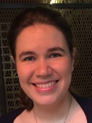 Sonia Nielles-Vallespin合作论文数National Heart & Lung Institute, Faculty of Medicine, Imperial College London;Royal Brompton & Harefield NHS Foundation Trust11
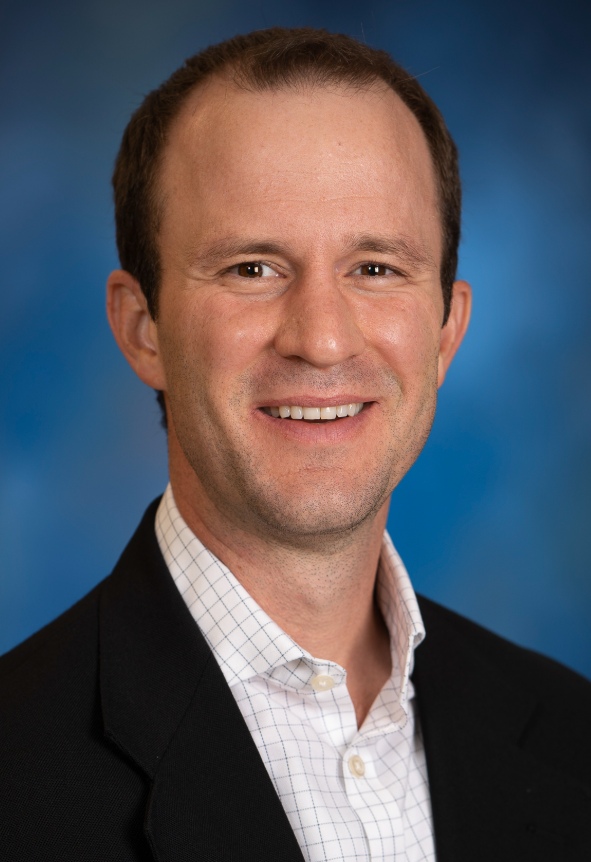 Craig Goergen合作论文数Weldon School of Biomedical Engineering, College of Engineering, Purdue University11
 Jose Luiz Figueiredo合作论文数Center for Molecular Imaging Research, Massachusetts General Hospital and Harvard Medical School11
 Farouc Jaffer合作论文数Cardiology Division, Department of Medicine, Massachusetts General Hospital;Cardiovascular Research Center, Massachusetts General Hospital;Harvard Medical School11
 Ronglih Liao合作论文数Medicine Department, Stanford University10Catana Ciprian合作论文数9
 Christian T Stoeck合作论文数Institute for Biomedical Engineering, ETH Zürich9
 Wei Chao合作论文数Center for Shock, Trauma and Anesthesiology Research, University of Maryland School of Medicine8Kostis William J合作论文数8
 Anthony Rosenzweig合作论文数Massachusetts General Hospital, Harvard Medical School8Peter Gatehouse合作论文数National Heart and Lung Institute, Faculty of Medicine, Imperial College London8
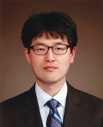 Hoonsung Cho合作论文数University of Cincinnati, University of Cincinnati8
 Maaike van den Boomen合作论文数7
 Peter Panizzi合作论文数Harrison School of Pharmacy, Auburn University7
 Sebastian Kozerke合作论文数Institute for Biomedical Engineering, Eidgenossische Technische Hochschule Zurich;Department of Information Technology and Electrical Engineering, Eidgenossische Technische Hochschule Zurich7
 Lawrence Wald合作论文数Athinoula A. Martinos Center for Biomedical Imaging, Massachusetts General Hospital, Harvard Medical School7
 Jeremy Ruskin合作论文数Telemachus & Irene Demoulas Family Foundation Center for Cardiac Arrhythmias, Massachusetts General Hospital;Harvard Medical School7
 Peter Libby合作论文数Brigham and Women's Hospital;Harvard Medical School7
 Niek Prakken合作论文数Department of Radiology, University of Groningen7
 Andrew D Scott合作论文数Imperial College London7
 Anand T.N. Kumar合作论文数Harvard Medical School;Mike Toth Head and Neck Cancer Research Center, Mass Eye and Ear7
 Ronald Borra合作论文数7
 R. H. J. A. (Riemer) Slart合作论文数UMCG;Faculty of Medical Sciences, University of Groningen7Anne Philip合作论文数6
 Marcello Panagia合作论文数6Velthuis B K合作论文数6
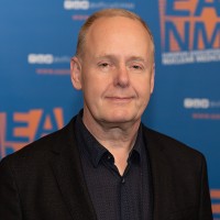 R.A.J.O. (Rudi) Dierckx合作论文数University Medical Center Groningen;Faculty of Medical Sciences, University of Groningen;International Atomic Energy Agency6
 Pedro Ferreira合作论文数Royal Brompton Hospital6
 James G. Miller合作论文数Laboratory of Ultrasonics, Department of Physics, Arts & Sciences, Washington University in St.Louis6
 Thomas Brady合作论文数Harvard Medical School;Division of Cardiac Imaging, Mass General Research Institute;Division of Nuclear Medicine and Molecular Imaging, Mass General Research Institute;Athinoula A. Martinos Center for Biomedical Imaging, Mass General Research Institute6
 David Izquierdo-Garcia合作论文数Mass General Research Institute, Massachusetts General Hospital;Universidad Carlos III de Madrid;Harvard Medical School6
 Jaume Coll-Font合作论文数B-Spiral Group, Northeastern University6
 Ahmed Tawakol合作论文数Massachusetts General Hospital, Harvard Medical School6




