Objective: To study the prevalence and characteristics of arterial thromboembolic events ( ATEE) in the course of AL amyloidosis.Methods: We report the case of a non- anticoagulated patient with AL amyloidosis restrictive cardiomyopathy who developed acute lower limb ischaemia. We then prospectively determined the prevalence of ATEE in all patients with AL amyloidosis who were evaluated in our institution for autologous peripheral stem cell transplantation.Results: Nine out of 15 non- anticoagulated patients ( 60%) developed ATEE: ischaemic stroke ( 3), transient cerebral ischaemic attack ( 2), multiple peripheral arterial emboli ( 1), bilateral iliac artery thrombosis ( 1), bilateral optic nerve ischaemia ( 1), and mesenteric ischaemia ( 1). Haemodynamic stasis seemed to play a leading role in the pathophysiology of ATEE, in that all patients were on sinus rhythm and only one had a thrombus on echocardiography. We identified possible contributing factors to ATEE occurrence: concomitant treatments with oestroprogestogen regimen, thalidomide, granulocyte- macrophage colony- stimulating factor ( GM- CSF) and extracellular volume disturbances related to the cytapheresis procedure.Conclusion: We report on an unusual frequency of ATEE among patients with AL cardiac amyloidosis. Despite its theoretical risks, anticoagulation should be discussed for patients with amyloid cardiomyopathy.
Systemic lupus erythematosus (SLE) is uncommon after the age of 50 years, and studies of elderly patients with SLE are scarce. We conducted the current study to analyze characteristics and outcome of patients with late-onset SLE in a French tertiary referral center, and to compare them with those of younger patients with SLE. From 1980 to 2000, 47 patients were identified as having late-onset SLE, defined as SLE diagnosed at or over the age of 50 years. These patients were compared with a group of 114 randomly selected patients aged younger than 50 years at SLE diagnosis. We compared clinical characteristics, laboratory data, therapy, and course.The female to male ratio was smaller in the late-onset SLE group (p = 0.0012). Some manifestations occurred less frequently in late-onset SLE: arthritis (p = 0.009), malar rash (p = 0.013), and nephropathy (p = 0.009). High-dose corticosteroids (p = 0.0016) and immunosuppressive drugs (p = 0.006) were less commonly used in the elderly. Deaths occurred more frequently in late-onset SLE (p = 0.019), with a 10-year survival rate of 71% versus 95% in early-onset SLE (p < 0.01). In patients with late-onset SLE, causes of death were usually unrelated to SLE.Analysis of pooled data from the literature, based on 714 old and 4700 young SLE patients, confirmed that late-onset SLE was characterized by a smaller female to male ratio (4.4:1 vs. 10.6:1; p = 3.10(-14)); a higher occurrence of serositis (36.7% vs. 28.6%; p = 7.10(-4)) and pulmonary involvement (21.2% vs. 11.3%,p = 6.10(-8)); and a lower occurrence of malar rash (31.1% vs. 62.4%; p = 10(-44)), photosensitivity (26.2% vs. 38.2%; p = 6.10(-6)), purpura/cutaneous vasculitis (13.4% vs. 25.9%; p = 9.10(-4)), alopecia/hair loss (24% vs. 44.9%; p = 3.10(-11)), Raynaud phenomenon (24.8% vs. 37.2%; p = 3.10(-7)), neuropsychiatric manifestations (15.3% vs. 20.2%; p = 0.025), lymphadenopathy (9.1% vs. 19.6%; p = 2.10(-4)), nephrotic syndrome (8.1 % vs. 24.3%; p = 0.015), and nephritis (28.6% vs. 42.7%; p = 2.10(-10)). Regarding laboratory features, rheumatoid factor positivity was more frequent (32.7% vs. 20.1%; p = 3.10(-5)), whereas anti-RNP positivity (10.4% vs. 20.9%; p= 9.10(-5)), anti-Sm positivity (9.1 % vs. 17.1 %; p = 0.001), and a low CH50 complement fraction (45% vs. 64.9%; p = 0.002) were less frequent in old compared with young SLE patients.In conclusion, the clinical pattern of late-onset SLE is characterized by a lower disease severity. The reduced survival observed in this group seems to result mainly from the consequences of aging.
This retrospective study concerned 18 female and 23 male patients with cardiac sarcoidosis (CS). The average age at CS diagnosis was 38 years. CS was observed in white (73% of cases) and in black or Caribbean patients (27% of cases). All patients had extracardiac histologic proof of sarcoid tissue. In 63% of cases, the CS arose during the follow-up of systemic sarcoidosis. Systemic sarcoidosis was not specific except for a high frequency of neurosarcoidosis. Revealing cardiac signs were clinical in 63% of cases and electrical in 22%. In most patients these signs were associated with an abnormal echocardiography (77%) and/or a defect on thallium-201 or sestamibi imaging (75%). Thirty-nine patients received steroid therapy (initial dose mostly equal to 1 mg/kg per day), associated in 13 cases with another immunosuppressive treatment. In 26% of cases the immunosuppressive treatment was associated with a specific cardiac treatment. In the long-term follow-up (average follow-up, 58 mo), 87% of the cases showed an improvement, and 54% were cured from a clinical and laboratory point of view (electrocardiogram, 24-hour monitoring, echocardiography, radionuclide imaging). There was no sudden death. Two patients worsened, which can be explained in 1 case by very late treatment and in the other case by lack of treatment, except for a pacemaker. Our experience leads us to treat CS with corticosteroids as soon as possible and to use another immunosuppressive treatment where there is an insufficient therapeutic response or where there are contraindications to corticosteroids.
This retrospective study concerned 18 female and 23 male patients with cardiac sarcoidosis (CS). The average age at CS diagnosis was 38 years. CS was observed in white (73% of cases) and in black or Caribbean patients (27% of cases). All patients had extracardiac histologic proof of sarcoid tissue. In 63% of cases, the CS arose during the follow-up of systemic sarcoidosis. Systemic sarcoidosis was not specific except for a high frequency of neurosarcoidosis. Revealing cardiac signs were clinical in 63% of cases and electrical in 22%. In most patients these signs were associated with an abnormal echocardiography (77%) and/or a defect on thallium-201 or sestamibi imaging (75%). Thirty-nine patients received steroid therapy (initial dose mostly equal to 1 mg/kg per day), associated in 13 cases with another immunosuppressive treatment. In 26% of cases the immunosuppressive treatment was associated with a specific cardiac treatment. In the long-term follow-up (average follow-up, 58 mo), 87% of the cases showed an improvement, and 54% were cured from a clinical and laboratory point of view (electrocardiogram, 24-hour monitoring, echocardiography, radionuclide imaging). There was no sudden death. Two patients worsened, which can be explained in 1 case by very late treatment and in the other case by lack of treatment, except for a pacemaker. Our experience leads us to treat CS with corticosteroids as soon as possible and to use another immunosuppressive treatment where there is an insufficient therapeutic response or where there are contraindications to corticosteroids.
Purpose. - Haptoglobin (H) and orosomucoid (O) are acute phase proteins that increase in a parallel manner. When hemolysis and inflammation are both present, study of the O-H couple on the protein profile may reveal an unknown hemolysis.Methods. -. To determine if hemolysis is more frequent during infectious endocarditis than during septicemia without valvulopathy or during valvulopathy without septicemia. Study of three groups of patients: 26 patients with infectious endocarditis, 13 patients with septicemia and 36 patients with valvulopathy without septicemia. Studied parameters were the O-H couple, hemoglobin and rate of O.Results. - Hemolysis is clear in patients with endocarditis. The difference O-H is significantly more important during endocarditis than during septicemia without valvulopathy (P < 0,001) and during valvulopathy without sepsis (P < 0.001).Conclusion. - Study of the O-H couple may be useful for the diagnosis of endocarditis showing a difficult-to-diagnose hemolysis. (C) 2002 Editions scientifiques et medicales Elsevier SAS.
Objective. To develop diagnostic imaging criteria for polymyositis (PM) and sporadic inclusion body myositis (sIBM).Methods. We investigated 220 patients with suspected inflammatory myopathies by magnetic resonance imaging (MRI). Findings were compared with the results of clinical andbiological examinations and muscle biopsy. PM and IBM were diagnosed in 25 patients each. Quantitative and qualitative MRI analysis of the 3 muscle groups of the 2 thighs included fatty infiltration, atrophy, inflammation, and the type and distribution of the lesions.Results. MRI was abnormal in all patients. Fatty infiltration and atrophy were more frequent in patients with sIBM (p < 0.05). Inflammation as the sole abnormality was preferentially encountered in PM (p = 0.05). Widespread abnormalities were more frequent in sIBM (p < 0.01). Abnormalities in PM tended to be distributed along the fascia. Involvement of the anterior group, an asymmetrical distribution, and a distal predominance were all more frequent in sIBM (p < 0.001).Conclusion. Despite some overlap in MRI findings between the 2 diseases, MRI was useful for distinguishing PM from sIBM.
Propos. – Les uvéites chroniques sévères représentent un défi diagnostique et thérapeutique permanent pour l’ophtalmologiste et l’interniste. Le développement des outils diagnostiques moléculaires appliqués aux liquides et tissus oculaires et des méthodes d’imagerie modernes a permis d’améliorer le rendement des bilans étiologiques. La mise en jeu du pronostic visuel au cours des uvéites chroniques impose une démarche diagnostique exhaustive et une intensification thérapeutique progressive.Actualités et points forts. – L’épidémiologie des uvéites chroniques sévères semble mieux précisée actuellement. Une étude portant sur 927 patients consécutifs, pris en charge sur une période de 5 ans, a permis de définir une orientation diagnostique dans 67 % des cas. Il a été possible d’individualiser 4 grands groupes d’atteinte : les uvéites infectieuses, les uvéites associées à une maladie systémique, les uvéites d’origine immunologique pure limitées à l’œil et les uvéites idiopathiques limitées à l’œil. Cette subdivision pourrait être appliquée à chacun des sous-groupes constituant la classification anatomique des inflammations intraoculaires. Le taux de cécité final semble corrélé à la rapidité de la prise en charge, l’orientation diagnostique, l’adaptation du traitement et à la sévérité de l’atteinte inflammatoire.Perspectives. – Il faut différencier l’uvéite aiguë, fréquente mais répondant généralement bien à une corticothérapie locale et l’uvéite chronique nécessitant une stratégie diagnostique souvent complexe et une conduite thérapeutique précise mettant en question les anciens dogmes. Tout échec d’une corticothérapie bien conduite doit poser le problème d’une participation infectieuse, qu’elle soit virale, bactérienne ou parasitaire. La mise en évidence d’arguments plus ou moins formels en faveur d’un agent pathogène impose un traitement spécifique et reste une étape majeure avant toute intensification thérapeutique fondée sur les immunosuppresseurs classiques et les nouvelles molécules disponibles dans le cadre de protocoles de recherche clinique.Purpose. – Severe chronic and refractory uveitis is a major diagnostic and therapeutic challenge for ophthalmologists and internists. Molecular tools, such as PCR but also new imaging techniques, have significantly changed the diagnostic approach during the last 10 years. Presumed and empirical diagnosis should be excluded in the face of atypical clinical presentations.Current knowledge and key points. – A retrospective study based on 927 consecutive patients presenting with severe uveitis between 1991–1996, has recently defined the epidemiological characteristics and the visual outcome of this group of patients. An associated condition was determined in 67.5% of cases, divided in 4 different subgroups: infectious uveitis; uveitis associated with a systemic disease; eye-limited, presumed immune-mediated disorder and idiopathic eye-limited disorder. The management of patients with sight-threatening forms of uveitis is efficiently performed in collaboration with internists and depends on a complete diagnostic procedure and a well-adapted treatment.Futures prospects and projects. – Extensive work-up is mandatory when the therapeutic response seems atypical with resistance to corticosteroids and classical immunosuppressive drugs. Infectious uveitis should be excluded in severe and intractable forms of uveitis. Thereafter, new therapeutic strategies based on type I interferon and anti-TNF molecules can be proposed in order to decrease the potential risk of blindness in this young group of patients.
Les malformations veineuses (MV) sont des dysembryogénies du système vasculaire veineux. Elles envahissent n'importe quel tissu ou type d'organe. Cliniquement, une MV cutanée se caractérise par une masse bleutée compressible à la palpation. Des phlébolithes sont fréquemment présents. Sa symptomatologie est fonction de sa localisation et de sa taille. Le plus souvent sporadique et isolée, la MV peut être associée à d'autres malformations et faire partie d'un syndrome ; le plus connu étant le syndrome de Klippel-Trenaunay ou malformation capillarolymphaticoveineuse associée à une hypertrophie du membre atteint. Les malformations glomuveineuses (MGV) représentent une autre forme d'anomalie veineuse que l'on retrouve chez 5 % des patients. À l'inverse des MV, les MGV sont très douloureuses à la palpation et non compressibles. Le diagnostic de MV est souvent évoqué suite à la coloration bleutée de la lésion. Néanmoins, d'autres anomalies tumorales ou malformatives peuvent présenter ce même symptôme. Les plus fréquents sont le naevus bleu, la malformation lymphatique hémorragique, l'hémangiome sous-cutané, la dilatation veineuse superficielle ou la présence d'un réseau veineux collatéral anormal stigmate d'une sténose sous-jacente. Ce chapitre détaille les caractéristiques cliniques des anomalies veineuses et leur diagnostic différentiel.Venous malformations (VM) are localized defects of blood vessels that are due to vascular dysmorphogenesis. These slow-flow lesions can affect any tissue or organ. Clinically, a cutaneous VM is characterized by a bluish mass that is compressible on palpation. Phleboliths are commonly present. Symptoms depend on location and size. VM are often sporadic and isolated, however, they can be associated with other malformations and be part of a syndrome; Klippel-Trenaunay (capillary-lymphatico-venous malformation with limb hypertrophy) is the most common. Glomuvenous malformation (GVM) is another type of venous anomaly. In contrast to VM, GVM is often painful on palpation and not compressible. Clinical diagnosis of VM is often made in the presence of a bluish cutaneous lesion: however, other lesions can mimick VM. The most frequent anomalies are a blue naevus, a hemorrhagic lymphatic malformation, a sub-cutaneous hemangioma or even the presence of dilated superficial normal veins due to underlying venous stenoses. This chapter will detail the clinical characteristics of venous anomalies and their differential diagnosis.
Central nervous system infections can be complications of neurosurgical procedures or can occur spontaneously, and occasionally lead to devastating neurological complications, increased rate of mortality, and lengthier stays in the hospital, subsequently increasing costs. The use of intrathecal antibiotics to bypass the blood brain barrier and provide effective concentrations to the central nervous system has been described as an adjunct treatment option. However, the regimens of antibiotics utilized intrathecally have not been standardized. Our review of the literature included all articles from MEDLINE/PubMed and Ovid from inception to 2017 and after removing duplicates and checking for relevancy, the final number of articles yielded was 200. This review summarizes the use of antibiotics intrathecally to treat CNS infections, the dosages, therapeutic efficacies, and highlights significant side effects. The current rates of mortality in patients suffering from CNS infections is high, thus intrathecal antibiotic therapy should be considered as a potential therapeutic strategy in this patient population. Multiple antibiotics have demonstrated safety and efficacy when used intrathecally, and further studies, including clinical trials, need to be performed to elucidate their full therapeutic potential and outline proper dosing regimens.
Acquired C1 inhibitor (C1-INH) deficiency with consequent angioedema is a rare condition that may indicate an underlying lymphoproliferative disorder. The defect is caused by increased catabolism, which is often associated with the presence of serum autoantibodies to C1-INH. The present report describes 3 patients with systemic lupus erythematosus who developed typical symptoms of acquired angioedema, characterized by recurrent swelling of subcutaneous and mucous tissues. The 3 patients demonstrated a major classical pathway-mediated complement consumption, with very low levels of C3 antigen and decreased levels of C1-INH antigen. Neither antibodies to C1-INH nor associated lymphoproliferative disease was found. No patient had clinical and biologic signs of lupus activity at the time the angioedema occurred. All patients were treated with steroids and exhibited a good response, without relapse of angioedema and with normalization of plasma levels of C1-INH. In lupus patients who present with an angioedema syndrome, acquired or hereditary angioedema must be sought by examining parameters of the classical pathway and levels of C1-INH. Our observations suggest the existence of a new form of acquired C1-INH deficiency associated with a major classical pathway-mediated complement consumption and systemic autoimmunity.
 J.C. Piette合作论文数324
 Bertrand Wechsler合作论文数Hôpital de La Pitié-Salpêtrière314Olivier Bletry合作论文数280
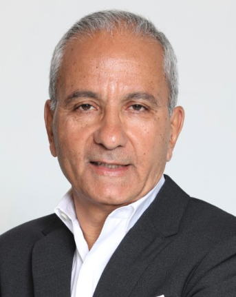 Patrice Cacoub合作论文数Departement de Medecine Interne et Immunologie Clinique, Hopital La Pitie Salpetriere153T. Papo合作论文数84C. Francès合作论文数82Serge Herson合作论文数53LTH DU合作论文数44C. De Gennes合作论文数44Du Le Thi Huong合作论文数43Jean Cabane合作论文数43Abdallah Sbai合作论文数41Edouard Kieffer合作论文数39
 Rachid Laraki合作论文数39J.Ch. Piette合作论文数38C Chapelon合作论文数34D. Le Thi Huong合作论文数34G Herreman合作论文数33
 Zahir Amoura合作论文数Hôpital Pitié-Salpêtrière33Guy Chomette合作论文数33Lucile Musset合作论文数Service d’immunochimie, groupe hospitalier Pitié-Salpêtrière32Patrick Cherin合作论文数28JM MOUTHON合作论文数27Anne Gatel合作论文数25C De Gennes合作论文数25Z Tazi合作论文数21J.M. Ziza合作论文数21
 Catherine Chapelon-Abric合作论文数Hopital Pitie Salpetriere21Gilles Raguin合作论文数20H. Beaufils合作论文数19Dlt Huong合作论文数17
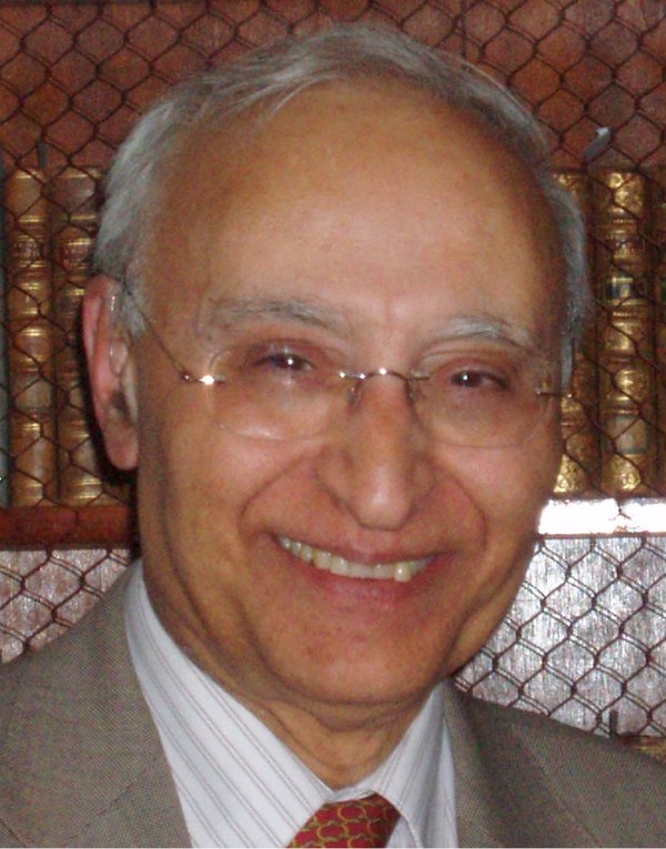 Iradj Gandjbakhch合作论文数Assistance Publique - Hôpitaux de Paris16
 Devulder Bernard合作论文数16F Lunel合作论文数15Kahn Marcel-Francis合作论文数15
 Olivier Chosidow合作论文数University Paris-Est Creteil15Etienne S D合作论文数15Pierre Opolon合作论文数15Sylvie Boisnic合作论文数15J.M. Huraux合作论文数15
 Dominique de Zuttere合作论文数14
 Didier Dormont合作论文数14F Sauvaget合作论文数14Julie Cosserat合作论文数14Didier Fassin合作论文数ORSTOM, IFEA14Yves Darbois合作论文数13
 Loic Guillevin合作论文数Hôpital Cochin;Université de Paris;Centre Médical Luxembourg Paris13P Langlois合作论文数12
 Pierre Hausfater合作论文数AH-HP Sorbonne Universités;Emergency Department, Centre Hospitalo-Universitaire Pitié-Salpêtrière Assistance-Publique Hôpitaux de Paris;Emergency Department, Université Pierre et Marie Curie Sorbonne-Universités12




