Der Nachweis von qualitativen und quantitativen VerÄnderungen im Bereich zellulÄrer Onkogene erlaubt Einblicke in die Mechanismen von Wachstum, Teilung und Differenzierung von gesunden und tumorÖsen Zellen. Derartige VerÄnderungen lassen sich auf der Ebene der DNS (Genamplifikation, -mutation und -rearrangement), der RNS und der Proteine (erhÖhte Genexpression) nachweisen. Eine klinische Relevanz fÜr Hals-Nasen-Ohren-Tumoren fand sich bisher bei einer Amplifikation und verstÄrkten Expression des c-mycGens (Korrelation mit fortgeschrittenen Tumorstadien und schlechter Prognose) sowie einer Amplifikation der Gene bcl-1, hst und int-2 (Korrelation mit Nikotinabusus und schlechter Prognose). Eine pathogenetische Bedeutung bei der Tumorentstehung scheint die verstÄrkte Expression des EGF-Rezeptors (EGF = epidermal growth factor) zu haben. Im Gegensatz zu normaler Schleimhaut ist die Expression dieses Rezeptors bei manifesten Karzinomen und bei chronischen EntzÜndungen der Schleimhaut infolge eines Alkohol- und Nikotinabusus deutlich erhÖht. Klinische Fortschritte durch den Nachweis dysregulierter Gene sind in den nÄchsten Jahren vor allem im Hinblick auf ein biologisches Tumorstaging zu erwarten. Erhofft werden dabei prospektive Aussagen zum Wachstumsverhalten eines Tumors, aber auch zu seiner Radio- und ChemosensibilitÄt.
Purpose This prospective phase II study was undertaken to assess the feasibility of a larynx preservation protocol with simultaneous radiochemotherapy. Patients and methods Between 3/1998 and 10/2000, 42 patients with moderately advanced cancer of the larynx (n = 25) and hypopharynx (n = 17) eligible for total laryngectomy (LE) were treated in a prospective larynx preservation study. The study protocol scheduled 66 Gy in 5 weeks using a concomitant boost technique and 70 mg/m2 Carboplatin on days 1–5 in weeks 1 and 5. Results The median follow-up time of the censored study patients was 41 months (9–95 months). The 5-year overall survival was 0.66 (95% CI 0.48–0.84), the 5-year laryngectomy-free survival 0.60 (95% CI 0.42–0.78), and the laryngeal preservation rate at 5 years 0.67 (95% CI 0.49–0.85). Cox multivariate regression analysis showed the total tumor volume to be the only statistically significant factor on locoregional failure-free survival. Six of 23 tumor-free long-term survivors received a tracheotomy because of late laryngeal toxicity associated with dysphagia 30–79 months after radiochemotherapy. Conclusions Due to the late laryngeal toxicity observed the value of this regimen for larynx preservation is limited.
Advanced unresectable head and neck cancer has a bad prognosis and concurrent chemoradiation (RCT) might be the first treatment of choice. To prove an expected benefit of simultaneously given chemotherapy a two arm randomized study with HF-ACC RCT versus HF-ACC-RT was performed between 1995 and 1999. In a subgroup analysis the influence of pretherapeutic Hb-level to survival and locoregional control was tested. The results after a median follow up of 57 months will be presented. The study included primarily untreated stage III/IV (UICC) oropharyngeal and hypopharyngeal carcinomas. Patients were randomized to receive either HF-ACC RCT with 2 cycles 5-FU(600mg/m2/d) and carboplatin (70mg/m2/d) on days 1–5 and 29–33 (arm A) or HF-ACC RT alone (arm B). Total RT-dose in both arms was 69.9 Gy in 38 days in concomitant boost technique (week 1–3: 1,8Gy/d/ week 4–6: bid RT with 1.8/1.5 Gy fractions). By May 1999 a total of 263 pts. have been randomized. Median age was 56 years, in 97% the tumor was stage IV, in 3% stage III. Average primary tumor volume (without lymph nodes) was 32.8 ml for RCT and 39.8 ml for RT. The actuarial analysis presented is based on an as treated population of 240 pts., qualified for protocol and beginning therapy. 113 pts. received RCT and 127 pts. RT. There were 178 oropharyngeal and 62 hypopharyngeal cancers. After a median follow up time of 57 months the survival in local control (SLC) is significantly better in RCT than in RT (p = 0,008), with median times of survival without local progression of 17 months and 11 months respectively. Also overall survival (OS) shows a benefit for RCT (p = 0,016), with a median survival of 23 months for RCT and 16 months for RT. However the benefit in SLC and OS is not seen in hypopharyngeal carcinomas. Pretherapeutical Hb levels up to 12,7 g/dl result in lower overall survival and survival in local control rates compared to higher pretherapeutical Hb-levels up to 13,8 g/dl. Pretherapeutical Hb-levels higher than 13,8 g/dl do not improve overall survival and survival in local control any further. Late toxicity in RCT and RT is not significantly different but there are more patients using gastral feeding tubes (PEG) in RCT than in RT. Concurrent chemoradiation with intensified hyperfractionated accelerated radiotherapy is superior to HF-ACC-RT alone. The benefit seems to be restricted to orpharyngeal carcinomas. Patients with pretherapeutical Hb-levels higher than 13,8 g/dl seem to have no further improvement of overall survival and survival in locoregional control seen in patients with pretherapeutic Hb-levels between 12,7 and 13,8 g/dl
5525 Background: Concurrent chemoradiation (RCT) might be first treatment of choice in advanced unresectable HNC. Between 1995 and 1999 a two arm multicentric randomized study with hyperfractionate...
Zusammenfassung Einleitung. Bezug nehmend auf die vielversprechenden Ergebnisse internationaler Studien wurde erstmals in Deutschland eine prospektive multizentrische Phase-II-Studie zum Thema Organerhalt durch eine primäre Radiochemotherapie bei nur durch Laryngektomie beherrschbarem Larynx- und Hypopharynxkarzinom durchgeführt. Methoden. Hierbei wurden 30 Patienten mit Larynx- bzw. Hypopharynxkarzinomen im Stadium II bzw. III (UICC) eingebracht, von denen 28 protokollgerecht behandelt wurden und im Follow-up evaluiert werden konnten. Als Ausschlusskriterien wurden neben den üblichen Kontraindikationen einer Radiochemotherapie die Larynxknorpelinfiltration, der N2C-Status und die über die Laryngektomie hinausgehende Notwendigkeit einer Lappenrekonstruktion herangezogen. Als primäre Radiochemotherapie kam eine akzelerierte “concomitant boost” Radiochemotherapie (GHD: 66 Gy) mit Carboplatin (70 mg/m 2 KO in der 1. und 5. Woche) zur Anwendung. Das Restaging wurde 1 Monat nach Therapieende durchgeführt und ggf. die Indikation zur Salvage-Laryngektomie gestellt. Ergebnisse. Nach einer Beobachtungszeit von 1 Jahr zeigte sich bei 20 der 28 Patienten (71%) eine stabile komplette Remission bei funktionell intaktem Larynx ohne Tracheostoma. Von diesen 20 Patienten entwickelten sich in 3 Fällen pulmonale Metastasen, in 1 Fall ein bronchiales Zweitkarzinom und in 3 Fällen zervikale Lymphknotenmetastasen, die eine Neck-dissection erforderlich machten. Von den 8 restlichen Patienten entstand bei 4 ein laryngektomiepflichtiges Rezidiv, 1 Patient musste ödembedingt tracheotomiert werden und 3 Patienten verstarben, wobei in 2 Fällen ursächlich Tumorprogress und in einem Fall therapiebedingte Komplikationen (Sepsis nach Wundheilungsstörungen in Folge der Salvage-Laryngektomie) auftraten. Schlussfolgerung. Das Protokoll war insgesamt gut durchführbar und wurde von den Patienten akzeptabel toleriert. Problematisch anzusehen ist allerdings die Rezidivdiagnostik, die in 2 Fällen inoperable Rezidive aufgrund therapiebedingter Veränderungen übersah, sowie die Salvage-Chirurgie, die in einem Fall zu tödlichen Komplikationen führte. Trotz des relativ guten 1-Jahres-Funktionserhaltungsergebnisses zeigt die vorliegende Studie auf, dass eine vorgeschaltete Induktionstherapie (vorzugsweise Chemotherapie) im Sinne einer besseren Patientenselektion Vorteile bieten könnte. Weitere Studien sollten diese Frage bearbeiten.
INTRODUCTION:Regarding the promising results of international trials we conducted the first German prospective multicentre phase II trial for organ preservation with primary simultaneous chemoradiation in advanced laryngeal and hypopharyngeal cancer.PATIENTS AND METHODS:28 of 30 recruited patients suffering from stage II and III (UICC) laryngeal and hypopharyngeal cancer were treated with primary simultaneous chemoradiation within an organ preservation program and monitored in follow-up of one year. Exclusion criteria included tumor infiltration of the laryngeal cartilage, bilateral neck nodes (N2c) and need for flap reconstruction in case of laryngectomy. The protocol included an accelerated concomitant boost chemoradiation (66 Gy) with Carboplatinum (70 mg/m2 1st and 5th week) and a restaging procedure one month after therapy. In case of residual disease, salvage laryngectomy and/or neck dissection were performed.RESULTS:After follow-up of one year 20 of 28 patients (71%) were presented with stable complete remission and functionally preserved larynx. Of these 20 patients 3 developed pulmonary metastases, 1 secondary primary carcinoma of the lung and 3 neck metastases which needed neck dissections. The other patients showed in 4 cases relapsing tumor which was indicated for laryngectomy. One patient needed tracheotomy because of persisting edema and 2 patients died due to tumor progress. One patient died after complications due to salvage surgery.CONCLUSION:The organ preservation protocol was feasible with well tolerated early toxicity. Problems of screening for recurrent disease, salvage surgery and late toxicity should be noted and pronounced in patient information. Further studies should focus on the improvement of patient selection which could be realized by induction Chemotherapy (using new components like taxan) and/or use of prediction factors such as tumor volume and hemoglobin levels.
BACKGROUNDEwing's sarcoma is a malignant tumour which usually arises from bony structures. It can be found in dia- or metaphysis of bones of the extremities but as well in 50% of cases in skeletal parts of the trunk. Clinically patients often present with various symptoms as there are pain, swelling and progredient loss of function if extremities are involved. But in many cases, Ewing's sarcoma shows a lack of specific symptoms and therefore stays inapparent for a long time.CASE REPORTA 14-year-old patient complained about intermittent nuchal pain and was sent to hospital because of an extensive retropharyngeal swelling. The preoperative imaging showed a tumour of the soft tissue of the neck. It had reached the cervical spine and after surgical treatment it was histologically and molecular biologically diagnosed as a Ewing's sarcoma arising from the transversal processes of the atlas.CONCLUSIONSince Ewing's sarcoma may have an immense component of soft tissue it has to be included in the range of differential diagnosis of soft tissue tumours of the head and neck, e.g., as there are extracranial schwannoma, inflammatory lymph nodes or lymph node metastases and lateral and medial cysts of the neck.
Purpose: To demonstrate the efficacy of radiochemotherapy (RCT) as the first choice of treatment for advanced unresectable head-and-neck cancer. To prove an expected benefit of simultaneously given chemotherapy, a two-arm randomized study with hyperfractionated accelerated radiochemotherapy (HF-ACC-RCT) vs. hyperfractionated accelerated radiotherapy (HF-ACC-RT) was initiated. The primary endpoint was 1-year survival with local control (SLC).Methods and Materials: Patients with Stage III and IV (UICC) unresectable oro- and hypopharyngeal carcinomas were randomized or HF-ACC-RCT with 2 cycles of 5-FU (600 mg/m(2)/day)/carboplatinum (70 mg/m(2)) on days 1-5 and 29-33 (arm A) or HF-ACC-RT alone (arm B). In both arms, there was a second randomization for testing the effect of prophylactically given G-CSF (263 mug, days 15-19) on mucosal toxicity. Total RT dose in both arms was 69.9 Gy in 38 days, with a concomitant boost regimen (weeks 1-3: 1.8 Gy/day, weeks 4 and 5: b.i.d. RT with 1.8 Gy/1.5 Gy). Between July 1995 and May 1999, 263 patients were randomized (median age 56 years; 96% Stage IV tumors, 4% Stage III tumors).Results: This analysis is based on 240 patients: 113 patients with RCT and 127 patients with RT, qualified for protocol and starting treatment. There were 178 oropharyngeal and 62 hypopharyngeal carcinomas. Treatment was tolerable in both arms, with a higher mucosal toxicity after RCT. Restaging showed comparable nonsignificant different CR + PR rates of 92.4% after RCT and 87.9% after RT (p = 0.29). After a median observed time of 22.3 months, 1- and 2-year local-regional control (LRC) rates were 69% and 51% after RCT and 58% and 45% after RT (p = 0.14). There was a significantly better 1-year SLC after RCT (58%) compared with RT (44%, p = 0.05). Patients with oropharyngeal carcinomas showed significantly better SLC after RCT (60%) vs. RT (40%, p = 0.01); the smaller group of hypopharyngeal carcinomas had no statistical benefit of RCT (p = 0.84). For both tumor locations, prophylactically given G-CSF was a poor prognostic factor (Cox regression), and resulted in reduced LRC (log-rank test: G-CSF, p = 0.0072).Conclusion: With accelerated radiotherapy, the efficiency of simultaneously given chemotherapy may be not as high as expected when compared to standard fractionated RT. Oropharyngeal carcinomas showed better LRC after HF-ACC-RCT vs. HF-ACC-RT; hypopharyngeal carcinomas did not. Prophylactic G-CSF resulted in an unexpected reduced local control and should be given in radiotherapy regimen only with strong hematologic indication. (C) 2001 Elsevier Science Inc.
Zusammenfassung Hintergrund und Fragestellung. Die pharyngokutane Fistel ist die häufigste Komplikation nach der Totalexstirpation des Kehlkopfs. In Deutschland wird postoperativ eine orale Nahrungskarenz und die Ernährung über eine Magensonde über 10–14 Tage empfohlen. International finden sich hierzu unterschiedliche Empfehlungen: die Mehrheit der Autoren postuliert eine postoperative Sondenernährung über 7–14 Tage, andere verzichten ganz auf eine Magensonde und beginnen bereits 1–4 Tage postoperativ mit einer oralen Ernährung. Wir untersuchten den Einfluss der Dauer einer postoperativen oralen Nahrungskarenz und Sondenernährung auf die Fistelinzidenz nach Laryngektomie. Patienten/Methodik. Im Rahmen einer prospektiven Studie wurde an unterschiedlichen Tagen bei 42 laryngektomierten Patienten zwischen dem 1. und 10. postoperativen Tag mit einer oralen Ernährung begonnen. Darüber hinaus wurde retrospektiv nach weiteren Risikofaktoren gesucht, die das Auftreten einer Pharynxfistel begünstigen. Ergebnisse. Insgesamt trat bei 5 Patienten eine Pharynxfistel auf (12%). Ein Zusammenhang zwischen dem Zeitpunkt des oralen Nahrungsbeginns und der Fistelinzidenz fand sich nicht. Bei 35 Patienten wurde innnerhalb der 1. postoperativen Woche mit der oralen Ernährung begonnen. Die Fistelinzidenz in dieser Gruppe betrug 9%. Von den retrospektiv analysierten Risikofaktoren für das Auftreten einer pharyngokutanen Fistel erwies sich nur die zusätzliche Pharynxteilresektion im Rahmen der Laryngektomie als statisch signifikanter Risikofaktor für das Auftreten einer Pharynxfistel ( p =0,018). Schlussfolgerungen. Eine orale Ernährung innerhalb der 1. Woche nach Laryngektomie hat keinen Einfluss auf die Entstehung einer pharyngokutanen Fistel.
BACKGROUND:Ewing's sarcoma is a malignant tumour which usually arises from bony structures. It can be found in dia- or metaphysis of bones of the extremities but as well in 50% of cases in skeletal parts of the trunk. Clinically patients often present with various symptoms as there are pain, swelling and progredient loss of function if extremities are involved. But in many cases, Ewing's sarcoma shows a lack of specific symptoms and therefore stays inapparent for a long time.CASE REPORT:A 14-year-old patient complained about intermittent nuchal pain and was sent to hospital because of an extensive retropharyngeal swelling. The preoperative imaging showed a tumour of the soft tissue of the neck. It had reached the cervical spine and after surgical treatment it was histologically and molecular biologically diagnosed as a Ewing's sarcoma arising from the transversal processes of the atlas.CONCLUSION:Since Ewing's sarcoma may have an immense component of soft tissue it has to be included in the range of differential diagnosis of soft tissue tumours of the head and neck, e.g., as there are extracranial schwannoma, inflammatory lymph nodes or lymph node metastases and lateral and medial cysts of the neck.
Background: Ewing's sarcoma is a malignant tumour which usually arises from bony structures. It can be found in dia- or metaphysis of bones of the extremities but as well in 50 % of cases in sceletal parts of the trunk. Clinically patients often present with various symptoms as there are pain, swelling and progredient loss of function if extremities are involved. But in many cases, Ewing's sarcoma shows a lack of specific symptoms and therefore stays inappearent for a long time. Case report: A 14-year-old patient complained about intermittend nuchal pain and was sent to hospital because of an extensive retropharyngeal swelling. The preoperative imaging showed a tumour of the soft tissue of the neck. It had reached the cervical spine and after surgical treatment it was histologically and molecularbiologically diagnosed as a Ewing's sarcoma arising from the transversal processus of the atlas. Conclusion: Since Ewing's sarcoma may have an immense component of soft tissue it has to be included in the range of differential diagnosis of soft tissue tumours of the head and neck, e.g., as there are extracranial schwannoma, inflammatory lymph nodes or lymph node metastases and lateral and medial cysts of the neck.
BACKGROUND AND OBJECTIVE:A pharyngocutaneous fistula is the most common complication after total laryngectomy. In Germany, a traditional recommendation is to use a nasogastric tube for feeding for 10-14 days postoperatively because many surgeons believe that oral feeding after surgery contributes to fistula development. However, there is no international agreement about when to begin oral feeding after total laryngectomy. Some authors begin oral feeding between the 1st and 4th postoperative day without any nasogastric tube, while others using a nasogastric tube delay oral feedings until 7-14 days after surgery. The aim of the present study was to investigate the relationship between the timing of oral feeding and the development of fistulas after total laryngectomy.PATIENTS/METHODS:In a prospective trial with 42 consecutive patients who underwent laryngectomy, oral feeding was started on different postoperative days between the 1st and the 10th. Most patients were selected randomly for the different postoperative days. Furthermore, other potential risk factors predisposing to fistula formation were analyzed retrospectively.RESULTS:Five fistulas occurred in the total group (12%). Early postoperative oral feeding does not increase the incidence of fistulas. The fistula rate was only 9% in patients fed orally in the 1st postoperative week. The analysis of further risk factors for fistula formation showed only a significant correlation between type of resection and fistula occurrence (extended laryngectomy with partial pharyngectomy vs standard laryngectomy; p = 0.018).CONCLUSIONS:Our results indicate that early oral feeding in the 1st postoperative week does not influence fistula formation after laryngectomy.
This study seeks to evaluate treatment modalities, mortality after surgery, survival, and local control rates for a consecutive cohort of patients with cancer of the hypopharynx treated according to a prospective protocol that favors surgery as an initial approach to the disease. The charts of 228 consecutive patients with previously untreated hypopharyngeal squamous cell carcinoma were reviewed. Outcome measures (overall survival, disease specific survival, and local control) were calculated using the Kaplan-Meier estimator. Of 228 consecutive patients, 136 (59.6%) were found suitable for initial surgical treatment. Of the remaining 92 patients, 18 (7.9%) had nonresectable lymph node metastases, 16 (7.0%) had unresectable primary tumors, 13 (5.7%) refused surgery, and 13 (5.7%) presented distant metastases during initial diagnostic evaluation. Of those who had surgery, 46 had larynx-sparing procedures, 54 had total laryngectomy, and 36 had total laryngo-pharyngectomy. None of the patients who had surgery died postoperatively. Actuarial 5-year overall survival was 27.2% for all 228 patients, 39.5% for the 136 patients with surgical treatment, and 61.1% for the 46 patients who were treated with larynx-sparing procedures.
Zusammenfassung Hintergrund und Fragestellung. Qualitätsmanagement ist für eine optimale Behandlung maligner Tumoren von besonderer Bedeutung. Im Gegensatz zu anderen Fachgebieten sind in der HNO-Heilkunde Qualitätssicherungsstandards bisher nicht erarbeitet worden. Der aktuelle Stand der Qualitätssicherung in der Kopf-Hals-Onkologie an den deutschen Hauptabteilungen des Fachgebiets wurde erfasst. Patienten/Methodik. In einer bundesweiten Fragebogenaktion wurden an 146 deutsche Hals-Nasen-Ohrenkliniken detaillierte Fragebögen ausgesandt. 110 Bögen wurden ausgefüllt zurückgesandt, das entspricht einer Rücklaufquote von 75%. Ergebnisse. 56% der Kliniken führen Qualitätssicherungsmaßnahmen bewusst durch, und in 38% ist ein Mitarbeiter zum Qualitätssicherungsbeauftragten bestimmt. Interdisziplinäre Tumorkonferenzen werden unter Beteiligung der Strahlentherapie in 86 Kliniken (78%), der medizinischen Onkologie in 84 (76%), der radiologischen Diagnostik in 82 (74%), der Pathologie in 73 (66%) und regionaler Tumorzentren in 47 Abteilungen (42%) abgehalten. Eine “Morbiditäts-Mortalitäts-Konferenz” ist lediglich in 7 Kliniken (6%) etabliert. Die benutzten Dokumentationssysteme sind vielfältig, größtenteils klinikintern und teilweise sehr zeit- und personalintensiv. In 95 Abteilungen (86%) erfolgt eine standardisierte Tumornachsorge für Patienten mit malignen Tumoren. In 53 Abteilungen (48%) werden die Nachsorgedaten in einer Datenbank aufgenommen; 78 Abteilungen (71%) führen regelmäßig eine Tumornachsorgesprechstunde durch. Schlussfolgerungen. Die Erhebung dokumentiert ein ausgeprägtes Problembewusstsein und ein insgesamt sehr reges Interesse an der praktischen Durchführung von Qualitätssicherungsmaßnahmen in der Onkologie neben einer Fülle von Einzelinitiativen. Allerdings ist derzeit kein einheitliches Qualitätssicherungskonzept erkennbar. Unverändert gibt es kein einheitliches Erfassungsinstrument (Datenbank). Die Ermächtigung zur Tumornachsorge ist ein wesentliches Instrument der Kliniken zur Qualitätssicherung, das auch in Zeiten wirtschaftlicher Zwänge im Gesundheitswesen unverzichtbar bleibt.
Quality control is of special importance in head and neck oncology since the quality of medical care constitutes a vital parameter for the diseased patient. In contrast to other medical specialties, no quality assurance program for head and neck cancer patients has yet been established in Germany. Therefore, a survey was conducted to assess the quality assurance instruments that are in use today in otorhinolaryngology-head and neck (ORL-HNS) centers.In a nationwide survey, questionnaires were sent out to 146 German ORL-HNS departments (the return rate was 75%).56% of all departments apply dedicated quality assurance processes, and 38% have appointed a formal quality assurance officer. Interdisciplinary oncological conferences are held in the vast majority of all departments with the participation of radiation oncologists in 86 (78%), medical oncologists in 84 (76%), diagnostic radiologists in 82 (74%), and pathologists in 73 (66%). Morbidity-mortality conferences are held in seven departments (6%). A standardized follow-up of oncological patients is carried out in 95 units (86%), and 53 departments use computer-assisted data bases to organize their follow-up data (48%). A wide variety of documentation systems is in use throughout the country: 78 units (71%) offer formal follow-up to their oncological patients.This survey documents a wide-spread interest in quality assurance procedures. Many individual efforts are being undertaken. However, no uniform quality assurance or auditing system is currently in use in Germany nor is a commonly accepted data base available. The ability to offer oncological follow-up within the national social security system is generally considered indispensable for the maintenance of high-quality oncological care in ORL-HNS departments.
STUDY DESIGN:A matched-pair study on the relationship between proliferation associated markers Ki-67, PCNA, and p53 and treatment failure in carcinomas of the oropharynx and oral cavity.MATERIAL AND METHOD:Fifty-six T1-T3 carcinomas of the oropharynx or oral cavity, treated uniformly with primary surgery and postoperative irradiation, were investigated. Twenty-eight patients had recurrent disease and were matched with 28 patients with nonrecurrent disease regarding stage and location of tumor as well as age and therapy. MIB1 staining was used to determine the Ki-67 labeling index. Immunohistochemical studies determined the p53 status and PCNA labeling index.RESULTS:Where treatment failed, the Ki-67 labeling index was significantly (p = 0.032) higher (mean = 59.1%) than in patients without recurrence (mean = 50.5%). Mean time to relapse was 45 months (n = 25) for carcinomas with a Ki-67 (MIB1) labeling index above the median (53.7%) of the general study population, compared to 61.7 months for those cases (n = 31) below this median (p = 0.029). The PCNA labeling index did not correlate significantly with tumor recurrence, nor with time to relapse. In 46% of all tumors, p53 overexpression was present. No significant correlation could be found between p53 overexpression and tumor recurrence or time to relapse.CONCLUSION:Examination of Ki-67 is thought to provide useful prognostic information concerning squamous cell carcinomas of the oropharynx and oral cavity. Overexpression of p53 or PCNA status is not of prognostic value, which is consistent with earlier results. We conclude that the detection of Ki-67 is an unfavorable prognostic factor for squamous cell carcinoma of the oropharynx and oral cavity, at least if treated with a combination of surgery and postoperative irradiation.
The pretherapeutic hemoglobin level (Hb) has been postulated to constitute a prognostic marker for outcome after primary chemoradiation of patients with advanced cancer of the head and neck. However, this hypothesis has not been tested systematically in large study samples. In the years 1992-1997, 125 patients with advanced head and neck cancer (stages III/IV UICC) were treated with primary chemoradiation in two different prospective multicentric trials, 62 patients in trial A (phase II, 1992-1995), and 63 in trial B (phase III, 1995-1997). Beside initial Hb, other pretherapeutic parameters with potential prognostic relevance were assessed and correlated with clinical outcome after 43-months follow-up: total tumor volume (TTV; calculated in initial CT scans), tumor oxygenation (polarographic measurements with Eppendorf histography), TNM, tumor localization, age, and performance status. The evaluation of the clinical end points (progression-free and overall survival and local tumor control) revealed that Hb and TTV were independent parameters with strong predictive character of outcome after primary chemoradiation in both trials (n = 125). Bivariate analysis showed < median (13.5 g/dl) a hazard ratio of 2.1 (P = 0.002) for Hb; and > median (98 ml) a Hazard ratio of 2.0 (P = 0.006) for TTV. Severe anemia (Hb < 10 g/dl) was an adverse factor in three patients. Hypoxia was associated with poorer initial therapeutical response but was not predictive of clinical outcome. Furthermore, tumor oxygenation showed no correlation with Hb. The other parameters examined failed to show prognostic significance. Our results indicate a high prognostic value of initial Hb for outcome after primary chemoradiation in advanced head and neck cancer and imply a therapeutic benefit of Hb substitution or erythropoietin administration. We propose to test this in randomized clinical trials.
Zusammenfassung Der prätherapeutische Hämoglobin-Wert (Hb) wird als einer der wichtigsten Einflussgrößen für den Erfolg einer Strahlentherapie diskutiert; wurde jedoch bislang im klinischen Kontext anderer Risikofaktoren bei fortgeschrittenen Kopf-Hals-Tumoren kaum systematisch untersucht. In den Jahren 1992–1997 wurden in Kooperation der Universitäts-HNO- und -Strahlenklinik Heidelberg 2 prospektive multizentrische Studien zur primären Radiochemotherapie bei nichtresektablen Stadium-III/IV-Kopf-Hals-Karzinomen durchgeführt, in deren Rahmen insgesamt 125 Patienten behandelt wurden (Studienleitung: Herr Prof. Dr. R.-P. Müller; Köln). Studie A (Phase II, 1992–1995) rekrutierte 62 und Studie B (Phase III, 1995–1997) 63 Patienten. Neben dem Ausgangs-Hb-Wert wurden weitere, als prognostisch wichtig eingestufte klinische Parameter wie die Tumoroxygenierung, das Gesamttumorvolumen (total tumor volume, TTV), TNM, Tumorlokalisation, Alter und “performance status” erfasst und bezüglich ihres prädiktiven Wertes überprüft (Nachbeobachtungszeit 43 Monate). In der Gesamtbetrachtung (125 Patienten) setzten sich der Hb-Wert und das TTV als die stärksten, von einander unabhängigen, prognostischen Parameter, bezogen auf die Endpunkte Überleben und lokoregionäre Kontrollzeit, durch (bivariate Analyse: HbMedian (98 ml): Hazard-Ratio: 2,0 [ p =0,006]. Bei nur 3 Patienten wurde eine Anämie mit Hb-Werten unter 10 g/dl gesehen. Die Tumoroxygenierung zeigte lediglich eine signifikante Korrelation mit dem initialen Therapieansprechen, die sich in den klinischen Verläufen nicht widerspiegelte. Eine Korrelation zwischen Hb-Werten und Tumoroxygenierungsparametern konnte nicht gesehen werden. Die Ergebnisse der Studie unterstreichen die hohe klinische Bedeutung des Ausgangs-Hb-Wertes für den Erfolg nach primärer Radiochemotherapie von fortgeschrittenen Kopf-Hals-Tumoren und unterstützen die Forderung nach randomisierten Studien zur Prüfung der Wertigkeit der prätherapeutischen Hb-Substitution bzw. -Anhebung durch Erythropoietin.
 M. Schröder合作论文数24Hans Edmund Eckel合作论文数21S Staar合作论文数19
 Olaf Ebeling合作论文数17E Stennert合作论文数14
 Dietz Andreas合作论文数13He Eckel合作论文数10Markus Jungehülsing合作论文数10
 Rolf-Peter Müller合作论文数Department of Radiation Therapy, University of Cologne10
 Volker Rudat合作论文数10Wolf Achterrath合作论文数8A. Kur合作论文数6M. Miemann合作论文数6G Bogan合作论文数6K. Pistorius合作论文数6H Kasparu合作论文数6T. Bimmler合作论文数6Detlev Latz合作论文数6A. Calavrezos合作论文数6P. Worst合作论文数6W. Sachsenheimer合作论文数6H Erdmann合作论文数5M. Flentje合作论文数5H. Kaden合作论文数5O Krieger合作论文数5U. Mende合作论文数5M Engelhard合作论文数5R. P. Mueller合作论文数5R Kuse合作论文数5Fauser A A合作论文数5Singelmann H合作论文数5L. Reichel合作论文数5Georg W Lohr合作论文数5
 Günther Steger合作论文数Department of Medicine I, Medical University of Vienna;Vienna General Hospital;Goldenes Kreuz Private Hospital5K. Lennert合作论文数5
 Wolfgang Kreisel合作论文数5
 Karl-Heinrich Link合作论文数Asklepios Tumortreatment Center Rhein5M Vössing合作论文数5
 Ludger Staib合作论文数Darmzentrums, Klinikum Esslingen;Klinik für Allgemein- und Viszeralchirurgie, Klinikum Esslingen5C Conradt合作论文数4H. Stuetzer合作论文数4
 Hannes Rainer合作论文数4M. Wannenmacher合作论文数4Michael Schroeder合作论文数4
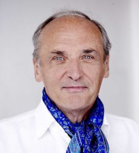 Raimund Jakesz合作论文数Abteilung für Allgemeinchirurgie, Chirurgische Universitatsklinik Wien4Eberhard Stennert合作论文数4u schroeder合作论文数3H Weidauer合作论文数3
 Olaf Michel合作论文数3




