Hans’ algorithm (HA) is the most frequently used surrogate biomarker scheme for subtyping Diffuse large B-cell lymphoma (DLBCL) by the cell-of-origin (COO) into GCB and non-GCB subtypes. The originally published positive and negative predictive value (PPV and NPV) against gene expression profiling (GEP) subtypes were less than perfect and were not fully reproducible in published literature. Furthermore, little is known about how the HA performs in clinical practice. The Canadian Association of Pathologists National Standard Committee for High Complexity Testing (CAP-ACP NSCHCT) initiated a Canada-wide project to assess the current diagnostic accuracy of the HA in clinical practice, harmonize the analytical phase of the IHC assays used for HA, and to optimize its overall diagnostic sensitivity and specificity against GEP subtypes. We divided a DLBCL cohort (n=96), where COO was defined by GEP (Lymph2CX, NanoString technologies) into training (TC, N=45) and validation cohort (VC, N=51) and tissue microarray (TMA)-TC and TMA-VC were constructed. Selected major Canadian laboratories applied their routine CD10, Bcl-6, and MUM1 IHC protocols and sent stained slides to the reference laboratory for review. The IHC protocols from laboratories were designated as “weak” (1/10), “moderate” (4/10), and “strong” (5/10) based on their overall analytical sensitivity. The results of the central review HA readouts were compared with GEP results. Furthermore, in TC, the original HA readout was modified to adjust the cutoff to overall IHC protocol sensitivity. The new readout criteria were also assessed in the VC set. The original HA readout showed good results against GEP for low sensitivity protocol only. For all other laboratories that had IHC protocols with moderate and high analytical sensitivity, a readout was adjusted to higher IHC protocol analytical sensitivity using a cutoff of >30% of >2+ staining intensity. This modification yielded significantly improved diagnostic accuracy against GEP even without any changes to the IHC protocols and was widely applicable to the range of analytical sensitivities of IHC protocols, which are currently in use. IHC biomarkers for HA can be highly accurate and harmonized across different laboratories for clinical application if the following criteria are met: (i) testing laboratories use standardized reference materials to set up and monitor analytical sensitivity, and (ii) the pathologist’s readout and cutoff are adjusted to the overall IHC protocol analytical sensitivity.
Clinical decision making in Acute Myeloid Leukemia (AML) critically relies on rapid genomic characterization. To better understand the AML diagnostic landscape in Canada, the Canadian Leukemia Study Group (CLSG) conducted a survey of laboratory hematology leadership (n = 18) at 16 laboratories across 10 provinces, administered using Google Forms in September 2024. Nearly all surveyed sites were equipped to deliver a full suite of testing platforms through existing on-site infrastructure or laboratory partnerships. Reporting practices varied in terms of genomic integration into bone marrow results and the use of AML classification systems. Turn-around-time (TAT) targets were predominantly determined through internal institutional consensus (62%) or recommendations by provincial cancer agencies/international groups (44%). TAT reduction was a top priority for 56% of laboratories, suggesting timely biomarker results to be an active area for improvement. Various treatment-determining biomarkers were frequently assessed as rapid-tests (defined as a 5-day TAT), including FLT3-ITD (69%), FLT3-TKD (56%), and NPM1 (56%), while others such as IDH1 and TP53 were rapid at a limited number of laboratories. Respondents demonstrated a strong shared interest in joint projects such as the validation of AML measurable residual disease (MRD) assays (56%). There was also unanimous support for establishing CLSG AML laboratory consensus guidelines. This survey documents the current state of Canadian AML laboratories and provides a foundation for future shared development projects.
BACKGROUND:Allogeneic hematopoietic cell transplantation (allo-HCT) for acute myeloid leukemia (AML) is associated with considerable morbidity and mortality. Machine learning (ML) techniques are increasingly applied to predict outcomes in medicine. OBJECTIVES:To evaluate the role of ML in predicting overall survival (OS) after allo-HCT for AML and compare ML with traditional Cox regression. STUDY DESIGN:Using an internal cohort of 2253 patients and 14 pre-allo-HCT variables, we developed three models: Cox regression with time-varying coefficients (Cox-TVC), Elastic-net Cox Regression (Cox-EN), and Random Survival Forest (RSF). Performance was evaluated using multiple metrics including C-index, net reclassification improvement (NRI) and decision curve analysis (DCA). Patients were stratified into tertiles of model predicted 24-month mortality. External validation was performed in 252 single-center patients with uniform measurable residual disease (MRD) assessment. RESULTS:Model-derived risk scores strongly correlated (r = 0.886 to 0.963). Across models, age ≥ 60 years, MRD positivity, and adapted European LeukemiaNet (aELN) adverse risk were the strongest predictors. Effects of age attenuated over time (HR for age ≥60: 2.59 [95% CI: 1.54 to 4.35] at 1 year, 1.92 [95% CI: 1.04 to 3.56] at 5 years). Compared with Hematopoietic Cell Transplant Comorbidity-Index (HCT-CI) and aELN, models improved risk stratification (NRI: 31% to 44%, p < .001). Discrimination remained modest but was higher in external cohort compared with internal cohort (0.69 to 0.71 versus 0.60 to 0.61), likely reflecting uniform MRD assessment. At a 25%, risk threshold for clinical decision-making, models identified approximately 1 additional high-risk patient per 100 versus HCT-CI, or aELN. At 2-years, 25% to 27% of patients categorized as low-risk had died, while 45% to 48% categorized as high-risk were alive. CONCLUSIONS:ML approaches improved risk stratification over HCT-CI and aELN but performed comparably with Cox model. Individual outcome prediction using static pre-transplant models remained modest. Progress will require MRD standardization, richer data, and dynamic peri- and post-transplant modeling.
FMS-like tyrosine kinase 3 (FLT3) mutations are detected in approximately 20–30% of patients with acute myeloid leukemia (AML), with the presence of a FLT3 internal tandem duplication (FLT3-ITD) mutation being associated with an inferior outcome. Assessment of FLT3 mutational status is now essential to define optimal upfront treatment in both newly diagnosed and relapsed AML, to support post-induction allogeneic hematopoietic stem cell transplantation (alloSCT) decision-making, and to evaluate treatment response via measurable (minimal) residual disease (MRD) evaluation. In view of its importance in AML diagnosis and management, the Canadian Leukemia Study Group/Groupe canadien d’étude sur la leucémie (CLSG/GCEL) undertook the development of a consensus statement on the clinical utility of FLT3 mutation testing, as members reported considerable inter-center variability across Canada with respect to testing availability and timing of use, methodology, and interpretation. The CLSG/GCEL panel identified key clinical and hematopathological questions, including: (1) which patients should be tested for FLT3 mutations, and when?; (2) which is the preferred method for FLT3 mutation testing?; (3) what is the clinical relevance of FLT3-ITD size, insertion site, and number of distinct FLT3-ITDs?; (4) is there a role for FLT3 analysis in MRD assessment?; (5) what is the clinical relevance of the FLT3-ITD allelic burden?; and (6) how should results of FLT3 mutation testing be reported? The panel followed an evidence-based approach, taken together with Canadian clinical and laboratory experience and expertise, to create a consensus document to facilitate a more uniform approach to AML diagnosis and treatment across Canada.
Background and ObjectivesThe practice regarding the selection and preparation of red blood cells (RBCs) for intrauterine transfusion (IUT) is variable reflecting historical practice and expert opinion rather than evidence-based recommendations. The aim of this survey was to assess Canadian hospital blood bank practice with respect to red cell IUT.Materials and MethodsA survey was sent to nine hospital laboratories known to perform red cell IUT. Questions regarding component selection, processing, foetal pre-transfusion testing, transfusion administration, documentation and traceability were assessed.ResultsThe median annual number of IUTs performed in Canada was 109 (interquartile range, 103-118). RBC selection criteria included allogeneic, Cytomegalovirus seronegative, irradiated, fresh units with most sites preferentially providing HbS negative, group O, RhD negative, Kell negative and units lacking the corresponding maternal antibody without extended matching to the maternal phenotype. Red cell processing varied with respect to target haematocrit, use of saline reconstitution (n = 4), use of an automated procedure for red cell concentration (n = 1) and incorporation of a wash step (n = 2). Foetal pre-transfusion testing uniformly included haemoglobin measurement, but additional serologic testing varied. A variety of strategies were used to link the IUT event to the neonate post-delivery, including the creation of a unique foetal blood bank identifier at three sites.ConclusionThis survey reviews current practice and highlights the need for standardized national guidelines regarding the selection and preparation of RBCs for IUT. This study has prompted a re-examination of priorities for RBC selection for IUT and highlighted strategies for transfusion traceability in this unique setting.
Allogeneic hematopoietic cell transplantation (HCT) is used increasingly to treat blood and immune-based disorders. Post-transplantation testing of HCT recipients can lead to unexpected molecular, cytogenetic, and other information in donor-derived cells, raising questions regarding the potential impact on donor health. This study was conducted to identify the breadth of donor-derived abnormalities identified by testing HCT recipients and to determine the extent to which disclosure and donor follow-up are described. A systematic search and scoping review were conducted following PRISMA (Preferred Reporting Items for Systematic Reviews and Meta-Analyses) extension for scoping review guidelines in OVID MEDLINE and Embase (1947 to May 24, 2021). We identified 38 studies (63 donor-recipient pairs) addressing nonleukemic abnormalities to complement existing literature describing donor cell leukemia and donor-derived myelodysplasia. Donors were unrelated adults (n = 20), related family members (n = 28), cord blood donors (n = 6), or not reported (n = 9). Acquired cytogenetic, molecular, and morphologic abnormalities were reported. Donor origin was confirmed by cytogenetic analysis via karyotyping, fluorescence in situ hybridization, single tandem repeat PCR, and other techniques. A disease in donor-derived cells was described in 35 recipients (56.5%). Despite the relevance for testing and disclosure to donors, only 22 cases (32%) mentioned donor follow-up, and in 5 cases the donor developed a disease associated with the identified abnormality. Unrelated donor disclosure was mentioned in 3 of 26 cases (12%), with the findings reported back to the registry. Incidental abnormalities identified in transplanted donor cells may contribute to the post-transplantation risk of illness in the recipient and may be relevant to donor health. A framework for donor disclosure is proposed that incorporates consideration of analytic validity of the testing, potential significance of the finding, and the extent to which the abnormality is actionable. Adoption of effective processes to safeguard both donor and recipient health outcomes related to this issue is needed. (C) 2022 The American Society for Transplantation and Cellular Therapy. Published by Elsevier Inc. All rights reserved.
ABSTRACT V392 Persei is a known dwarf nova (DN) that underwent a classical nova eruption in 2018. Here we report ground-based optical, Swift UV and X-ray, and Fermi-LAT γ-ray observations following the eruption for almost three years. V392 Per is one of the fastest evolving novae yet observed, with a t2 decline time of 2 d. Early spectra present evidence for multiple and interacting mass ejections, with the associated shocks driving both the γ-ray and early optical luminosity. V392 Per entered Sun-constraint within days of eruption. Upon exit, the nova had evolved to the nebular phase, and we saw the tail of the supersoft X-ray phase. Subsequent optical emission captured the fading ejecta alongside a persistent narrow line emission spectrum from the accretion disc. Ongoing hard X-ray emission is characteristic of a standing accretion shock in an intermediate polar. Analysis of the optical data reveals an orbital period of 3.230 ± 0.003 d, but we see no evidence for a white dwarf (WD) spin period. The optical and X-ray data suggest a high mass WD, the pre-nova spectral energy distribution (SED) indicates an evolved donor, and the post-nova SED points to a high mass accretion rate. Following eruption, the system has remained in a nova-like high mass transfer state, rather than returning to the pre-nova DN low mass transfer configuration. We suggest that this high state is driven by irradiation of the donor by the nova eruption. In many ways, V392 Per shows similarity to the well-studied nova and DN GK Persei.
A 54-year-old man presented in profound obstructive shock. Investigations revealed a right atrial mass causing severe right ventricular inflow obstruction and compromised cardiac output. The patient was treated with emergency balloon catheter intervention to relieve the obstruction, with resulting hemodynamic stability. The pathology report later returned a positive result for diffuse large B-cell lymphoma. (Level of Difficulty: Intermediate.).
TransfusionVolume 62, Issue 2 p. E14-E16 REPORT OF NEW ALLELES OR ANTIGENS An intron c.149-2632T>A change in RHD is associated with aberrant transcription and very weak D phenotype Aline Floch, Aline Floch orcid.org/0000-0003-3238-4566 Immunohematology and Genomics Laboratory, New York Blood Center, New York, New York, USA Etablissement francais du sang Ile-de-France, Creteil, France INSERM U955 Equipe « Transfusion et maladies du globule rouge », IMRB, Univ Paris Est Creteil, Creteil, FranceSearch for more papers by this authorSunitha Vege, Sunitha Vege orcid.org/0000-0003-0243-266X Immunohematology and Genomics Laboratory, New York Blood Center, New York, New York, USASearch for more papers by this authorPhilip Berardi, Philip Berardi National Immunohematology Reference Laboratory, Canadian Blood Services, Brampton, Ontario, CanadaSearch for more papers by this authorJudith Hannon, Judith Hannon Department of Laboratory Medicine and Pathology, University of Alberta, Edmonton, Alberta, CanadaSearch for more papers by this authorGorka Ochoa-Garay, Gorka Ochoa-Garay orcid.org/0000-0003-3817-9416 Immunohematology and Genomics Laboratory, New York Blood Center, New York, New York, USASearch for more papers by this authorChristine Lomas-Francis, Christine Lomas-Francis Immunohematology and Genomics Laboratory, New York Blood Center, New York, New York, USASearch for more papers by this authorConnie M. Westhoff, Corresponding Author Connie M. Westhoff cwesthoff@nybc.org orcid.org/0000-0002-1929-4593 Immunohematology and Genomics Laboratory, New York Blood Center, New York, New York, USA Correspondence Connie M. Westhoff, Laboratory of Immunohematology and Genomics, New York Blood Center, 310 East 67th St, New York, NY 10065, USA. Email: cwesthoff@nybc.orgSearch for more papers by this author Aline Floch, Aline Floch orcid.org/0000-0003-3238-4566 Immunohematology and Genomics Laboratory, New York Blood Center, New York, New York, USA Etablissement francais du sang Ile-de-France, Creteil, France INSERM U955 Equipe « Transfusion et maladies du globule rouge », IMRB, Univ Paris Est Creteil, Creteil, FranceSearch for more papers by this authorSunitha Vege, Sunitha Vege orcid.org/0000-0003-0243-266X Immunohematology and Genomics Laboratory, New York Blood Center, New York, New York, USASearch for more papers by this authorPhilip Berardi, Philip Berardi National Immunohematology Reference Laboratory, Canadian Blood Services, Brampton, Ontario, CanadaSearch for more papers by this authorJudith Hannon, Judith Hannon Department of Laboratory Medicine and Pathology, University of Alberta, Edmonton, Alberta, CanadaSearch for more papers by this authorGorka Ochoa-Garay, Gorka Ochoa-Garay orcid.org/0000-0003-3817-9416 Immunohematology and Genomics Laboratory, New York Blood Center, New York, New York, USASearch for more papers by this authorChristine Lomas-Francis, Christine Lomas-Francis Immunohematology and Genomics Laboratory, New York Blood Center, New York, New York, USASearch for more papers by this authorConnie M. Westhoff, Corresponding Author Connie M. Westhoff cwesthoff@nybc.org orcid.org/0000-0002-1929-4593 Immunohematology and Genomics Laboratory, New York Blood Center, New York, New York, USA Correspondence Connie M. Westhoff, Laboratory of Immunohematology and Genomics, New York Blood Center, 310 East 67th St, New York, NY 10065, USA. Email: cwesthoff@nybc.orgSearch for more papers by this author First published: 22 December 2021 https://doi.org/10.1111/trf.16774Read the full textAboutPDF ToolsRequest permissionExport citationAdd to favoritesTrack citation ShareShare Give accessShare full text accessShare full-text accessPlease review our Terms and Conditions of Use and check box below to share full-text version of article.I have read and accept the Wiley Online Library Terms and Conditions of UseShareable LinkUse the link below to share a full-text version of this article with your friends and colleagues. Learn more.Copy URL Share a linkShare onFacebookTwitterLinked InRedditWechat No abstract is available for this article. Volume62, Issue2February 2022Pages E14-E16 RelatedInformation
T-Acute lymphoblast leukemia (T-ALL) accounts for 25-30% of adult ALL and occurs more frequently in young adults. Up to 35% of adults with T-ALL have a high-risk subtype called Early T-cell precurs...
Background Alloimmunisation and haemolytic transfusion reactions (HTRs) can occur in patients with sickle cell disease (SCD) despite providing phenotype-matched red blood cell (RBC) transfusions. Variant RBC antigen gene alleles/polymorphisms can lead to discrepancies in serological phenotyping. We evaluated differences between RBC antigen genotyping and phenotyping methods and retrospectively assessed if partial antigen expression may lead to increased risk of alloimmunisation and HTRs in SCD patients at a tertiary centre in Canada. Methods RBC antigen phenotyping and genotyping were performed by a reference laboratory on consenting SCD patients. Patient demographic, clinical and transfusion-related data were obtained from a local transfusion registry and chart review after research ethics board approval. Results A total of 106 SCD patients were enrolled, and 91% (n = 96) showed additional clinically relevant genotyping information when compared to serological phenotyping alone. FY*02N.01 (FY*B GATA-1) (n = 95; 90%) and RH variant alleles (n = 52, 49%; majority accompanied by FY*02N.01) were common, the latter with putative partial antigen expression in 25 patients. Variability in genotype-phenotype antigen prediction occurred mostly in the Rh system, notably with the e antigen (kappa: 0.17). Fifteen (14.2%) patients had a history of alloimmunisation, with five having HTR documented; no differences in clinical outcomes were found in patients with partial antigen expression. Genotype/extended-phenotype matching strategies may have prevented alloimmunisation events. Conclusion We show a high frequency of variant alleles/polymorphisms in the SCD population, where genotyping may complement serological phenotyping. Genotyping SCD patients before transfusion may prevent alloimmunisation and HTRs, and knowledge of the FY*02N.01 variant allele increases feasibility of finding compatible blood.
We report a case of a 45-year-old female who developed an ALK-positive anaplastic large cell lymphoma (ALCL) 9 years after renal transplant. The patient underwent a cadaveric renal transplant for diabetic nephropathy, and presented 9 years later with fever and multiorgan dysfunction. The initial CT scans showed multiple enlarged supra- and infradiaphrgamatic lymph nodes. A CT-guided core needle biopsy of a retroperitoneal lymph node revealed ALK positive ALCL. She received six cycles of cyclophosphamide, adriamycin, vincristine, etoposide, and prednisone, and has been in remission for over 3 years. Monomorphic T-cell posttransplant lymphoproliferative disorder (PTLD) is an established but rare entity of PTLD and generally carries poor prognosis. This is a case report of a late PTLD with pathology reporting an aggressive T-cell lymphoma that has been successfully treated with multiagent chemotherapy.
In this contribution we introduce our photometric and spectroscopic observations of the newly (August 9, 2018) discovered outburst of the emission-line star, HBHA 1704-05, whose photometric variability and the spectrum during the outburst are both characteristic for a symbiotic star.
 Francois Teyssier合作论文数10David Boyd合作论文数10Olivier Garde合作论文数9J. Guarro合作论文数9Y. Buchet合作论文数8S. Charbonnel合作论文数7C. Buil合作论文数7C. Boussin合作论文数6
 Karl T. Riabowol合作论文数Department of Biochemistry & Molecular Biology, Cumming School of Medicine, University of Calgary/Department of Oncology, Cumming School of Medicine, University of Calgary6U. Sollecchia合作论文数5J. Edlin合作论文数5J. Montier合作论文数5r leadbeater合作论文数5Terry Bohlsen合作论文数5
 Antonio Giulivi合作论文数5
 Mindy Goldman合作论文数Department of Obstetrics and Gynecology, University of California San Francisco5F. Sims合作论文数4d mirsky合作论文数4steven n shore合作论文数4
 Susan Faye Dent合作论文数Department of Medicine, Duke University School of Medicine4Cote Jacqueline合作论文数4
 Hannon Judith L合作论文数Canadian Blood Services4Gwen Clarke合作论文数4Nathaniel Bouganim合作论文数4
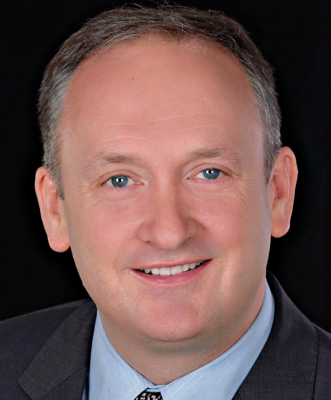 Mark Clemens合作论文数Division of Medical Oncology, The Ottawa Hospital Cancer Centre;Department of Medicine, University of Ottawa;Cancer Therapeutics Program, Ottawa Hospital Research Institute4Vandermeer Lisa合作论文数4Arnaout Angel合作论文数4Saeed Samiee合作论文数4
 Jean-Michel Caudrelier合作论文数Department of Radiology, University of Ottawa4J. N. Terry合作论文数3
 T. Lemoult合作论文数3
 Olivier Thizy合作论文数3M. Chasen合作论文数3E. Pollmann合作论文数3E. Barbotin合作论文数3K. B. Alton合作论文数3
 Kim Page合作论文数UK Swift Science Data Centre, School of Physics & Astronomy, University of Leicester3
 Irmgard Irminger-Finger合作论文数3
 Anis Feki合作论文数Department of Gynecology and Obstetrics, Geneva, Switzerland3D Neurath合作论文数3
 David Allan合作论文数Ottawa Hospital Research Institute, University of Ottawa;Department of Biochemistry, Microbiology and Immunology, University of Ottawa3D. Lane合作论文数2P. Cazzato合作论文数2B. Mauclaire合作论文数2Paul Luckas合作论文数2Allam Shawwa合作论文数2Brian Olsen合作论文数2Connie M. Westhoff合作论文数2Antonio Maietta合作论文数2




