Kashin-Beck disease (KBD) is an endemic osteoarthropathy characterized by chondrocyte death and extracellular matrix (ECM) degradation; however, the underlying molecular mechanisms remain poorly understood. This study aimed to explore whether glutathione peroxidase 6 (GPx6) is involved in regulating ferroptosis and matrix degradation in KBD cartilage injury. In articular cartilage from children with KBD, GPx6 expression was markedly reduced, particularly in the deep zone where chondrocyte death and matrix loss were most severe. Similarly, downregulation of GPx6 was observed in a T-2 toxin‑induced rat model and in C28/I2 chondrocytes, accompanied by decreased COL2A1 and ACAN levels and increased expression of MMP13 and ADAMTS4. Molecular docking indicates a potential interaction between T-2 toxin and GPx6. Gpx6 knockout mice exhibited signs of ECM degradation in articular cartilage, along with an enhanced susceptibility to T-2 toxin exposure. Mechanistically, Gpx6 deficiency led to elevated MDA levels, reduced SOD activity and GSH/GSSG ratio, as well as mitochondrial shrinkage, cristae loss, and lipid peroxidation. Knockdown of GPX6 suppressed the expression of SLC7A11, SLC3A2, and GPx4, while Ferrostatin‑1 partially reversed these ferroptosis‑related alterations but failed to restore GPx6 expression. Conversely, overexpression of GPX6 partially restored the function of the SLC7A11/GPx4 axis, improved redox balance, attenuated mitochondrial lipid peroxidation, and mitigated matrix degradation. Collectively, these findings suggest that downregulation of GPx6 may be closely associated with ferroptosis in KBD chondrocytes, and that GPx6 may serve as a potential upstream regulator of this process. Furthermore, dysregulation of the GPx6‑SLC7A11/GPx4 pathway may also be involved in the pathological process of cartilage injury in KBD.
Selenoproteins serve as the primary carriers for the biological functions of selenium (Se) within the body, which can harm health if deficient. Se deficiency is related to cartilage damage and osteoarticular diseases, wherein compromised chondrocyte function leads to disturbed cartilage homeostasis. Preliminary in vitro research showed that low selenium levels reduced chondrocyte selenoprotein S (SelS) expression, but the precise function of SelS in the terminal differentiation of chondrocytes, along with the signaling pathways by which SelS deficiency influences this process, has remained to be thoroughly clarified. This study, focusing on SelS deficiency, was the inaugural investigation into the mechanism by which SelS deficiency activates the Wnt/β-catenin pathway, resulting in impaired terminal differentiation of chondrocytes, and aims to provide an early intervention target for the prevention and treatment of bone and joint diseases associated to low selenium levels. SelS expression was initially evaluated in cartilage samples from rats fed a low-selenium diet and from patients with Kashin-Beck disease (KBD), and reduced SelS expression was found to be associated with cartilage degradation under selenium-deficient conditions. Using in vivo SelS knockout mice and in vitro chondrocyte models, it was found SelS deficiency reduced cartilage thickness and increased terminal differentiation markers OCN and Col1α1. RNA sequencing implicated the Wnt/β-catenin pathway, confirmed by functional rescue and inhibition experiments. SelS deficiency activates Wnt/β-catenin, impairing terminal differentiation and contributing to selenium-deficiency cartilage injury. This study offers critical insights into the pathogenesis of selenoprotein-related diseases and presents new experimental evidence regarding the aetiology of cartilage terminal differentiation-associated osteoarthritis, while also establishing a significant strategy for targeted interventions against selenium related diseases.
Bone scaffolds designed based on the Voronoi-tessellation algorithm have been increasingly studied owing to their structural similarity with natural cancellous bone. The irregularity of pore morphology (IPM) influences the osteogenesis efficiency of Voronoi scaffolds since it may alter the static and hydromechanical microenvironments for the initial adhesion and mechano-regulated osteoblast differentiation (MrOD) of mesenchymal stem cells (MSCs). In this work, animal experiments were conducted to explore the relationship between IPM and osteogenesis efficiency in Voronoi scaffolds. A computational fluid dynamics (CFD) analysis based on discrete phase models was performed to predict the efficiency of MSC adhesion in different IPMs. Another combined finite element and CFD analysis based on the mechano-regulation algorithm was performed to predict the influence of IPM on the MrOD of the adhesive MSCs. The results showed that the osteogenesis efficiency of the Voronoi scaffolds increased as the IPM rose from low to moderate and then dropped as the IPM further rose. Same trends were also found in the MSC adhesion and MrOD, which caused by the changes of strain tensors on the strut surface and the tortuosity and fluid velocity of the fluid pathway. Moderate IPM induced the highest osteogenesis efficiency owing to its highest efficiencies of MSC adhesion and MrOD. This work identified the optimal IPM for the osteogenesis of Voronoi scaffolds and clarified its biomechanical mechanisms from the adhesion and mechano-regulated differentiation of MSCs, which is of great importance for guiding Voronoi scaffold design when it is used for bone defect repair.
老龄化、大数据、精准医疗等在近些年成为热门,人们经常在新闻或者专业的文献期刊上发现他们的身影.这些看似"时髦"的名词,却是在社会进步与发展的同时,给人们带来的挑战.老龄化一定程度代表了社会经济的发展,使拥有更高质量的生活和更长的生存时间.
Background and objective: Conventional method for evaluating the biomechanical effects of a specific elastic modulus of cage (cage-E) on spinal fusions requires establishing a "one-on-one" biomechanical model, which seems laborious and inefficient when dealing with the emergence of numerous cage materials with various cage-Es. We aim to offer a much convenient method to instantly predicting the biomechanical effects of any targeted cage-E on transforaminal lumbar interbody fusion (TLIF) by using a parametric finite element (FE) analysis to determining the regression relationship between cage-E and biomechanical properties of TLIF. Materials and methods: A L4/5 FE TLIF construct was modeled. Cage-E was linearly increased from 0.1 GPa (cancellous bone) to 110 GPa (titanium alloy). The function equations for assessing the influence of cage-E on the biomechanical indexes of TLIF were established using a logarithmic regression analysis. Experimental results: As cage-E increased from 0.1 GPa to 110 GPa, all the biomechanical indexes initially increased or decayed rapidly, and then slowed over time. Logarithmic regression models and functional equations were successfully established between cage-E and these indexes (P<0.0001). Their determination coefficients ranged from 0.72 to 0.99. The range of motions decreased from 0.37-1.10 degrees to 0.201.07 degrees. The mean stresses of the central and peripheral grafts reduced from 0.10-0.41 and 0.25-0.42 MPa to 0.03-0.04 and 0.19-0.27 MPa, respectively. In addition, the maximum stresses of the screw-bone interface and posterior instrumentation reduced from 11.76-25.04 and 8.91-84.68 MPa to 9.71-18.92 and 6.99-70.59 MPa, respectively. Finally, the maximum stresses of the cage and endplate increased from 0.28-1.35 MPa and 3.90-8.63 MPa to 14.86-36.16 MPa and 11.01-36.55 MPa, respectively. Conclusions: The decrease of cage-E reduces the risks of cage subsidence, cage breakage, and pseudarthrosis, while increasing the risk of instrumentation failure. The logarithmic regression models optimally demonstrate the relationship between cage-E and biomechanical properties of TLIF. The functional equations based on these models can be adopted to predict the biomechanical effects of any targeted cage-Es on TLIF, which effectively simplifies the procedures for the biomechanical assessments of cage materials. (C) 2021 Elsevier B.V. All rights reserved.
A potential load-bearing bone substitution and repair material, that is, carbon fiber (CF)-reinforced magnesium-doped hydroxyapatite (CF/Mg-HAs) composites with excellent mechanical performance and tailored biological properties, was constructed via the hydrothermal method and spark plasma sintering. A high-resolution transmission electron microscopy (TEM) was employed to characterize the nanostructure of magnesium-doped hydroxyapatite (Mg-HA). TEM images showed that the doping of Mg-induced distortions and dislocations in the hydroxyapatite lattice, resulting in decreased crystallinity and enhanced dissolution. Compressive strengths of 10% magnesium-doped hydroxyapatite (1Mg-HAs) and CF-reinforced 1Mg-HAs (CF/1Mg-HAs) were within the range of that of cortical bone. Compared with 1Mg-HAs, the fracture toughness of CF/1Mg-HAs increased by approximately 38%. The bioactivity, biocompatibility, and osteogenic induction properties of Mg-HAs and CF/Mg-HAs composites were evaluated in vitro using simulated body fluid (SBF) immersion, cell culture, osteogenic differentiation of bone marrow mesenchymal stem cells (BMSCs), and expression of genes associated with osteogenesis. When Mg-HAs were immersed in SBF, Mg2+ continued to release for up to 21 days. Mg-HAs demonstrated a satisfactory ability to induce apatite formation in comparison with HAs. The cell proliferation and morphology on CF/1Mg-HAs were similar to those of 1Mg-HAs, suggesting that adding CF had no adverse effect on cellular activity. The expression levels of osteogenesis-related genes [osteocalcin (OPN), osteopontin (OCN), and runt-related transcription factor 2 (Runx2)] on 1Mg-HAs were significantly higher at days 3 and 7 than those on HAs and 0.5Mg-HAs groups. This finding suggests that a certain amount of Mg doping had beneficial influences in the different stages of osteogenic differentiation and could induce osteogenic differentiation of BMSCs. The new bone volume to total volume ratio of implanted 1Mg-HAs (30.9% ± 4.1%) and CF/1Mg-HAs (25.4% ± 5.4%) was remarkably higher than that of HAs (21.6% ± 3.9%). 1Mg-HAs and CF/1Mg-HAs tailored an ideal effect of new bone information and implant osseointegration. The excellent mechanical performance and tailored biological properties of CF/Mg-HAs were attributed to nano Mg-doped HA, CF reinforcing, refined microstructure, and controlled composition.
Pore irregularity, which significantly affects pore morphology, plays a critical role in the osteogenesis of Voronoi scaffolds. However, the biomechanical mechanisms of this influence and the optimal values of pore irregularity remain unknown. In this study, finite element and computational fluid dynamics analyses were conducted to predict the permeabilities, mesenchymal stem cells (MSCs) adhesion, and distribution of mechanical stimulation on the strut surfaces of titanium Voronoi scaffolds with five different pore irregularities (low to high). The results revealed that all the titanium Voronoi scaffolds satisfied the permeability requirements of natural cancellous bones. Low pore irregularity had the highest ratio of the strut surface that yielded the mechanical stimulation required for the osteogenic differentiation of MSCs. In contrast, moderate pore irregularity induced the maximum efficiency of MSCs adhesion. By combining the effects of MSCs adhesion and mechanically-induced osteogenic differentiation, moderate pore irregularity was considered to optimal for inducing maximum osteogenesis in the titanium Voronoi scaffolds since it yielded the highest ratio of MSCs that simultaneously adhered to the strut surface and received the mechanical stimulation required for osteogenic differentiation. In conclusion, moderate pore irregularity is recommended for the future design of titanium Voronoi scaffolds to effectively improve the osteogenesis efficiency.
Mg-doped hydroxyapatite (MH) with oriented microchannels was prepared by hot-pressing sintering and pore forming heat treatment using continuous carbon fibres (CFs) as the pore-forming agent to achieve balanced mechanical and biological properties. The proportion of MH and (Ca, Mg)(3)(PO4)(2) in the microporous bioceramic with inter-microchannel spacing of 400 mu m obtained by sintering at 900 degrees C (900-2P-MH) was 43.9/56.1 (wt%), which could promote degradation. The compressive strength of the MH bioceramics containing oriented microchannels with a suitable pore size (5-14 mu m) did not decrease, but increased in comparison with that of a dense MH sample without microchannels. In particular, the compression strength of the MH bioceramic with oriented microchannels formed using single CF units was 53.31% higher than that of the dense ones. The fracture toughness of the MH bioceramics with oriented microchannels increased to 177.42% of that of the dense ones. The strengthening and toughening mechanism includes contributions from the combination of heating and pressing, uniform distribution of microchannels, and in situ formation of continuous micro/nano-MH ceramic tubes. Moreover, the microchannel-containing MH showed noticeably improved apatite mineralisation in simulated body fluid (SBF). Analysis of the rat tibial bone defect model revealed that the relative bone volumes in the cases of the dense MH without microchannels and the MH with microchannels increased by 23.18 and 40.14%, respectively, compared with that of hydroxyapatite. Furthermore, the MH bioceramics with oriented microchannels displayed a moderate reduction in strength owing to degradation after 8 weeks of implantation. The satisfactory osteogenic properties and degradability of the microporous bioceramic can be attributed to Mg2+ doping and oriented microchannels.
Objective To explore the clinical efficacy of one-stage anterolateral-posterior approach debridement,bone graft and internal fixation in treatment of thoracolumbar spinal tuberculosis.Methods From January 2010 to December 2014,56 cases of thoracolumbar spinal tuberculosis were retrospectively analyzed, including 31 males and 25 females,aged 18 -72 years (mean 43.1 years).All patients were managed by standard courses of chemotherapy with quadruple anti-TB drugs for 2 - 4 weeks.Patients were treated by anterolateral debridement,autologous iliac bone graft fixed by absorbable screw fixation,and posterior pedicle screw fixation via multi-split muscle gap (Wiltse approach).We recorded the operation time,the amount of bleeding,bone graft fusion,postoperative erythrocyte sedimentation rate (ESR),Cobb angle,VAS score,and American Spinal Injury Association (ASIA)score to evaluate the surgical results.Results The average operation time was 175-290 min, with an average of (248±42)min.The bleeding volume was 300 -900 mL with an average of (420 ±68)mL.The average follow-up time was (24 ± 5.2 )months,bone fusion rate was 100%,and fusion time was (4.7 ± 0.5 ) months.At the last follow-up,the average Cobb angle was (8.2±3.1)°,VAS was (2.1±0.8),and ESR was (17± 4.2)mm/h.The ASIA neurological functions were all classified as Grade E except for 3 cases of Grade D.All these were significantly different from the preoperative ones.Six patients had complications of different degree but without serious complications.Conclusion One-stage anterolateral debridement,autologous iliac bone graft fixedby absorbable screw fixation,and posterior pedicle screw fixation via multi-split muscle gap (Wiltse approach)can completely remove the tuberculosis lesions and achieve ideal kyphosis correction,high bone graft fusion,and satisfactory neurological function recovery.Absorbable screws can be safely applied to the bone graft site after debridement.
Study Design: This study tested the biomechanics of artificial atlantoodontoid joint replacement (AAOJR) in a dog model. Dogs were divided into the artificial AAOJR group (n=10), the decompression group (n=10), and the healthy control group (n=10) using a random number table. Objective: To evaluate whether the use of AAOJR for repair of atlantoaxial instability retains rotation and restores stability. Summary of Background data: Atlantoaxial instability is characterized by excessive movement or laxity at the junction between the atlas (C1) and axis (C2). Pure decompression can lead to considerable loss of head and neck rotation and postoperative impairment. A series of biomechanical tests on cadavers found that the artificial AAOJR might rebuild the stability and retain the rotation function. Methods: We designed the AAOJ based on the radiologic and anatomic data of the dog atlas and axis, and established an animal model by resecting the odontoid and implanting the AAOJ into dogs. The biomechanical experiments measured the range of motion (ROM), neutral zone (NZ), and stiffness of flexion, extension, lateral bending, and axial rotation in the intact state, the decompressed state, after AAOJR, and after a fatigue test. Results: Compared with the intact state, after decompression operation, ROM and NZ in all directions, and stiffness during flexion were increased, and stiffness in all other directions was decreased. Compared with the after decompression state, AAOJR before and after the fatigue test resulted in decreased ROM in all directions (all P<0.05), decreased NZ during flexion/extension and lateral bending (all P<0.05), an increased NZ during axial rotation (both P<0.05), and increased stiffness in all directions (all P<0.05). Conclusions: These results indicate that AAOJR could reconstruct the vertebral stability of the C1–C2 segment and retain some axial rotation function.
In order to provide effective options for minimally invasive treatment of spinal metastases, the present study retrospectively evaluated the efficacy and safety of image-guided minimally invasive percutaneous treatment of spinal metastases. Image-guided percutaneous vertebral body enhancement, radiofrequency ablation (RFA) and tumor debulking combined with other methods to strengthen the vertebrae were applied dependent on the indications. Percutaneous vertebroplasty (PVP) was used when vertebral body destruction was simple. In addition, RFA was used in cases where pure spinal epidural soft tissue mass or accessories (spinous process, vertebral plate and vertebral pedicle) were destroyed, but vertebral integrity and stability existed. Tumor debulking (also known as limited RFA) combined with vertebral augmentation were used in cases presenting destruction of the epidural soft tissue mass and accessories, and pathological vertebral fractures. A comprehensive assessment was performed through a standardized questionnaire and indicators including biomechanical stability of the spine, quality of life, neurological status and tumor progression status were assessed during the 6 weeks-6 months follow-up following surgery. After the most suitable treatment was used, the biomechanical stability of the spine was increased, the pain caused by spinal metastases within 6 weeks was significantly reduced, while the daily activities and quality of life were improved. The mean progression-free survival of tumors was 330±54 days, and no associated complications occurred. Therefore, the use of a combination of image-guided PVP, RFA and other methods is safe and effective for the treatment of spinal metastases.
Objectives:To analyze the causes of postoperative revision surgery of atlantoaxial dislocation,and to discuss the therapeutic strategies.Methods:15 patients with atlantoaxial dislocation revision surgeries(11 male,4 female;15-68 years,mean 46.60±14.95 years) were analyzed in this retrospective study.The intervals of two operations ranged from 2 to 120 months(28.73±38.59 months).The reasons were analyzed for revision surgery according to the image data and intraoperative findings.All patients received 8-10kg high dose skull traction and posterior release during revision surgery.For patients with integral posterior structure,the option of anterior release depended on the degree of reduction.After decompression and reduction,all patients received posterior internal fixation.One patient who received posterior decompression in the primary operation was performed with anterior decompression and fixation.Assisted atlantoaxial screw placement under 3D navigation template was performed;autogenous iliac cancellous bone was placed.The atlantoaxial reduction,screw position,bone graft fusion and surgical efficacy were evaluated at follow-up.Results:Reasons for revision were as following:10 cases of insufficient decompression/reduction,3 cases of failed internal fixation,3 cases of unfused bone graft(including 1 case of internal fixation failure caused by unfused bone graft).Among all the 15 revision surgeries,14 cases received posterior fixation surgery,1 case received anterior fixation surgery.By intraoperative skull traction and full release,13 cases achieved anatomical reduction,the other 2 cases did not reach anatomical reduction due to extensive bony fusion and were fully decompressed after odontoidectomy.42 atlantoaxial screws were implanted with 3D template-assisted navigation,the accuracy of screw implantation was 97.6%.The follow-up time ranged from 3 to 36 months(16.0±4.2 months).All patients got bone fusion,the fusion time ranged from 3-6 months(3.7±0.5 months).The final JOA score ranged from 11 to 16(mean,13.8±3.1),which improved compared with the preoperative(range form 6 to 11,mean 8.1± 2.3),the improvement rate was (64.0±21.2)%(45.4%-88.8%).Conclusions:Inadequate decompression/reduction,failed internal fixation,and unfused bone graft are the common reasons of C1-2 dislocation needing revision surgery.Intraoperative full release,8-10kg skull traction,and proper bony structure resection are leading to the reduction of atlantoaxial dislocation,3D navigation template is beneficial to the accuracy of screw placement.
OBJECTIVE:To evaluate curative effects of intramedullary nail and volar plate internal fixation for distal radius fractures.METHODS:The studies concerning about randomized controlled trial of intramedullary nail and volar plate internal fixation for distal radius fractures from create database to May, 2016 were searched from PubMed, Cochrane Library, Web of Science, CNKI, Wan Fang data and VIP. Information was screen and taken out according to inclusion and exclusion criteria, quality of literature was evaluated according to Cochrane Handbook evaluating system. Meta analysis was performed by RevMan 5.2 software. Postoperative grip strength, clinical effects(results of flexion and extension, pronation, supination, deflection of ulnar and radialis) , imaging evaluation (ulnar variance, radial height, volar tilting angle and radial inclination) and the incidence of postoperative complications were analyzed.RESULTS:Totally 6 RCT including 370 patients were chosen. According to the Meta analysis, there were no statistical differences in extension[MD=5.63, 95%CI(-7.01, 18.27), P=0.38], flexion[MD=3.10, 95%CI(-0.67, 6.86), P=0.11], pronation[MD=2.58, 95%CI(-0.49, 5.65), P=0.10], supination[MD=0.82, 95%CI(-1.89, 3.54), P=0.55], radial deviation[MD=-5.16, 95%CI(-14.42, 4.11), P=0.28], ulnar deviation[MD=0.19, 95%CI(-2.39, 2.77), P=0.88], ulnar variance[MD=-0.01, 95%CI(-0.43, 0.42), P=0.97], ulnar inclination[MD=-0.31, 95%CI(-1.37, 0.73), P=0.56], radial height[MD=-0.27, 95%CI(-0.98, 0.44), P=0.45], radial inclination[MD=0.29, 95%CI(-0.41, 0.99), P=0.42] and the incidence of postoperative complications[RR=0.71, 95%CI(0.46, 1.09), P=0.12] between intramedullary nail and volar plate internal fixation.CONCLUSIONS:Compared with volar plate internal fixation, intramedullary nail has the same effect in grip strength, clinical effect, but could not reduce the occurrence of postoperative complications. However, a large number of prospective randomized controlled study should be carried out to prove clinical effect of intramedullary nail for distal radius fractures.
Objective To compare advantages and disadvantages,clinical efficacy of one-stage anterior debridement,bone fusion combined with dual screw-rod construct fixation versus one-stage posterior debridement and bone fusion with pedicle screws in treatment of lumbosacral spinal tuberculosis.Methods Clinical data of 56 patients suffering from lumbosacral tuberculosis from January 2004 to June 2015 were reviewed retrospectively.Among 56 patients,29 patients were treated with one-stage anterior debridement,bone fusion combined with dual screw-rod construct fixation(group A),and 27 patients were treated with one-stage posterior debridement,bone fusion with pedicle screws (group B).The operation time,bleeding volume,hospitalization time,preoperative,postoperative and last followup VAS scores,erythrocyte sedimentation rate (ESR),lumbosacral lordosis angle,ASIA grade,bone graft fusion time were recorded and compared between the two groups.The complications were recorded.Results There was no significant difference between the two groups in operation time,bleeding volume,hospitalization time and bone graft fusion time (P > 0.05).The ESR,VAS scores were decreased and the lordosis angle was increased in the two groups at one week and the last follow-up after operation(P <0.05).The ASIA grades at the last follow-up were significantly improved in the two groups after operation(P < 0.01),and all patients achieved grade E.There was no significant difference in ESR,VAS scores and the lordosis angle between the two groups.There were no serious complications in both groups.Conclusion The one-stage anterior debridement,bone fusion combined with dual screw-rod construct fixation and one-stage posterior debridement,bone fusion with pedicle screws can achieve satisfactory clinical outcome,but the two surgical programs have their own advantages and disadvantages,so the surgery decision should be based on the disease and the experience of the surgeons for specific cases.
Objective To evaluate the clinical effect of transarticular approach in treating thoracic spinal stenosis caused by multi-segmental ossification of ligamentumflavum (MSOLF).Methods From January 2008 to December 2014,in Second Affiliated Hospital of Xi'an Jiaotong University,the clinical data of 26 MSOLF patients with thoracic spinal stenosis were retrospective analyzed.Postoperative neurological status was evaluated by the modified JOA score system.The Frankel grade and Cobb angle of kyphosis were measured at postoperative 1 week and final followed up respectively,the complications during and after operation were recorded.Results All operations were performed under theuse of spinal cord evoked potential (SEP) monitoring.The operation time was 2.7-5.2 h,mean was (4.1±1.2) h and bleeding volume was 650-2900 mL,mean was (1940±824) mL.2 patients had abnormalitiesin intraoperative monitoring in SEP,4 cases suffered cerebrospinal fluid leakage,1 case suffered pneumothorax.The follow-up time was 24-96 months,mean was (40.0±4.8) months.At final follow-up:6 cases were grade C,8 cases were grade D,12 cases were grade E.JOA at final follow-up improved than before operation,the difference was statistically significant (P < 0.05),improvement rate was 70.3%;the bone graft fusion rate was 100%,there was no internal fixation failure;the Cobb angle at postoperative 1 week compared with final follow-up,the difference was not statistically significant (P > 0.05).Conclusion Treating MSOLF thoracic spinal stenosis through transarticular approach with pedicle screws can decompress the spinal cord completely,the complicationsare few,the clinical outcomes are favorite.Intraoperative SEP monitoringis of high sensitivity,it can improve the safety of intraoperative manipulatation.The method of persistent drainage for 1 week followed by the close the sinus of drainage tubeis can effectively treat cerebrospinal fluid leakage.
Objective To evaluate the clinical efficacy and safety of a new 3D anatomical titanium cage in single-segment anterior cervical corpectomy fixation (ACCF) and bone grafting.Methods From June 2014 to December 2015,fifty-six cases of cervical spondylotic myelopathy were analyzed retrospectively.All patients underwent C5 ACCF and titanium cage implantation.Among these cases,27 cases were included in 3D anatomical titanium cage group (observation group) and 29 cases in traditional titanium cage group(control group).The time of operation and bleeding volume were compared between the two groups.The changes of JOA score and the improvement rate were compared before and after operation.The cage subsidence degree,bone fusion and Cobb angle changes were also studied in the X-ray at 3 d after operation and final follow-up.Results Fifty-six patients were followed up,the average follow-up time were (15.34±3.61)months and (15.42± 2.98)months in the observation group and control group respectively.All patients achieved bone fusion,and the fusion rate was 100%.Cage subsidence and mean subsidence in observaion/control group were 21/25 patients,0.3-0.7 (0.52±0.21)mm/2.3-4.2 (2.93±0.42)mm respectively,there was no severe subsidence in observation group while severe subsidence was found in 3 patients in control group.The distance of cage subsidence in observation group was smaller than that in the control group (P <0.05).The improvement rate of JOA score at the final follow-up was 69.5% in the observation group and 67.0% in the control group,there was no significant difference between the two groups (P >0.05).The Cobb angle of the fusion segment in the observation group was smaller than that in the control group,the difference was statistically significant (P <0.05).Conclusion Compared with the use of traditional titanium cage in treatment of cervical spondylotic myelopathy with single segment ACCF,the 3D anatomical titanium cage can achieve similar clinical efficacy and bone fusion rate,can reduce the incidence of cage subsidence effectively,it's clinical use is safe.
Objective To design a bionic artificial atlanto-odontoid joint,which can keep the motion of the atlantoaxial axis in all directions,and explore the feasibility of its clinical use.Methods 30 cases of normal atlantoaxial CT data were collected and reconstructed with Mimcs15.0 software.The anatomical parameters were measured and statistically analyzed.Design the joint to retain the activities of all directions according to the anatomical data.Joint arthroplasty were performed on 10 fresh cadaver specimens;X-ray and CT were performed to evaluate joint and screws' placement.Results The distance between the midpoint of the atlas lateral mass was (32.53 ± 1.38) mm,the height of the lateral mass was (13.48±1.60) mm,the distance between the medial margin of the vertebral artery hole was (51.12±2.31) mm,the width of the lateral mass was (16.23± 1.01) mm,the distance between the anterior tuberosity and the posterior margin of the odontoid process was (18.48 ± 1.77) mm.The axial height ofodontoid process was (14.82±1.81) mm,the basal diameter was (9.53±0.65) mm,the anteroposterior diameter was (10.91±0.91) mm,The vertebral height was (19.33± 1.77) mm and the vertebral width was (18.19± 1.59) mm.The artificial joint is composed of atlas components,axial components and screws.After operation,the plate and bone are in good condition.The spinal cord is not compressed and the directions of screws were well.The function of flexion,extension and rotation of the atlantoaxial joint can be preserved after joint replacement.Conclusion The anatomical design of the bionic artificial atlanto-odontoid joint is fine;After the joint replacement,the atlantoaxial joint can not only maintain the stability,but also can keep the activities of all directions;This joint has the possibility of clinical replacement.
BACKGROUND:Traumatic neuromas are rare benign tumors, which are common in trauma or post-operation and accompanied with obvious symptoms of pain. This study will show the superficial peroneal nerve neuroma occurring after resection of hemangioma.CASE PRESENTATION:A 44-year-old male had an operation of the right leg cavernous hemangioma resection in 1995. Half a year after the operation, pain around the wound appeared and gradually aggravated. The patient had the lesion exploration resection in 2013, and the pathological result showed traumatic neuroma. Within half a year of the second operation, severe pain showed up again, so neuroma resection proceeded in May 2015. The postoperative pathological and immunohistochemical results showed traumatic neuroma. According to the postoperative follow-up, there were no symptoms of pain appearing again.LITERATURE REVIEW:The pain is obvious, and B ultrasonography is the most efficient way to find neuromas. Both conservative and operative therapy have their advantages and disadvantages.CONCLUSIONS:There remain many unanswered questions in relation to the treatment of traumatic neuromas, and further research is required, although we have already had adequate understanding of traumatic neuromas.
We aimed to provide evidence for clinical choice of surgical approach in treating spinal tuberculosis, including anterior, posterior and combined approaches (combined anterior and posterior approach).
The aim of the present study was to determine whether hyaluronic acid (HA) or physical therapy agents (PTA) can improve functional parameters in patients with knee Kashin-Beck disease (KBD). For 2 years, patients (n=55) were treated with HA weekly for 5 weeks, then received 6th and 7th injections on the 3rd and 6th month, respectively, for 7 injections in total. Patients (n=53) were treated with PTA five times a week for 3 weeks every month for 6 months. The patients were evaluated with the Western Ontario and McMaster University Osteoarthritis Index (WOMAC) and the visual analog pain scale (VAS). Trial registration, ChiCTR-TRC-12002189 (http://www.chictr.org/). During the study, following treatment interruption, pain increased in the PTA group (from a mean value of 85.7±83.8 mm at month 12 to 145.2±128.8 mm at month 18 and 201.3±150.5 mm at month 24), while it remained stable in the HA group (from a mean value of 80.7±70.6 mm at month 12 to 90.1±95.2 mm at month 18 and 82.6±85.3 mm at month 24), with a statistically significant difference in favor of HA at month 18 (P<0.05) and month 24 (P<0.05). Joint stiffness, physical function and total WOMAC showed the same trend as pain. The global efficacy judgments by the patients and the investigators showed a statistically significant difference in favor of HA at month 18 (P<0.05) and month 24 (P<0.05). In conclusion, although all the patients improved in terms of pain and function, HA was superior to PTA alone for pain relief and lasting effect.
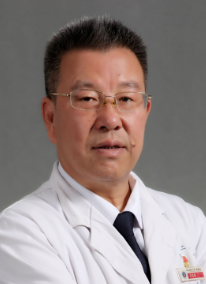
 Xijing He (贺西京)合作论文数Health Science Center, Xi'an Jiaotong University44
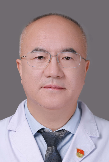
 Haopeng Li (李浩鹏)合作论文数The Second Affiliated Hospital of Xi'an Jiaotong University33
 Quanjin Zang (臧全金)合作论文数21GuoYu Wang (王国毓)合作论文数15Cheng-Chung Lin合作论文数9Ming-Zhang Kuo合作论文数9
 Sang Dhong合作论文数8Baohui Yang (杨保辉)合作论文数8
 Puwei Yuan (袁普卫)合作论文数6BinShang Lan合作论文数6Min-Jer Wang合作论文数5SiYue Xu (徐思越)合作论文数5
 Yiheng Liu (刘亦恒)合作论文数中南大学附属海口医院骨科中心5
 lu Teng合作论文数5osamu takahashi合作论文数4Wen-long YANG (杨文龙)合作论文数4DeYu Liu (刘德玉)合作论文数4Hui Liang (梁辉)合作论文数4
 Jianqiang Qu (屈建强)合作论文数The Second Affiliated Hospital of Xi'an Jiaotong University4Jiakun Ren合作论文数3
 Zhongwei Sun合作论文数School of Civil Engineering, Southeast University3
 Wei Hwang (黃威)合作论文数Department of Electronics Engineering, National Yang Ming Chiao Tung University3
 Jialiang Li合作论文数2Cunwei Jia合作论文数2Sensen Wei合作论文数2Zhiyuan Ma合作论文数2Dong Jun合作论文数2Jian Kang合作论文数2Ao Liu合作论文数2Qiang Li (历强)合作论文数2XueNi Zhao (赵雪妮)合作论文数2Zhi Yang (杨治)合作论文数2Henry Hsieh合作论文数2
 Jie Li (李杰)合作论文数School of Information and Communications Engineering, Xi'an Jiaotong University2
 Jie Qin (秦杰)合作论文数School of Basic Medical Sciences, Fudan University2Jing Zhang合作论文数2
 Wei Shi (师蔚)合作论文数The Second Affiliated Hospital of Xi'an Jiaotong University2
 Dong Wang (王栋)合作论文数Center of Microstructure Science, Frontier Institute of Science and Technology, Xi'an Jiaotong University2Fei Wu合作论文数1Jing-Jing Zhai合作论文数1Zhai Xu合作论文数1jiarong hsu合作论文数1yuhuai Liu合作论文数1
 Pei Fan合作论文数1Zhao Shan合作论文数1Zhijian Cheng合作论文数1Yanming Liu合作论文数1




