Management options for carotid stenosis include medical management, carotid endarterectomy, carotid stenting with distal filter protection, or stenting with either flow arrest or flow reversal techniques.1 Flow reversal with transcarotid artery revascularization (TCAR) technique represents a hybrid approach with surgical access to the common carotid for endovascular placement of a stent in the internal carotid artery. This direct access to the carotid artery avoids navigating the challenging anatomy of the aortic arch with endovascular devices.2 Compared to transfemoral stenting, TCAR possesses lower risk of transient ischemic attack and stroke, and compared to carotid endarterectomy, there is less risk of cranial neuropathy.3,4 We present the case of an 87-yr-old man with recurrent severe stenosis (85%) of the right internal carotid artery. The patient had a remote history of bilateral endarterectomies for asymptomatic stenosis and was found with recurrence and progression of right internal carotid artery stenosis. Options were discussed and decision was made to proceed with TCAR after he consented for the procedure. The patient tolerated the procedure well with satisfactory revascularization. Exam remained unremarkable prior to discharge on postoperative day 1 and during follow-up at 1 mo. Patient consented to the publication of their image.
Mechanical thrombectomy as a treatment for large vessel occlusion to achieve rapid revascularization is supported in the literature. 1-3 The presenting symptoms will localize to functions of the ischemic area. The middle cerebral artery (MCA) supplies areas of the frontal, temporal, and parietal cortices, as well as the basal ganglia. Occlusion of the MCA will present with contralateral hemiplegia, sensory loss, and, if the dominant hemisphere is involved, language deficits. We present a right-hand-dominant 79-yr-old female with right MCA syndrome—her last known well time was 1.5 h prior to presentation. Her NIH (National Institutes of Health) Stroke Scale was 16, most notable for left hemiplegia. Although the patient presented early in the clinical time course, as part of our institution protocol, a computed tomography (CT) head, CT perfusion, and CT angiogram (CTA) were performed. CT head did not demonstrate acute hemorrhage, so she received intravenous tissue plasminogen activator. CTA demonstrated a right MCA occlusion and CT perfusion suggested a large area of salvageable tissue, so she was taken to the angiography suite for mechanical thrombectomy. Angiography of the right internal carotid artery (ICA) showed MCA occlusion (insular segment). A thrombectomy device was deployed over the area of occlusion and allowed to engage for 5 min. An aspiration catheter was advanced over the stentriever up against the clot. The stentriever device was withdrawn under continuous aspiration and follow-up angiography showed complete reperfusion. The patient demonstrated improvement and was eventually discharged to an inpatient rehabilitation center. Patient provided consent for photography per university protocol. Institutional review board (IRB) approval was not needed for the single-patient data included in this report.
As capabilities for endovascular treatment of intracranial vascular pathologies continue to expand, the need for access to the distal internal carotid artery with rigid support catheter systems continues to increase. One of the dominant factors limiting this access is patient anatomy, specifically vessel tortuosity. Increased tortuosity of the carotid siphon is a frequently encountered anatomic variant and may complicate endovascular procedures in adults and children. 1,2 Failed attempts to navigate the carotid siphon with a distal access catheter carry a risk of vessel injury and treatment failure. For this reason, techniques that aid in supporting safe advancement of a distal access catheter across a tortuous carotid siphon are essential. 3,4 In this video, we demonstrate 2 ways in which this may be accomplished. The first technique uses a larger diameter microcatheter, such as the AXS Offset catheter (Stryker, Kalamazoo, Michigan), to increase support for the distal access catheter, while the second uses a buddy wire technique to accomplish this increased support. Both of these techniques can help increase the safety of navigating a tortuous carotid siphon and increase the likelihood of successful treatment. The procedures shown were performed with the informed consent of the patients.
Transfemoral access has long been the main access site for cerebral angiography and neurointerventional procedures. Radial access is accepted as an alternative to the traditional transfemoral approach. Ulnar access may be undertaken if the radial artery is occluded or small caliber, or when radial artery preservation is needed. The safety and feasibility of ulnar access for neuroangiographic procedures has been demonstrated. 1-3 In this operative video, we demonstrate ulnar artery access in a patient in whom radial artery preservation was desired. We further elaborate on the technical nuances of this access. This nontraditional access site offers the same advantages as radial access, avoiding the need to switch to femoral artery access. A preoperative Allen's test is not necessary. Ultrasound imaging is used to aid in the identification and successful puncture of the ulnar artery. A medial to lateral approach for ulnar artery puncture is advised to avoid injury to the ulnar nerve. Careful application of wrist closure bands avoids hematoma accumulation. The patient gave informed consent for the procedure and video recording. Institutional review board approval was deemed unnecessary. Video. © University at Buffalo, May 2021. Used with permission.
BackgroundCavernous carotid artery aneurysms (CCAs) represent a unique subset of intracranial aneurysms due to their distinct natural history and the anatomy of the cavernous sinus. Enlarging CCAs can cause elastic compression of the parent internal carotid artery (ICA). We suggest defining aneurysms that cause luminal stenosis of their parent vessels as ‘matricidal aneurysms.’Though many patients are asymptomatic, presenting symptoms of CCAs include ophthalmoplegia with resulting diplopia, vision changes, pain, ptosis, facial numbness, and cavernous-carotid fistula. Less commonly, patients with CCAs can present with epistaxis, subarachnoid hemorrhage, and—in cases of matricidal aneurysms—ischemia due to stenosis. The proper management of stenosis caused by a matricidal CCA is not well established and may not be intuitive.MethodsWe present a multicenter retrospective case series of patients with matricidal CCAs.ResultsForty patients with matricidal aneurysms presented with both asymptomatic and symptomatic stenosis. These patients were either treated with conservative medical management, coiling, flow diversion, or endovascular sacrifice of the parent artery. Planned treatment modalities were not executed in 11 cases (28% treatment failure rate). Presenting symptoms, patient outcomes, and follow-up data are presented for all cases.ConclusionMatricidal aneurysms require careful consideration and planning. The restricted anatomy of the cavernous sinus can make successful execution of endovascular interventions more difficult. Direct elastic compression of the parent artery does not respond to angioplasty and stenting in the same way atherosclerotic stenosis does. Because of this, planning for the possibility of parent vessel sacrifice is important.
Introduction: A potential source of recanalization failure with stent retrieval devices is the consistency of the occlusive material during mechanical thrombectomy/embolectomy for ultraacute stroke. The devices that are currently in use, have been evaluated pre-clinically using an autologous blood clot model of stroke, which may not accurately reflect the subset of organized and/or calcified emboli such as carotid plaque and ultra-hard cardiac thrombus or cardiac vegetations. The purpose of the study was to develop a porcine model of stroke using ethylene alcohol mixed with powder (Onyx (R) embolic agent) to model organized/calcified emboli.Materials and methods: Twenty five total vascular occlusions were created in three pigs using Onyx (R) 18 and 500, as well as autologous clot preparations. Onyx emboli with a diameter of 3mm ranging in length of 5-12 mm were created. The non-deformable emboli were delivered to the target vasculature using a novel carotid cutdown technique. The Solitaire FR (R) device and a novel stent retrieval device were evaluated for efficacy.Results: Onyx (R) 18 and 500 were reliably used to produce thrombolysis in cerebral infarction 0 occlusions in a variety of vascular locations. Onyx (R)-device interaction was easily visualized because of tantalum within the ethylene-alcohol co-polymer. A clear difference in efficacy was seen while comparing the devices with the hard occlusions. The Solitaire FR (R) failed to remove Onyx (R) obstructions in five occlusions.Conclusion: The use of Onyx (R) to model organized or calcified hard vascular occlusions provides a method to evaluate current and emerging technology regarding their ability to remove these types of ultra-hard obstructions.
OBJECTIVE:The Cerebral Aneurysm Multicenter European Onyx trial was the first multicenter prospective case series using liquid embolization for cerebral aneurysms. It suggested a possible decreased risk for recanalization of smaller aneurysms compared with coil embolization. After publication of that trial, the Onyx HD embolic agent, injection devices, and treatment protocols have all been updated and improved to increase efficacy and decrease adverse events. We present the results from a multicenter registry of liquid embolization treatments using the current method and materials. We hypothesize that the evolution of this technique will result in lower complication and recanalization rates when compared with earlier series.METHODS:Surgeons performing liquid embolization submitted data about consecutive patients and procedures. The data provided were used to determine what characteristics were associated with durable occlusion and recanalization.RESULTS:One hundred thirteen patients at 25 medical centers throughout the United States underwent liquid embolization. Of these, 110 had follow-up data of at least 6 months. Seventeen patients (15%) experienced a complication and 4 (3.5%) experienced procedure-related ischemic stroke or death. Nine patients (8.1%) had aneurysm recanalization and 5 (4.5%) required retreatment. All retreatments were accomplished with further liquid embolization and were stable at the last follow-up angiogram available. Those with durable occlusion had smaller aneurysm size (mean, 10 mm vs. 17 mm) (P = 0.03) and were de novo (P = 0.02).CONCLUSIONS:Recent advances in liquid embolization have led to decreased recanalization and complications. Liquid embolization is more likely to achieve a durable result in smaller aneurysms that have not been previously treated.
OBJECT:Foramen ovale (FO) puncture allows for trigeminal neuralgia treatment, FO electrode placement, and selected biopsy studies. The goals of this study were to demonstrate the anatomical basis of complications related to FO puncture, and provide anatomical landmarks for improvement of safety, selective lesioning of the trigeminal nerve (TN), and optimal placement of electrodes.METHODS:Both sides of 50 dry skulls were studied to obtain the distances from the FO to relevant cranial base references. A total of 36 sides from 18 formalin-fixed specimens were dissected for Meckel cave and TN measurements. The best radiographic projection for FO visualization was assessed in 40 skulls, and the optimal trajectory angles, insertion depths, and topographies of the lesions were evaluated in 17 specimens. In addition, the differences in postoperative pain relief after the radiofrequency procedure among different branches of the TN were statistically assessed in 49 patients to determine if there was any TN branch less efficiently targeted.RESULTS:Most severe complications during FO puncture are related to incorrect needle placement intracranially or extracranially. The needle should be inserted 25 mm lateral to the oral commissure, forming an approximately 45° angle with the hard palate in the lateral radiographic view, directed 20° medially in the anteroposterior view. Once the needle reaches the FO, it can be advanced by 20 mm, on average, up to the petrous ridge. If the needle/radiofrequency electrode tip remains more than 18 mm away from the midline, injury to the cavernous carotid artery is minimized. Anatomically there is less potential for complications when the needle/radiofrequency electrode is advanced no more than 2 mm away from the clival line in the lateral view, when the needle pierces the medial part of the FO toward the medial part of the trigeminal impression in the petrous ridge, and no more than 4 mm in the lateral part. The 40°/45° inferior transfacial-20° oblique radiographic projection visualized 96.2% of the FOs in dry skulls, and the remainder were not visualized in any other projection of the radiograph. Patients with V1 involvement experienced postoperative pain more frequently than did patients with V2 or V3 involvement. Anatomical targeting of V1 in specimens was more efficiently achieved by inserting the needle in the medial third of the FO; for V2 targeting, in the middle of the FO; and for V3 targeting, in the lateral third of the FO.CONCLUSIONS:Knowledge of the extracranial and intracranial anatomical relationships of the FO is essential to understanding and avoiding complications during FO puncture. These data suggest that better radiographic visualization of the FO can improve lesioning accuracy depending on the part of the FO to be punctured. The angles and safety distances obtained may help the neurosurgeon minimize complications during FO puncture and TN lesioning.
INTRODUCTION: Outcomes studies utilize patient reported outcome (PRO) measurements to assess treatment effectiveness but can lack direct clinical meaning. Minimum clinical important difference (MCID) is a tool which measures the critical threshold needed to achieve clinically- relevant treatment effectiveness. MCID remains uninvestigated for Percutaneous Stereotactic Rhizotomy (PSR), a common surgical procedure for Trigeminal Neuralgia (TN). We aimed to determine MCID for the most commonly used PRO measures of pain after PSR: Visual Analog Scale (VAS) and Barrow Neurological Institute Pain Scale (BNI-PS). METHODS: 43 consecutive patients with TN who decided to undergo PSR by a single surgeon were prospectively assessed with VAS and BNI-PS preoperatively and 2 years postoperatively. Three anchors were utilized to assign each patient's outcome. Three well-established, anchor-based methods were then used to calculate MCID. RESULTS: Patients experienced significant improvement in both VAS (10.0 vs 3.35, P < 0.001) and BNI-PS (5.0 vs 2.44, P < 0.001) after PSR. The area under the receiver operating characteristic curve was greater for BNI-PS compared to VAS for all three anchors, indicating BNI-PS is probably better-suited for calculating MCID. The three MCID calculation methods generated a range of MCID values for each of the PROs (VAS: 4.13-8.20, BNI-PS: 1.03-3.30). CONCLUSION: PSR-specific MCID is highly variable based on calculation technique. Some of these calculations appear to either overestimate or underestimate the patients' preoperative expectations. When the different MCID methods are averaged, the results are clinically appropriate and consistent with preoperative expectations. The average MCID for VAS is 5.98 and for BNI-PS is 2.29.
Aim. Cerebral vasospasm is a leading cause of death and disability following aneurysmal subarachnoid hemorrhage (SAH). Nitric oxide (NO) is a potent mediator of vasodilation, and citrulline is a known contributor to NO production. The leukocytosis inflammatory response can increase vasoconstrictive compounds that may also contribute to vasospasm. Dexamethasone is a glucocorticosteroid commonly administered after SAH, which may alter the production of leukocytes and citrulline. The goal of this project was to study the effects of dexamethasone on leukocytosis, citrulline, and angiographic vasospasm.Methods. Experimental SAH was induced in 18 New Zealand white rabbits. Intravenous dexamethasone was administered to one group (N.=9) at 2 mg/kg/day. A placebo group (N.=9) was given a saline infusion with otherwise identical procedures. CSF citrulline, leukocytes, protein, and glucose, as well as plasma citrulline were measured at baseline and 3 days post-SAH in a blinded fashion. Basilar artery angiography was performed at baseline and repeated 3 days post-SAH.Results. The change in CSF citrulline from day 0 to day 3 was significantly lower in the dexamethasone group compared to controls (P=0.002). The change in CSF white blood cells was also significantly lower (P=0.005). There was no significant change in plasma citrulline levels or angiographic vasospasm.Conclusion. Dexamethasone significantly decreases CSF citrulline and CSF leukocytosis after experimental SAH. It is possible this could lead to a relative vasoconstriction and vasodilation, respectively. These processes could cancel-out opposing effects of dexamethasone on cerebral vasospasm, partially contributing to the recognized, multifactorial, inconsistent effects of glucocorticoids on vasospasm.
OBJECTIVE:A steel-reinforced and a nitanol-reinforced microcatheter are both approved for use with Onyx HD-500 embolization of intracranial aneurysms. The biomechanical behavior of these catheters when used with high viscosity embolic liquids is poorly understood. We performed biomechanical laboratory testing and examined our clinical experience to identify situations where one catheter might have an advantage over the other.METHODS:The catheters were tested for detachment force from aneurysm cast, burst pressure, burst location, and pressure under dynamic delivery pressure. The results were compared using ANOVA.RESULTS:The average detachment forces for the Echelon 10, 14, and Rebar 14 catheters were 97.6, 76.825, and 62.6 g, respectively (p=0.023). The average burst pressures for the Echelon 10, 14, and Rebar 14 were 1108, 1213, and 1365 psi, respectively (p=0.003). The average burst location was 26.0, 20.0, and 4.5 mm, respectively, from the tip (p=0.035). There was no significant difference regarding burst location (p=0.39). The delivery pressures of the catheters were not significant (p=0.98). Two cases are presented that illustrate the importance of these findings and how they can be incorporated into practice.CONCLUSION:The lower detachment force of the Rebar 14 makes it ideal for liquid embolization, but its stiffness makes it less desirable for accessing smaller aneurysms or navigating tortuous anatomy. The Echelon 10 should be avoided unless it is the only catheter that can access an aneurysm because of small size or tortuous anatomy. In such cases, the higher detachment force suggests a stent should be in place to prevent the cast from being destabilized.
在美国,卒中是引起成年人残疾的首位原因,也是人口死亡的第三大原因,已成为一个重要的公共健康问题。卒中每年危及近80万人的健康,其治疗费用位居所有疾病之首。10%的卒中患者在发病后1个月内死亡,约1乃的卒中患者遗留严重残疾并在发病后6个月内死亡。这些数据提示,卒中的死亡率高于癌症。
BACKGROUND:There is a considerable variety of management practices for nontraumatic subarachnoid hemorrhage (ntSAH) across high-volume centers in the United States. We sought to design a survey which would highlight areas of controversy in the modern management of ntSAH and identify specific areas of interest fo further study.METHODS:A questionnaire on management practices in ntSAH was formulated using a popular web-based survey tool (SurveyMonkey(™), Palo Alto, CA) and sent to endovascular neurointerventionists and cerebrovascular surgeons who manage a high volume of these patients annually. Two-hundred questionnaires were delivered electronically, and after a period of 2 months, the questionnaire was resent to nonresponders.RESULTS:Seventy-three physicians responded, representing a cross-section of academic and other high-volume centers of excellence from around the country. On average, the responding interventionists in this survey each manage approximately 100 patients with ntSAH annually. Over 57% reported using steroids to treat this patient population. Approximately 18% of the respondents use intrathecal thrombolytics in ntSAH. Over 90% of responding physicians administer nimodipine to all patients with ntSAH. Over 40% selectively administer antiepileptic drugs to patients with ntSAH. Several additional questions were posed regarding the methods of detecting and treating vasospasm, as well as the indications for CSF diversion in patients with ntSAH further demonstrating the great diversity in management.CONCLUSION:This survey illustrates the astonishing variety of treatment practices for patients with ntSAH and underscores the need for further study.
Object Postprocedural rebleeding is a significant source of morbidity following endovascular treatment of ruptured intracranial aneurysms. Previous large-scale reports include the Cerebral Aneurysm Rerupture After Treatment trial, the International Subarachnoid Aneurysm Trial, and the study on Early Rebleeding after Coiling of Ruptured Cerebral Aneurysms, which reported nonprocedural rebleeding rates within 30 days of treatment of 2.7%, 1.9%, and 1.4%, respectively. However, coiling of intracranial aneurysms is in a state of continual change due to advancing device design and evolving techniques. These studies included only patients initially treated prior to 2004. In the present study the authors assess the most recent short-term results with endovascular treatment of ruptured aneurysms. Methods A multicenter retrospective chart review was conducted of patients undergoing endovascular treatment for ruptured intracranial aneurysms between July 2004 and October 2009. The technique used, including the use of stent or balloon assistance, was evaluated. Demographic and clinical factors, such as sex, age, initial clinical presentation, aneurysm size, aneurysm location, and modified Raymond Classification following initial treatment, were also evaluated and compared between the groups in which rebleeding did and did not occur. Results A total of 469 patients underwent endovascular treatment for a ruptured aneurysm; nonprocedural rehemorrhage occurred within 30 days of the initial coiling in 4 cases (0.9%). Two patients (50%) died after rehemorrhage. Stent-assisted coiling was used during the original treatment in 1 (25%) of the 4 patients with a rerupture. However, no technical, clinical, or demographic factors were found to be statistically significant in association with rebleeding. Conclusions Recent data suggest that the periprocedural rebleeding rate may be improving over time.
BACKGROUND:Based on numerous reports citing high sensitivity and specificity of non-invasive imaging [e.g. computed tomography angiography (CTA) or magnetic resonance angiography (MRA)] in the detection of intracranial aneurysms, it has become increasingly difficult to justify the role of conventional angiography [digital subtraction angiography (DSA)] for diagnostic purposes. The current literature, however, largely fails to demonstrate the practical application of these technologies within the context of a "real-world" neurosurgical practice. We sought to determine the proportion of patients for whom the additional information gleaned from 3D rotational DSA (3DRA) led to a change in treatment.METHODS:We analyzed the medical records of the last 361 consecutive patients referred to a neurosurgeon at our institution for evaluation of "possible intracranial aneurysm" or subarachnoid hemorrhage (SAH). Only those who underwent non-invasive vascular imaging within 3 months prior to DSA were included in the study. For asymptomatic patients without a history of SAH, aneurysms less than 5 mm were followed conservatively. Treatment was advocated for patients with unruptured, non-cavernous aneurysms measuring 5 mm or larger and for any non-cavernous aneurysm in the setting of acute SAH.RESULTS:For those who underwent CTA or MRA, the treatment plan was changed in 17/90 (18.9%) and 22/73 (30.1%), respectively, based on subsequent information gleaned from DSA. Several reasons exist for the change in the treatment plan, including size and location discrepancies (e.g. cavernous versus supraclinoid), or detection of a benign vascular variant rather than a true aneurysm.CONCLUSIONS:In a "real-world" analysis of intracranial aneurysms, DSA continues to play an important role in determining the optimal management strategy.
Background Embolization of intracranial arteriovenous malformations (AVMs) is generally a preoperative adjunctive procedure in the USA. However, sometimes embolization can result in complete angiographic obliteration of the AVM. There is significant controversy regarding the best management strategy for this subset of patients. There is a scarcity of literature predicting which embolized, angiographically obliterated AVMs are likely to recur and which ones are cured. We present our series of patients with complete obliteration of their AVMs from embolization.Methods A prospectively maintained database identified 122 patients who underwent embolization of an intracerebral pial AVM with liquid embolics. Eighteen patients (15%) achieved complete angiographic obliteration of the AVM with embolization. We followed several parameters to assess possible predictors of recurrence.Results Fifteen of 18 patients (83%) had angiographic/anatomical follow-up to assess for AVM recurrence and 3 (17%) refused angiographic follow-up. Three patients underwent surgical resection with intraoperative angiography despite complete AVM obliteration with embolization alone. Thirteen of the 15 (87%) patients with follow-up remained obliterated at time of follow-up, and all of these patients had an embolic cast that had a similar morphology to the AVM nidus. Two of 15 patients (13%) had AVM recurrence, both of whom had incomplete embolic nidal opacification (proximal pedicle embolization).Conclusions A minority of intracranial AVMs can be safely obliterated with stand-alone embolization. Proximal occlusion of feeding arteries appears to be associated with recurrence. Prospective studies with longer follow-up and larger patient numbers are necessary.
OBJECT:Vasospasm is one of the leading causes of morbidity and death following aneurysmal subarachnoid hemorrhage (SAH). Many patients suffer devastating strokes despite the best medical therapy. Endovascular treatment is the last line of defense for cases of medically refractory vasospasm. The authors present a series of patients who were treated with a prolonged intraarterial infusion of verapamil through an in-dwelling microcatheter.METHODS:Over a 1-year period 12 patients with medically refractory vasospasm due to aneurysmal SAH were identified. Data were retrospectively collected, including age, sex, Hunt and Hess grade, Fisher grade, aneurysm location, aneurysm treatment, day of the onset of vasospasm, intracranial pressure, mean arterial pressures, intraarterial treatment of vasospasm, dosages and times of verapamil infusion, presence of a new ischemic area on CT scan, modified Rankin scale score at discharge and at the last clinical follow-up, and discharge status.RESULTS:Twenty-seven treatments were administered. Between 25 and 360 mg of verapamil was infused per vessel (average dose per vessel 164.6 mg, range of total dose per treatment 70-720 mg). Infusion times ranged from 1 to 20.5 hours (average 7.8 hours). The number of treated vessels ranged from 1 to 7 per patient. The number of treatments per patients ranged from 1 to 4. There was no treatment-related morbidity or death. Blood pressure and intracranial pressure changes were transient and rapidly reversible. Among the 36 treated vessels, prolonged verapamil infusion was completely effective in 32 cases and partially effective in 4. Only 4 vessels required angioplasty for refractory vasospasm after prolonged verapamil infusion. There was no CT scanning evidence of new ischemic events in 9 of the 12 patients treated. At last clinical follow-up 6-12 months after discharge, 8 of 11 patients had a modified Rankin Scale score ≤2.CONCLUSIONS:Prolonged intraarterial infusion of verapamil is a safe and effective treatment for medically refractory severe vasospasm and reduces the need for angioplasty in such cases.
 Lee R Guterman合作论文数Catholic Health20
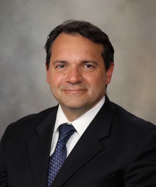 Giuseppe Lanzino合作论文数Department of Neurologic Surgery, Mayo Clinic;Department of Radiology, Mayo Clinic;Department of Neurosurgery, Mayo Clinic15
 Ajay K. Wakhloo合作论文数Department of Radiology, University of Massachusetts Medical School15
 Demetrius Lopes合作论文数Advocate Aurora Health15
 Swadeshmukul Santra合作论文数Department of Chemistry, College of Sciences, University of Central Florida;NanoScience Technology Center, University of Central Florida14
 L Nelson Hopkins合作论文数11LN Hopkins合作论文数9Scott D. Simon合作论文数8Reig Adam S合作论文数8Eric Eskioglu合作论文数8Arthur J. Ulm合作论文数7
 Ricardo A Hanel合作论文数6James Robert F合作论文数6Turner Raymond D合作论文数6
 Scott Simon合作论文数Department of Pediatrics, Baylor College of Medicine6Alan S. Boulos合作论文数6Erol Veznedaroglu合作论文数6Chris Batich合作论文数Department of Materials Science and Engineering, University of Florida6Matthew V. Burry合作论文数6
 Babu Guai Welch合作论文数6Peter Kan合作论文数5JT Stanley合作论文数5Eric Saugaveau合作论文数5Mocco J合作论文数5
 Aditya S. Pandey合作论文数Medical School, University of Michigan;Comprehensive Stroke Center University of Michigan5
 Adnan Siddiqui合作论文数Department of Neurosurgery, Jacobs School of Medicine and Biomedical Sciences, University at Buffalo;Canon Stroke and Vascular Research Center, University at Buffalo5
 Schirmer M. Clemens合作论文数Geisinger5Replogle Robert E合作论文数5
 Elad I. Levy合作论文数Department of Neurosurgery, Jacobs School of Medicine & Biomedical Sciences, University at Buffalo5Hoit Daniel合作论文数Department of Neurosurgery, University of Tennessee5
 Brij M. Moudgil合作论文数Department of Materials Science and Engineering, University of Florida;Particle Engineering Research Center, University of Florida5Jay U. Howington合作论文数5Courtney Watkins合作论文数5
 William J. Mack合作论文数Keck School of Medicine, University of Southern California5
 Rabih G. Tawk合作论文数Department of Neurosurgery, Mayo Clinic Florida5
 Alexander Khalessi合作论文数Department of Neurological Surgery, School of Medicine, University of California, San Diego5
 Howard A. Riina合作论文数Department of Neurology, Grossman School of Medicine, New York University;Department of Radiology, Grossman School of Medicine, New York University;Langone Health Center for Stroke and Neurovascular Diseases, New York University5
 Michael Levitt合作论文数University of Washington5
 Adam S. Arthur合作论文数Semmes Murphey5
 Louis J. Kim合作论文数University of Washington5
 Mandy J Binning合作论文数5David J. Langer合作论文数4Stanley Kim合作论文数4
 R. Webster Crowley合作论文数Department of Neurosurgery, Rush Medical College, Rush University;Medical Center, Rush University4
 Brian Thomas Jankowitz合作论文数Department of Neurological Surgery, University of Pittsburgh;Bayshore Medical Center, Hackensack Meridian Health4Bain Mark合作论文数Cerebrovascular Center, Cleveland Clinic4
 Spiotta Alex合作论文数4
 Christopher S. Ogilvy合作论文数Beth Israel Deaconess Medical Center;Harvard Medical School4DK Lopes合作论文数3




