Patent foramen ovale is increasingly diagnosed in patients who are undergoing clinical study for cryptogenic stroke or migraine. In addition, patent foramen ovale is often suspected as a cause of paradoxical embolism in patients who present with arterial thromboembolism. The femoral venous approach to closure has been the mainstay. When the femoral approach is not feasible, septal occluder devices have been deployed via a transjugular approach.Herein, we describe 2 cases of patent foramen ovale in which the transhepatic approach was used for closure. To our knowledge, this is the 1st report of a transhepatic approach to patent foramen ovale closure in an adult patient. Moreover, no previous case of patent foramen ovale closure has been reported in a patient with interrupted inferior vena cava.
Thoracic pseudoaneurysms are a rare variety of aortic disorders that are potentially fatal. Traditionally, these are treated surgically. False aneurysms are usually a late complication of a previous surgical procedure. Surgical management is often complicated by poor outcomes with high morbidity and mortality. We report a patient with recurrence of an aortic pseudoaneurysm after closure with an Amplatzer (AGA Medical Corp, Plymouth, NH) septal occluder that was successfully treated with a second Amplatzer device.
Background: Refractory angina(Ang) effects 150 –250,000 pts in the US alone. These individuals have exhausted options for revasc and continue to have lifestyle limiting ang despite optimal medical Rx. Pre-clinical studies provided evidence that human CD34+ cells(CD34) can stimulate new blood vessel formation in ischemic myocardium(Myo), improving perfusion and function. We performed a phase 2 study of intraMyo injection of autologous CD34 in pts with CCS Class 3 and 4 Ang to obtain evidence for feasibility, safety and bioactivity. Methods: A phase 2 randomized, double-blind, placebo-controlled clinical trial was performed at 26 centers in the United States. There were 3 treatment groups: placebo, low dose (1×10^5 CD34/kg body wt) and high dose (5×10^5 CD34/kg). All pts underwent mobilization with GCSF 5 mcg/kg/day SC for 5 d, followed by apheresis on d5 to collect mononuclear cells. On day 6 CD34 were purified from the collected cells using the Isolex 300i device. After lot release testing, CD34 were injected endocardially at 10 locations in the ischemic Myo using the Myostar catheter following NOGA mapping. Placebo injections consisted of identical volumes of the diluent only. Results: A total of 167 pts were randomized and completed the injection procedure and 162 completed the 6 mo evaluation. The mean age of subjects was 61.0±8.9, there were 22 female and 145 male subjects. Trop was elevated in 26 pts at baseline and in 71 at some point following GCSF or injection. At 6 mo CD34 treated pts showed more reduction in ang frequency (−8.6 placebo vs 14.2 low dose) and improved ETT time (+69 sec placebo vs. +138 low dose). At 12m CD34 treated continue to show more reduction in angina (−8.2 placebo vs. −15.0 low dose) and improved ETT (+58 sec placebo vs. + 139 sec low dose). For key endpoints the high dose group results were inferior to low dose. The 12m data set has been locked and is being analyzed and the results of the completed analysis will be presented. Conclusions: Autologous CD34 cell therapy was associated with improved exercise tolerance and reduced angina in no-option pts with intractable angina. Ongoing analysis will determine endpoints, sample size and suitability of this therapy for a phase III study in no-option pts with refractory angina.
Thoracic pseudoaneurysms are rare variety of aortic disorders. Although mostly asymptomatic, they represent potentially fatal conditions (Sullivan et al., Chest 1988;93:138–143; Razzouk et al., Ann Surg 1993;59:818–823) that are traditionally treated surgically. False aneurysms of aorta are usually a late complication of previous surgical procedure (Sullivan et al., 1988)‐especially reconstructive surgery, trauma (Razzouk et al., 1993), and rarely infection (Sanchez‐Recalde et al., J Am Coll Cardiol 2003;41:152–154). Surgical management is often complicated by poor outcomes with high morbidity and mortality (Mulder et al., Arch Surg 1998;133:45–49). Endovascular treatment is emerging as promising options for aortic diseases with fewer complications. We report a series of six cases at a single center where Amplatzer devise was used to treat thoracic aortic pseudo aneurysm. To our knowledge only one series of three cases (Kanani et al., Catheter Cardiovasc Interv 2007;69:146–153) and few case reports (Bashi et al., Catheter Cardiovasc Interv 2005;65:547–551) of successful closure have been published previously. Our case series is the largest, so far, including mortality and specifically the preprocedural and postprocedural CT angiographic assessment of the pseudoaneurysms. © 2008 Wiley‐Liss, Inc.
OBJECTIVES:Our aim was to investigate the safety and efficacy of intravenous allogeneic human mesenchymal stem cells (hMSCs) in patients with myocardial infarction (MI). BACKGROUND:Bone marrow-derived hMSCs may ameliorate consequences of MI, and have the advantages of preparation ease, allogeneic use due to immunoprivilege, capacity to home to injured tissue, and extensive pre-clinical support. METHODS:We performed a double-blind, placebo-controlled, dose-ranging (0.5, 1.6, and 5 million cells/kg) safety trial of intravenous allogeneic hMSCs (Prochymal, Osiris Therapeutics, Inc., Baltimore, Maryland) in reperfused MI patients (n=53). The primary end point was incidence of treatment-emergent adverse events within 6 months. Ejection fraction and left ventricular volumes determined by echocardiography and magnetic resonance imaging were exploratory efficacy end points. RESULTS:Adverse event rates were similar between the hMSC-treated (5.3 per patient) and placebo-treated (7.0 per patient) groups, and renal, hepatic, and hematologic laboratory indexes were not different. Ambulatory electrocardiogram monitoring demonstrated reduced ventricular tachycardia episodes (p=0.025), and pulmonary function testing demonstrated improved forced expiratory volume in 1 s (p=0.003) in the hMSC-treated patients. Global symptom score in all patients (p=0.027) and ejection fraction in the important subset of anterior MI patients were both significantly better in hMSCs versus placebo subjects. In the cardiac magnetic resonance imaging substudy, hMSC treatment, but not placebo, increased left ventricular ejection fraction and led to reverse remodeling. CONCLUSIONS:Intravenous allogeneic hMSCs are safe in patients after acute MI. This trial provides pivotal safety and provisional efficacy data for an allogeneic bone marrow-derived stem cell in post-infarction patients. (Safety Study of Adult Mesenchymal Stem Cells [MSC] to Treat Acute Myocardial Infarction; NCT00114452).
Myocardial infarction in the absence of significant atherosclerotic coronary artery disease is not uncommonly encountered in clinical practice. This has been more often seen with the current sensitive biomarker assays for myocardial necrosis. Acute illnesses, spontaneous coronary dissection, sepsis, pulmonary embolism and coagulation disorders are some of the common clinical situation where elevated cardiac markers are noted. We describe two brothers presenting with acute myocardial infarction due to thrombus without any obvious cause.
We describe the management of a large iliac arteriovenous fistula in a 48-year-old patient presenting with heart failure and pulmonary hypertension. The presumptive etiology was a left-lower back stab wound approximately 20 years earlier. A large communication between the left internal iliac artery and the left internal iliac vein was successfully treated with a septal occlusion device. (J Vasc Surg 2008;48:1597-9.)
Ascending pseudoaneurysm is an infrequent complication of ascending aortic surgery. Redo operations are often associated with a high surgical morbidity and mortality. Endovascular management of ascending aortic pathologies with endoluminal graft therapies are challenging due to short landing zones and the fear of flow obstruction to the coronaries and brachiocephalic circulation. We report mid-term follow-up of the management of an ascending aortic pseudoaneurysm using a an Amplatz septal occluder (AGA Medical Corp, Golden Valley, MN) in a 51-year-old man considered at high risk for conventional open surgical repair.
The purpose of this prospective, multicenter trial was to evaluate the safety and effectiveness of the TRAP Vascular Filtration System (VFS) to reduce embolic complications during stenting of diseased saphenous vein grafts (SVGs). Patients with SVG lesions were randomly assigned to undergo stenting with or without the TRAP device. The trial was designed to enroll 752 randomized patients. However, the sponsor terminated the study after a total of 467 patients (358 randomized) were enrolled because of poor recruitment once another distal protection device was approved for clinical use. The primary study endpoint, major adverse cardiac events at 30 days, occurred in 17.3% of control patients and 12.7% of patients treated with the TRAP device (P = 0.24). There was a trend toward a lower incidence of myocardial infarction in the TRAP group compared with the control group (16.2% vs 10.5%, P = 0.12). This difference was predominantly due to a lower incidence of moderate-large infarction (CKMB >5x) in the TRAP group. Use of the TRAP VFS during SVG intervention was safe and was associated with a trend toward a lower incidence of adverse events, however, due to low enrollment the study lacked sufficient power to detect a significant benefit with the device.
Coronary angioscopy was used in an attempt to visualize the internal architecture of cardiac vessels before and after deployment of Palmaz-Schatz stents in 50 patients. The vessel was successfully visualized in 48 (96%) of these patients. In 24 patients, angioscopy was performed both after preliminary balloon angioplasty and then again after stent deployment. In all 24 patients the diameter of the lumen appeared larger after stent deployment as compared to after balloon angioplasty. In 16 of these 24 patients a dissection was documented by angioscopy after balloon angioplasty. The dissection was absent after stent deployment in all 16 patients. In seven patients, thrombus that was not apparent by angiography was visualized by angioscopy. Moreover, in four patients, thrombus that was suggested by angiography could not be confirmed by angioscopy.
 Warren Sherman合作论文数5
 Edward B. Diethrich合作论文数Arizona Heart Hospital5
 Gary L. Schaer合作论文数Med Ctr, Rush Univ5Timothy D. Henry合作论文数4Jamal Hussain合作论文数4
 Jay Traverse合作论文数3
 Steven P. Schulman合作论文数School of Medicine, Johns Hopkins University;Johns Hopkins Hospital3
 Gary Gerstenblith合作论文数Department of Otolaryngology-Head and Neck Surgery, School of Medicine, Johns Hopkins University3
 Grayson H Wheatley合作论文数3Denktas Ali E合作论文数3
 Joshua M. Hare合作论文数Interdisciplinary Stem Cell Institute, Miller School of Medicine, University of Miami;Division of Cardiovascular Medicine, Department of Medicine, Miller School of Medicine, University of Miami3
 Anthony N. DeMaria合作论文数University of California San Diego Health3Nabil Dib合作论文数3Marco Costa合作论文数2roger gammon合作论文数2Aslan Ghandforoush合作论文数2Peter A. Soukas合作论文数2Ayman Jamal合作论文数2Mendelsohn Farrell O合作论文数2
 Kenneth Rosenfield合作论文数Massachusetts General Hospital;Mass General Research Institute2Lee Joon Sup合作论文数2
 Daniel I. Simon合作论文数The Department of Physiology and Biophysics, School of Medicine, Case Western Reserve University;Division of Cardiovascular Medicine, University Hospitals Case Medical Center;Heart & Vascular Institute, University Hospitals Cleveland2
 Dean J. Kereiakes合作论文数The Christ Hospital Heart and Vascular Institute, The Christ Hospital Health Network;The Christ Hospital Research Institute, The Christ Hospital Health Network;The Ohio State University;Medpace2
 Karen Pieper合作论文数Pieper Consulting2
 Theodore A Bass合作论文数2James Hermiller合作论文数2
 Richard A. Schatz合作论文数Ralph H. Johnson Veterans Affairs Medical Center2
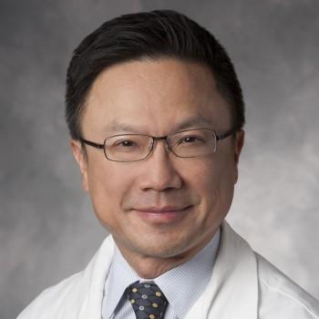 Alan C. Yeung合作论文数Medicine Department, Stanford University2
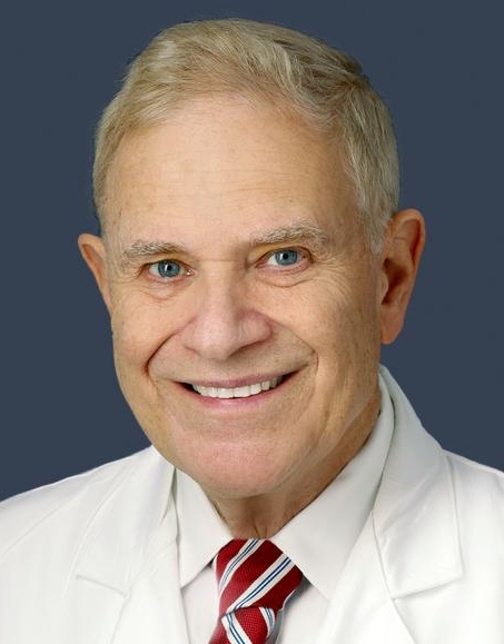 Ron Waksman合作论文数Division of Cardiology, MedStar Heart Institute;Georgetown University2
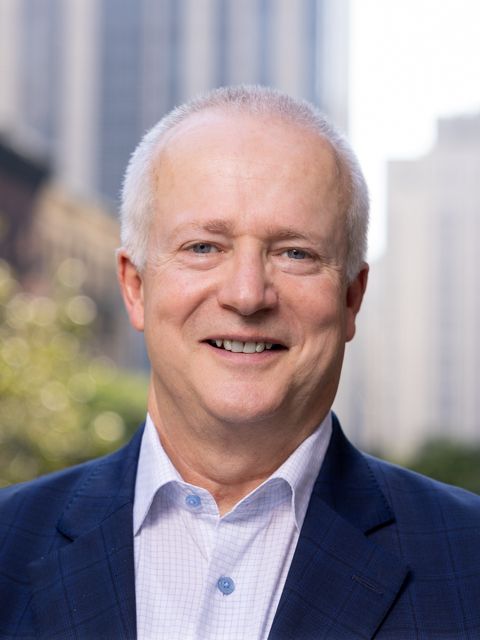 Robert A. Harrington合作论文数Weill Cornell Medicine;Cornell University2Neal L. Weintraub合作论文数2Steven C. Port合作论文数Sinai Samaritan Medical Center, University of Wisconsin Medical School2
 Paul P. Huang合作论文数Department of Neurosurgery, NYU Langone Health2
 Nicolas Chronos合作论文数Lake Country Medical Group2
 Shing-Chiu Wong合作论文数Greenberg Division of Cardiology, Department of Medicine, Weill Cornell Medical College, Cornell University;Cardiac Catheterization Laboratories, NewYork-Presbyterian Hospital2F. David Fortuin合作论文数2
 Juan P Zambrano合作论文数2Leonardo Tamariz合作论文数2Andrew S. Taussig合作论文数2
 Mark Reisman合作论文数Swedish Medical Center, Seattle, Washington2Tim Henry合作论文数1Alan Niederman合作论文数1kerry barker合作论文数1Peter Adams合作论文数1Curtis A Anderson合作论文数1
 Venkatesh Ramaiah合作论文数1William W. O'Neill合作论文数1
 Charles J Davidson合作论文数Department of Medicine, Feinberg School of Medicine, Northwestern University1Story Kenneth合作论文数1




