High occupancy target (HOT) regions, genomic sites bound by many transcription factors, are found across eukaryotes, but their regulatory roles remain unclear. In Caenorhabditis elegans , transcription primarily relies on promoter-proximal elements, and HOT regions frequently coincide with constitutively active promoters. Examining the dlg-1 locus, we identify a putative HOT region essential for embryonic expression. A small sequence within this region, far upstream of the transcription start site, is essential for dlg-1 transcriptional levels. In addition, the first intron modestly enhances transcription. These findings reveal that HOT regions in non-canonical positions can play critical roles in gene regulation in C. elegans . ### Competing Interest Statement The authors have declared no competing interest. University of Basel, https://ror.org/02s6k3f65 Swiss National Science Foundation, https://ror.org/00yjd3n13, SNF 310030_197713
A key output of the NIH-Common Fund 4D Nucleome (4DN) project 1,2 is the open publication of datasets related to the structure of the human cell nucleus and the genome. Recent years have seen a rapid expansion of multiplexed Fluorescence In Situ Hybridization (FISH) or FISH-omics methods, which quantify the spatial organization of chromatin in single cells, sometimes together with RNA and protein measurements, and provide an expanded understanding of how 3D higher-order chromosome structure relates to transcriptional activity and cell development in both health and disease. Despite this progress, results from Chromatin Tracing FISH-omics experiments are difficult to share, reuse, and subject to third-party downstream analysis due to the lack of standard specifications for data exchange. Following up on the recent publication of Microscopy Metadata specifications 3,4, we present the 4DN FISH Omics Format - Chromatin Tracing (FOF-CT), a community-developed data standard for processed results derived from a wide variety of imaging techniques for Chromatin Tracing, with the most recent studies falling roughly into two categories: ball-and-stick and volumetric based on whether they represent the targeted genomic segment as individual fluorescence spots or as clouds of single-molecule localizations. To demonstrate the value and potential use of FOF-CT to promote the FAIR sharing of the results obtained from Chromatin Tracing techniques, this manuscript will focus on ball-and-stick Chromatin Tracing techniques, including those described by the pioneering Chromatin Tracing study of Wang et al. 5 as well as Optical Reconstruction of Chromatin Architecture (ORCA) 6, microscopy-based chromosome conformation capture (Hi-M) 7, Multiplexed Imaging of Nucleome Architectures (MINA) 8, DNA Sequential Fluorescence In Situ Hybridization (DNA seqFISH/seqFISH+) 9-11, Oligopaint Fluorescent In Situ Sequencing (OligoFISSEQ) 12, DNA Multiplexed error-robust fluorescence in situ hybridization (DNA-MERFISH) 13, and In-situ Genomic Sequencing (IGS) 14. The manuscript will describe the structure of the format and present a collection of FOF-CT datasets that were recently deposited to the 4DN Data Portal 15 and the Open Microscopy Environment (OME) Image Data Resource (IDR) platform 16 and are ideally suited for promoting reuse, exchange, further processing, and integrative modeling. Furthermore, the manuscript will present examples of analysis pipelines that could be applied more widely due to the existence of the FOF-CT exchange data format and provide examples of biological conclusions that could be drawn thanks to the availability of such datasets.
The gastrointestinal tract is subjected to extensive mechanosensory stimulation during food ingestion. However, the identities of mechanosensory receptors in the enteric nervous system remain largely unknown. The pharynx of C. elegans is structurally and functionally analogous to the vertebrate foregut, but it contains only 20 neurons embedded among the muscles and epithelial cells of the organ. Here, we report that the DEG/ENaC family ion channel DEGT-1 is a proprioceptor of pharynx movement. DEGT-1 protein is expressed in four pharyngeal neurons (MI, M3, I4, and M5) and localized to their neuronal soma in direct contact with the collagenous pharyngeal basement membrane. degt-1 mutants display abnormally rapid feeding in the presence of food, causing global changes in lipid accumulation. degt-1 mutants also pump rapidly when pumping is induced by the presence of serotonin alone, suggesting that DEGT-1 is required for proprioception of pharyngeal pumping itself rather than for sensing ingested food. DEGT-1 is required in only two pharyngeal neurons (I4 and M5) to control pumping rate. I4 and M5 neurons show a DEGT-1-dependent calcium response. Taken together, these results suggest that DEGT-1 modulates pharyngeal pumping rate by relaying proprioceptive feedback generated by the shear force of the pharynx against its own basement membrane. Thus, mechanosensors in the enteric nervous system modulate organ function by detecting not only the forces from ingested contents but also the movements of the organ itself.
Recent advances in microscopy have enabled studying chromosome organization at the single-molecule level, yet little is known about inherited chromosome organization. Here we adapt single-molecule chromosome tracing to distinguish two C. elegans strains (N2 and HI) and find that while their organization is similar, the N2 chromosome influences the folding parameters of the HI chromosome, in particular the step size, across generations. Furthermore, homologous chromosomes overlap frequently, but alignment between homologous regions is rare, suggesting that transvection is unlikely. We present a powerful tool to investigate chromosome architecture and to track the parent of origin.
Differentiated cells rely on segregation of the genome into heterochromatin and euchromatin, but the mechanisms that establish these domains in vivo are incompletely understood. The current models suggest that heterochromatin is marked by histone H3 lysine 9 methylation (H3K9me), which recruits heterochromatin protein 1 (HP1), to compact chromatin and repress transcription. In C. elegans, the SETDB1 homolog MET-2 is essential for H3K9me-mediated gene silencing; however, MET-2 also localizes to heterochromatic foci independently of H3K9me and exhibits non-catalytic functions that support both germline and somatic development. Here we extend these findings by examining the classical H3K9me reader HP1. We show that HPL-2/HP1 deficient in methyl-lysine binding or in the absence of H3K9me can still repress transcription, promote organogenesis and localize to heterochromatin foci. Whereas activity remains in the absence of the HPL-2:H3K9me interaction, complete loss of met-2 and hpl-2 shows synergistic phenotypes for organogenesis and gene de-repression, underscoring the existence of additional, H3K9me-independent functions. HPL-2 and MET-2 require distinct binding partners for their localization and activity: MET-2 associates with the disordered protein LIN-65/ATF7IP, while HPL-2 depends on the multi-zinc finger protein LIN-13, which interacts with the HPL-2 chromoshadow domain. Our findings suggest that HPL-2 and MET-2 operate in parallel pathways, localizing to heterochromatic foci and promoting gene silencing and organogenesis, with H3K9me acting as a reinforcing, but non-essential, element in these processes. ### Competing Interest Statement The authors have declared no competing interest.
Live imaging of RNA molecules constitutes an invaluable means to track the dynamics of mRNAs, but live imaging in Caenorhabditis elegans has been difficult to achieve. Endogenous transcripts have been observed in nuclei, but endogenous mRNAs have not been detected in the cytoplasm, and functional mRNAs have not been generated. Here, we have adapted live imaging methods to visualize mRNA in embryonic cells. We have tagged endogenous transcripts with MS2 hairpins in the 3' untranslated region (UTR) and visualized them after adjusting MS2 Coat Protein (MCP) expression. A reduced number of these transcripts accumulates in the cytoplasm, leading to loss-of-function phenotypes. In addition, during epithelial morphogenesis, MS2-tagged mRNAs for dlg-1 fail to associate with the adherens junction, as observed for untagged, endogenous mRNAs. These defects are reversed by inactivating the nonsense-mediated decay pathway. RNA accumulates in the cytoplasm, mutant phenotypes are rescued, and dlg-1 RNA associates with the adherens junction. These data suggest that MS2 repeats can induce the degradation of endogenous RNAs and alter their cytoplasmic distribution. Although our focus is RNAs expressed in epithelial cells during morphogenesis, we find that this method can be applied to other cell types and stages.
Spatiotemporal patterns of gene expression underlie embryogenesis. Despite progress in single-cell genomics, mapping these patterns across whole embryos with comprehensive gene coverage and at high resolution has remained elusive. Here, we introduce a whole-embryo imaging platform using multiplexed error-robust fluorescent in-situ hybridization (weMERFISH). We quantified the expression of 495 genes in whole-mount zebrafish embryos at subcellular resolution. Integration with single-cell multiomics data generated an atlas detailing the expression of 25,872 genes and the accessibility of 294,954 chromatin regions, explorable with an online interface MERFISHEYES (beta version). We found that temporal gene expression aligns with cellular maturation and morphogenetic movements, diverse expression patterns correspond to composites of tissue-specific accessible elements, and changes in gene expression generate sharp boundaries during gastrulation. These results establish a novel approach for whole-organism spatial transcriptomics, provide a comprehensive spatially resolved atlas of gene expression and chromatin accessibility, and reveal the diversity, precision and emergence of embryonic patterns. ### Competing Interest Statement The authors have declared no competing interest.
Eukaryotic genome organization is ordered and multilayered, from the nucleosome to chromosomal scales. These layers are not static during development, but are remodeled over time and between tissues. Thus, animal model studies with high spatiotemporal resolution are necessary to understand the various forms and functions of genome organization in vivo. In C. elegans, sequencing- and imaging-based advances have provided insight on how histone modifications, regulatory elements, and large-scale chromosome conformations are established and changed. Recent observations include unexpected physiological roles for topologically associating domains, different roles for the nuclear lamina at different chromatin scales, cell-type-specific enhancer and promoter regulatory grammars, and prevalent compartment variability in early development. Here, we summarize these and other recent findings in C. elegans, and suggest future avenues of research to enrich our in vivo knowledge of the forms and functions of nuclear organization.
AbstractRecent advances in high-throughput microscopy have paved the way to study chromosome organization at the single-molecule level and have led to a better understanding of genome organization in space and time. During development, distinct maternal and paternal contributions ensure the formation of an embryo proper, yet little is known about the organization of chromosomes inherited from mothers versus fathers. To tackle this question, we have modified single-molecule chromosome tracing to distinguish between the chromosomes of two well-studied strains ofC. eleganscalled Bristol and Hawai’ian. We find that chromosomes from these two strains have similar folding patterns in homozygous hermaphrodites. However, crosses between Bristol and Hawai’ian animals reveal that the paternal chromosome adopts the folding parameters of the maternal chromosome in embryos. This is accomplished by an increase in the polymer step size and decompaction of the chromosome. The data indicate that factors from the mother impact chromosome folding in trans. We also characterize the degree of intermixing between homologues within the chromosome territories. Sister chromosomes overlap frequently inC. elegansembryos, but pairing between homologues is rare, suggesting that transvection is unlikely to occur. This method constitutes a powerful tool to investigate chromosome architecture from mothers and fathers.
Mothers contribute cytoplasmic components to their progeny in a process called maternal provisioning. Provisioning is influenced by the parental environment, but the molecular pathways that transmit environmental cues between generations are not well understood. Here, we show that, in Caenorhabditis elegans, social cues modulate maternal provisioning to regulate gene silencing in offspring. Intergenerational signal transmission depends on a pheromone-sensing neuron and neuronal FMRFamide (Phe-Met-Arg-Phe)-like peptides. Parental FMRFamide-like peptide signaling dampens oxidative stress resistance and promotes the deposition of mRNAs for translational components in progeny, which, in turn, reduces gene silencing. This study identifies a previously unknown pathway for intergenerational communication that links neuronal responses to maternal provisioning. We suggest that loss of social cues in the parental environment represents an adverse environment that stimulates stress responses across generations.
mRNA localization is an evolutionarily widespread phenomenon that can facilitate subcellular protein targeting. Extensive work has focused on mRNA targeting through 'zip-codes' within untranslated regions (UTRs), whereas much less is known about translation-dependent cues. Here, we examine mRNA localization in Caenorhabditis elegans embryonic epithelia. From an smFISH-based survey, we identified mRNAs associated with the cell membrane or cortex, and with apical junctions in a stage- and cell type-specific manner. Mutational analyses for one of these transcripts, dlg-1/discs large, revealed that it relied on a translation-dependent process and did not require its 5' or 3' UTRs. We suggest a model in which dlg-1 transcripts are co-translationally localized with the nascent protein: first the translating complex goes to the cell membrane using sequences located at the C-terminal/3' end, and then apically using N-terminal/5' sequences. These studies identify a translation-based process for mRNA localization within developing epithelia and determine the necessary cis-acting sequences for dlg-1 mRNA targeting.
Understanding the coordination of cell division timing is one of the outstanding questions in the field of developmental biology. One active control parameter of the cell cycle duration is temperature, as it can accelerate or decelerate the rate of biochemical reactions. However, controlled experiments at the cellular-scale are challenging due to the limited availability of biocompatible temperature sensors as well as the lack of practical methods to systematically control local temperatures and cellular dynamics. Here, we demonstrate a method to probe and control the cell division timing in Caenorhabditis elegans embryos using a combination of local laser heating and nanoscale thermometry. Local infrared laser illumination produces a temperature gradient across the embryo, which is precisely measured by in-vivo nanoscale thermometry using quantum defects in nanodiamonds. These techniques enable selective, controlled acceleration of the cell divisions, even enabling an inversion of division order at the two cell stage. Our data suggest that the cell cycle timing asynchrony of the early embryonic development in C. elegans is determined independently by individual cells rather than via cell-to-cell communication. Our method can be used to control the development of multicellular organisms and to provide insights into the regulation of cell division timings as a consequence of local perturbations.
Current models suggest that chromosome domains segregate into either an active (A) or inactive (B) compartment. B-compartment chromatin is physically separated from the A compartment and compacted by the nuclear lamina. To examine these models in the developmental context of C. elegans embryogenesis, we undertook chromosome tracing to map the trajectories of entire autosomes. Early embryonic chromosomes organized into an unconventional barbell-like configuration, with two densely folded B compartments separated by a central A compartment. Upon gastrulation, this conformation matured into conventional A/B compartments. We used unsupervised clustering to uncover subpopulations with differing folding properties and variable positioning of compartment boundaries. These conformations relied on tethering to the lamina to stretch the chromosome; detachment from the lamina compacted, and allowed intermingling between, A/B compartments. These findings reveal the diverse conformations of early embryonic chromosomes and uncover a previously unappreciated role for the lamina in systemic chromosome stretching.
This protocol describes a high-throughput and multiplexed DNA fluorescence in situ hybridization method to trace chromosome conformation in Caenorhabditis elegans embryos. This approach generates single-cell and single-chromosome localization data that can be used to determine chromosome conformation and assess the heterogeneity of structures that exist in vivo. This strategy is flexible through modifications to the probe design steps to interrogate chromosome structure at the desired genomic scale (small-scale loops to whole-chromosome organization). For complete details on the use and execution of this protocol, please refer to Sawh et al. (2020).
During the first hours of embryogenesis, formation of higher-order heterochromatin coincides with the loss of developmental potential. Here, we examine the relationship between these two events, and we probe the processes that contribute to the timing of their onset. Mutations that disrupt histone H3 lysine 9 (H3K9) methyltransferases reveal that the methyltransferase MET-2 helps terminate developmental plasticity, through mono- and di-methylation of H3K9 (me1/me2), and promotes heterochromatin formation, through H3K9me3. Although loss of H3K9me3 perturbs formation of higher-order heterochromatin, embryos are still able to terminate plasticity, indicating that the two processes can be uncoupled. Methylated H3K9 appears gradually in developing C. elegans embryos and depends on nuclear localization of MET-2. We find that the timing of H3K9me2 and nuclear MET-2 is sensitive to rapid cell cycles, but not to zygotic genome activation or cell counting. These data reveal distinct roles for different H3K9 methylation states in the generation of heterochromatin and loss of developmental plasticity by MET-2, and identify the cell cycle as a crucial parameter of MET-2 regulation.
H3K9me3 (histone H3 modified with tri-methylation at lysine 9) is a hallmark of transcriptional silencing and heterochromatin. However, its global effects on the genome, including euchromatin, are less well understood. Here we develop Formaldehyde-Assisted Identification of Regulatory Elements (FAIRE) for C. elegans to examine the chromatin configuration of mutants that lack virtually all H3K9me3, while leaving H3K9me1 and H3K9me2 intact. We find that nucleosomes are mildly disrupted, and levels of H3K9me2 and H3K27me3 rise in mutant embryos. In addition to these expected changes, the most dramatic change occurs in euchromatin: many regions encompassing transcription start sites (TSSs) gain an average of two nucleosomes in mutants. The affected regions normally lack H3K9me3, revealing a locus non-autonomous role for H3K9me3. Affected TSSs are associated with genes that are active in epithelia and muscles, and implicated in development, locomotion, morphogenesis and transcription. Mutant embryos develop normally under ideal laboratory conditions but die when challenged, with defects in morphogenesis and development. Our findings reveal that H3K9me3 protects transcription start sites within euchromatin from nucleosome deposition. These results may be relevant to mammals, where diseases that disrupt the nuclear lamina and heterochromatin can alter epithelial and muscle gene expression.
Heterochromatin formation during early embryogenesis is timed precisely, but how this process is regulated remains elusive. We report the discovery of a histone methyltransferase complex whose nuclear accumulation and activation establish the onset of heterochromatin formation in Caenorhabditis elegans embryos. We find that the inception of heterochromatin generation coincides with the accumulation of the histone H3 lysine 9 (H3K9) methyltransferase MET-2 (SETDB) into nuclear hubs. The absence of MET-2 results in delayed and disturbed heterochromatin formation, whereas accelerated nuclear localization of the methyltransferase leads to precocious H3K9 methylation. We identify two factors that bind to and function with MET-2: LIN-65, which resembles activating transcription factor 7-interacting protein (ATF7IP) and localizes MET-2 into nuclear hubs, and ARLE-14, which is orthologous to adenosine 5'-diphosphate-ribosylation factor-like 14 effector protein (ARL14EP) and promotes stable association of MET-2 with chromatin. These data reveal that nuclear accumulation of MET-2 in conjunction with LIN-65 and ARLE-14 regulates timing of heterochromatin domains during embryogenesis.
During the first hours of embryogenesis, formation of higher-order heterochromatin coincides with the loss of developmental potential. Here we examine the relationship between these two processes, and we probe the determinants that contribute to their onset. Mutations that disrupt histone H3 lysine 9 (H3K9) methyltransferases reveal that the methyltransferase MET-2 helps terminate developmental plasticity, likely through mono- and di- methylation of H3K9 (me1/me2), and promotes heterochromatin formation, likely through H3K9me3. We examine how MET-2 is regulated and find that methylated H3K9 appears gradually and depends on the accumulated time of embryogenesis. H3K9me is independent of zygotic genome activation or cell counting. These data reveal how central events are synchronized during embryogenesis and distinguish distinct roles for different H3K9 methylation states. Summary Statement During early embryogenesis, heterochromatin formation and loss of developmental plasticity are coordinately regulated by distinct Histone H3 Lysine 9 (H3K9) methylation states, by the methyltransferase MET-2.
To establish the animal body plan, embryos link the external epidermis to the internal digestive tract. In Caenorhabditis elegans, this linkage is achieved by the arcade cells, which form an epithelial bridge between the foregut and epidermis, but little is known about how development of these three epithelia is coordinated temporally. The arcade cell epithelium is generated after the epidermis and digestive tract epithelia have matured, ensuring that both organs can withstand the mechanical stress of embryo elongation; mistiming of epithelium formation leads to defects in morphogenesis. Using a combination of genetic, bioinformatic, and imaging approaches, we find that temporal regulation of the arcade cell epithelium is mediated by the pioneer transcription factor and master regulator PHA-4/FoxA, followed by the cytoskeletal regulator and kinesin ZEN-4/MKLP1 and the polarity protein PAR-6. We show that PHA-4 directly activates mRNA expression of a broad cohort of epithelial genes, including junctional factor dlg-1 Accumulation of DLG-1 protein is delayed by ZEN-4, acting in concert with its binding partner CYK-4/MgcRacGAP. Our structure-function analysis suggests that nuclear and kinesin functions are dispensable, whereas binding to CYK-4 is essential, for ZEN-4 function in polarity. Finally, PAR-6 is necessary to localize polarity proteins such as DLG-1 within adherens junctions and at the apical surface, thereby generating arcade cell polarity. Our results reveal that the timing of a landmark event during embryonic morphogenesis is mediated by the concerted action of four proteins that delay the formation of an epithelial bridge until the appropriate time. In addition, we find that mammalian FoxA associates with many epithelial genes, suggesting that direct regulation of epithelial identity may be a conserved feature of FoxA factors and a contributor to FoxA function in development and cancer.
 Judith Kimble合作论文数Department of Biochemistry, College of Agricultural and Life Sciences, University of Wisconsin-Madison7
 Stephen E Von Stetina合作论文数7
 Ahilya N Sawh合作论文数6Jeb Gaudet合作论文数6Beste Mutlu合作论文数4Tocchini Cristina合作论文数4
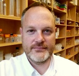 Dustin L. Updike合作论文数Department of Oncological Sciences;Huntsman Cancer Institute;University of Utah;Huntsman Cancer Institute, University of Utah4Reynaldo Sequerra合作论文数4
 Ahmed M. Elewa合作论文数Program in Gene Function and Expression and Program in Molecular Medicine, University of Massachusetts Medical School4Silvia Gutnik合作论文数3Sabine Keppler-Ross合作论文数3
 Huei-Mei Chen合作论文数3Me Domeier合作论文数3Bart Deplancke合作论文数School of Life Sciences, École Polytechnique Fédérale de Lausanne3
 Marc Vidal合作论文数Dana-Farber Cancer Institute;Harvard Medical School;Division of Medical Sciences, Harvard Kenneth C. Griffin Graduate School of Arts and Sciences, Harvard University3Lynn Doucette-Stamm合作论文数3Trisha J. Brock合作论文数3David H. Hall合作论文数3Eric J. Lambie合作论文数3
 Michel Labouesse合作论文数Institut de Biologie Paris-Seine3Jennifer R Saam合作论文数2Michèle Rohner合作论文数2Md Cole合作论文数2S Strome合作论文数2
 Stuart Milstein合作论文数Alnylam Pharmaceut2Julie C. Kiefer合作论文数2
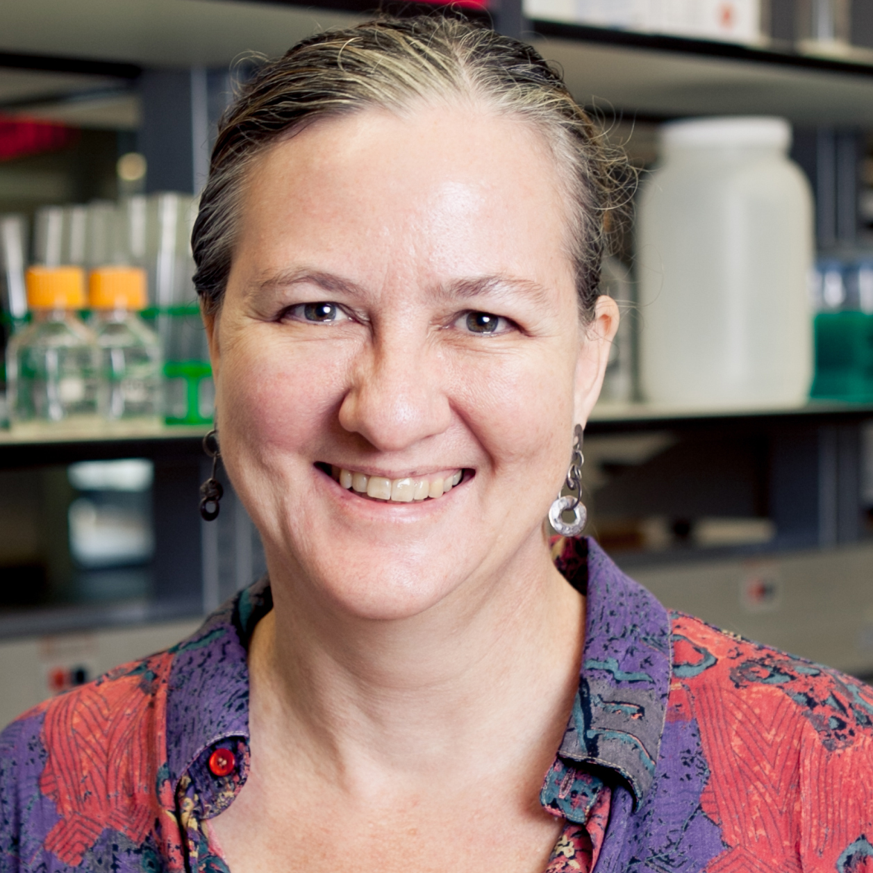 Kristin Gunsalus合作论文数College of Arts & Science, New York University;CGSB, NYU Abu Dhabi2
 Muneesh Tewari合作论文数Department of Biomedical Engineering, College of Engineering, University of Michigan-Ann Arbor2
 Siyuan Wang (王思远)合作论文数Department of Genetics, Yale School of Medicine, Yale University;Department of Cell Biology, Yale School of Medicine, Yale University;Translura Inc2
 Kenneth S Zaret合作论文数Department of Cell and Developmental Biology, Perelman School of Medicine, University of Pennsylvania2
 Alexander F. Schier合作论文数The Center for Molecular Life Sciences, University of Basel2
 Wade Harper合作论文数Department of Cell Biology, Harvard Medical School;Blavatnik Institute, Harvard Medical School2
 Albertha J.M. Walhout合作论文数Walhout lab, Department of Systems Biology, University of Massachusetts Chan Medical School2
 Mark B Gerstein合作论文数School of Engineering & Applied Science, Yale University2Alder Matthew N合作论文数2
 Karyn Sheaffer合作论文数2Wong Sharyl L合作论文数2Jon E. Levine合作论文数2
 Hao Tong (童浩)合作论文数Huazhong University of Science and Technology2Lai Xu合作论文数2
 Mike Boxem合作论文数Massachusetts General Hospital Cancer Center;Center for Cancer Systems Biology (CCSB) and Department of Cancer Biology, Dana-Farber Cancer Institute;Massachusetts General Hospital Cancer Center, Dana-Farber Cancer Institute2Gabriel F. Berriz合作论文数Harvard Medical School, Department of Biological Chemistry and Molecular Pharmacology, 250 Longwood Avenue, SGMB-322, Boston, MA 02115, USA2
 Frederick Roth合作论文数Department of Molecular Genetics, Temerty Faculty of Medicine, University of Toronto;Donnelly Centre for Cellular & Biomolecular Research, Temerty Faculty of Medicine, University of Toronto;Lunenfeld-Tanenbaum Research Institute, Mt. Sinai Hospital;BioSymetrics;Constantiam Biosciences;Ranomics;SeqWell;Center for Cancer Systems Biology, Dana Farber Cancer Institute2William M. Saxton合作论文数Department of Biology;Indiana University;Department of Biology, Indiana University2Jérôme Reboul合作论文数2
 Haiyuan Yu合作论文数Department of Computational Biology, Cornell University;Center for Innovative Proteomics, Cornell University;Weill Institute for Cell and Molecular Biology, Cornell University;Meinig School of Biomedical Engineering, Cornell University2Qian-Ru Li合作论文数2
 Harrison Gabel合作论文数Massachusetts General Hospital, Harvard Medical School2Stephanie Bosak合作论文数2




