(1) Background: Genomic medicine—i.e., the use of laboratory-based biomarkers that measure the expression, function and regulation of genes and gene products to aid healthcare decision making is a rapidly emerging technology. Readiness to consider and adopt new testing programs effectively and avoid critical challenges requires health systems to harbor a number of key conditions that address infrastructural, as well operational and other needs. This assessment re-examines Canada’s state of readiness since a previously published 2023 assessment. (2) Methods: A mixed-methods approach of a review of the literature and key informant interviews with a purposive sample of experts was used. Health system readiness was assessed using a previously published set of conditions. (3) Results: This updated analysis of Canada’s state of readiness for genetic and genomic testing reveals Canada is only partially ready for a future of genomic medicine, although some progress has been made since 2023. The most established conditions were the use of appropriate service models and the integration of innovation and healthcare delivery functions. They suggest that Canada’s major healthcare regions are moving closer to a state of readiness for the consideration and adoption of new testing required for genomic medicine, although using different approaches and at different rates. These findings should be seen as generalizable to other regions internationally—health systems need to have functions that promote responsiveness and resilience, i.e., are able to recognize valuable innovation and quickly shift priorities and create conditions necessary to enable it.
Surgical resection of brain tumors is guided by radiology, anatomical relationships to critical neurological structures, and clinical metrics including patient age and neurological status. Intraoperative decision-making relies on histological assessment through smear and frozen section analysis of tissue; however, such approaches may be insufficient in the era of precision neuro-oncology. Molecular characterization now informs diagnosis, prognosis, and therapeutic response - factors that may directly influence surgical decisions. The integration of novel and rapid intraoperative diagnostic modalities holds the potential to enhance neurosurgical precision, reduce procedure-related morbidity, and maximize the overall effectiveness of modern multimodal brain tumor management.
BACKGROUND:Targeted treatment or immunotherapy may yield increased, durable responses for melanoma patients. Whether patient-level benefits translate to population health is unknown. This study sought to estimate patient and population impacts of a cancer control policy that reimbursed multigene panel testing and pembrolizumab for metastatic melanoma in British Columbia, Canada. METHODS:This retrospective study examined a population-based cohort of 721 adults diagnosed with metastatic melanoma in British Columbia who received single or multigene testing between 2013 and 2018. We determined patient-level policy impacts using 1:1 genetic algorithm matching of policy-affected patients with historical control patients and Kaplan-Meier analysis and inverse probability of censoring weighted regression of 2-year health-care costs and survival times. For population-level effects, we applied interrupted time-series analysis on monthly health-care system expenditures and mortality rates, estimating autoregressive integrated moving average and generalized least squares Poisson regressions. RESULTS:Matched cohort analysis (control patients, n = 154; intervention patients, n = 154) found mean cumulative patient-level cost increases of CAD$53 963 (95% confidence interval [CI] = $35 641 to $72 621; P < .001) and increased survival times of 111 days (95% CI = 44 to 166 days; P < .001) over 2 years. Higher patient-level systemic therapy spending of CAD$48 890 (95% CI = $31 110 to $66 910; P < .001) drove overall cost differences. Population-interrupted time-series analysis detected an immediate, sustained increase in mean monthly health-care expenditures of CAD$1921 (95% CI = $935 to $2908; P < .001) per patient. Higher overall spending did not coincide with population-level mortality changes. CONCLUSIONS:The policy of reimbursing multigene testing and pembrolizumab produced patient survival improvements, but selectivity of response prevented population mortality improvement. Health-care system costs statistically significantly increased at the patient and population levels.
Abstract Background: Determining the parent of origin (PofO) of a variant in hereditary cancer guides counseling, risk management, recurrence risk assessment, and variant classification. In conditions involving genes with PofO effects, such as SDHD, SDHAF2 and MAX, this information can determine whether disease will manifest. Current approaches rely on family-based testing, yet uptake among eligible first-degree relatives remains low, with fewer than 30% undergoing testing, creating a major barrier in hereditary cancer.To address this gap, we developed Parent-of-Origin-Aware Genomic Analysis (POAga), a method that integrates chromosome-scale haplotyping with DNA methylation at differentially imprinted regions to assign PofO without parental data. To validate POAga, we applied it to individuals with hereditary cancer and known segregation of their pathogenic variants, and compared the predicted PofO with the established segregation to assess concordance and limitations. Methods: Blood samples are being collected from individuals with pathogenic variants in hereditary cancer genes, representing broad ranges of ages, ancestries, and cancer histories. Parental segregation was previously known or established through confirmatory testing. Predicted PofO is compared with true segregation to assess concordance. All samples undergo Strand-seq and long-read sequencing under an REB-approved protocol. Results: To date, 285 samples with 290 pathogenic variants have been analyzed across the following genes: BRCA2 (n=46), BRCA1 (n=42), MSH2 (n=34), SDHD (n=29), MLH1 (n=27), MSH6 (n=26), PMS2 (n=20), PALB2 (n=15), TP53 (n=14), ATM (n=14), CDH1 (n=9), CHEK2 (n=3), EPCAM (n=2), SDHAF2 (n=2), MUTYH (n=2), CDKN2A (n=1), POT1 (n=1), RAD51D (n=1), and SDHC (n=1). PofO was assigned for 250 variants, with 98.4% concordance (246/250). PofO could not be determined for 40 variants (13.8%, 40/290), mainly due to insufficient allele-specific methylation at imprinted regions or extended homozygosity that impeded phasing. Misassignments were rare and mainly due to stochastic phasing errors, unresolved inversions, or random allelic methylation at imprinted regions. Conclusion: POAga achieves clinical-grade accuracy in assigning PofO from a single blood sample in hereditary cancer. This directly addresses a major barrier in clinical genetics, particularly when parental samples are unavailable. For genes with PofO effects, this information can determine whether disease will manifest. By enabling reliable segregation without parental testing, POAga helps direct clinical efforts toward those truly at risk and improves the clinical interpretation of variants. Ongoing analyses will refine its performance and support its adoption as a transformative tool in hereditary cancer genomics. Citation Format: Lilian Cordova, Vahid Akbari, Tiffany Leung, Kieran O’Neill, Katherine Dixon, Alexandra Roston, Eugene Cheung, Chuyi Zheng, Millicent Sharman, Alshanee Sharma, Steve Bilobram, Yaoqing Shen, Janine Senz, Yanni Wang, Daniel Chan, Alexandra Fok, Jennifer Nuk, Quang Hong, Robin Coope, Eric Chuah, Simon Chan, Hyun-Wu Lee, Yongjun Zhao, Miruna Bala, Karen Mungall, Andrew Mungall, Richard Moore, Nur Diana Binte Ishak, Siao Ting Chong, Ee Ling Chew, Ashley McDonald, Anna Martinez, Gregory Kelly, Rosella Delgado, Caitlin Orr, Joanne Yuen Yie Ngeow, Kara N. Maxwell, Stephen B. Gruber, Dean Regier, Alice Virani, Louis Lefebvre, Fabio Feldman, Marco Marra, Sophie Sun, Stephen Yip, Peter Lansdorp, Steven John Jones, Kasmintan Schrader. Parent-of-Origin-Aware genomic analysis in hereditary cancer: identifying the side of the family at risk using only the proband’s blood sample [abstract]. In: Proceedings of the American Association for Cancer Research Annual Meeting 2026; Part 1 (Regular Abstracts); 2026 Apr 17-22; San Diego, CA. Philadelphia (PA): AACR; Cancer Res 2026;86(7 Suppl):Abstract nr 5287.
The sequence of the human genome provides a foundation for understanding cellular processes in health and disease1. The organisation of this primary genetic information into cell-specific structure and function is critical to understanding the cell type-specific interpretation and execution of the genome. Epigenetic processes are essential for packaging and higher-level functional organisation of the genome, and changes therein are increasingly recognised as contributors to human disease. Building on primary data generated by multinational consortia, the International Human Epigenome Consortium2 (IHEC) has uniformly processed a collection of more than 2000 comprehensive human reference epigenomes, collectively referred to as EpiATLAS. This effort involved the development of standardised molecular and bioinformatics protocols, metadata models, and analytical tools to manage, integrate, display, and share vast amounts of epigenomic data. This includes the creation of a publicly available Epigenome Reference Registry, which provides a system for accessing protected human subject datasets and facilitates open searching of de-identified samples and experimental data. The integrated EpiATLAS ecosystem and its comprehensive human reference epigenome maps provide an unprecedented resource for the biosciences, expanding the annotated epigenomic landscape while uncovering previously unappreciated relationships among regulatory layers and revealing how epigenetic inputs underpin fundamental cellular functions and disease associations.
Background/Objectives: Genomic alterations play a central role in diagnosis, prognostication, and therapeutic planning for hematolymphoid malignancies. At our tertiary care center, frontline genomic testing relies on optical genome mapping, karyotyping, and fluorescence in situ hybridization, with targeted next-generation sequencing (NGS) performed externally. To provide more comprehensive genomic profiling, we evaluated two large-panel NGS platforms and subsequently performed a clinical validation of the selected assay. Methods: The Illumina PanHeme DNA panel and the SOPHiA Genetics Community Myeloid Solution were compared using 24 bone marrow aspirate specimens with previously characterized alterations, including single nucleotide variants (SNVs), insertions/deletions (indels), and copy number variants (CNVs). The selected panel underwent full analytical validation using 60 specimens. Results: Both panels demonstrated excellent concordance for SNVs and indels, with comparable analytical performance and workflow. CNV calling with SOPHiA was notably strong. Platform selection was influenced by practical considerations, including panel content and cost, leading to a preference for further evaluation of the Illumina assay. Clinical validation of the Illumina PanHeme DNA panel, along with a complementary RNA Exome panel, was subsequently performed. Sequence variant detection showed 100% concordance with orthogonal testing, while CNV detection was variable, reflecting known limitations of targeted NGS. The RNA panel detected all expected fusion transcripts. Conclusions: These findings demonstrate robust analytical performance of both evaluated DNA panels. Clinical validation of the Illumina PanHeme DNA and RNA Exome assays supports their use for comprehensive molecular profiling of hematologic malignancies.
Diagnostic classification criteria of canine meningioma remain poorly codified and have not been linked to outcome data in large scale studies employing standardized review of slides by a board of multiple pathologists. The National Cancer Institute-led multidisciplinary Comparative Brain Tumor Consortium (CBTC) convened a meningioma pathology board, comprising both veterinarian pathologists and physician neuropathologists, and conducted a comprehensive review of 190 cases of canine meningioma. Comparative histologic and cellular features were assessed and discussed by the board for their similarities and differences to human meningioma. All cases were morphologically classified by histologic subtypes defined by the 2016 World Health Organization (WHO) and by histologic grading system for human meningioma. The majority of canine meningiomas were classified as meningothelial (140/190); most cases (109/190) were diagnosed as grade 2, 79 as grade 1, and two cases as grade 3. The histologic features with the highest agreement between reviewers were necrosis and central nervous system (CNS) invasion (intraclass correlation (ICC) of 0.44 for both features). Although morphologic features of the human grading scheme could be applied to canine tumors, there was no statistically significant difference in outcomes between grade 1 and grade 2 tumors in our patient subset with either detailed progression and cause of death information (n = 37) or data restricted to live/dead status (n = 84). While similarities exist between human and canine meningioma, a revised grading framework must be utilized for canine cases, distinct from the World Health Organization classification scheme for humans. Additional studies evaluating larger numbers of similarly treated dogs are necessary, and a framework for studies to enable future meta-analysis is suggested.
Abstract Purpose Brain metastases (BM) far exceed primary CNS tumours and constitute the majority workload for neuro-oncology care providers. Currently, the cancer registries only capture synchronous BMs, which is only a small proportion of all BMs. We aim to develop and validate a natural language processing (NLP) algorithm that identifies brain metastases in radiology reports, enabling scalable surveillance of asynchronous BMs. Methods Using population-based cancer registry data in Alberta, Canada, we identified a cancer cohort diagnosed between 2012–2019 with follow-up to 2022. All brain/head radiology reports at and post-cancer diagnosis were identified. Reports were sampled through a multi-phase approach and manually labeled for BM presence. We trained two Bio_ClinicalBERT models on the “Findings” and “Impressions” sections, respectively, and took the maximum predicted probability as the report-level prediction. Internal and external validation used reports from the Canadian provinces of Alberta, Ontario, and British Columbia. Results The models were trained on 1,879 samples. For internal validation, 1,833 reports from 357 patients were tested. At a probability threshold of 0.4, the model achieved a sensitivity of 0.888 and precision of 0.499. The ensemble substantially outperformed single-section models, which achieved sensitivities of only 67.8% (Findings) and 74.2% (Impressions). On external validation, sensitivity was 0.918 in Ontario and 0.726 in British Columbia, demonstrating robustness across diverse data distributions. Conclusions An NLP-based pipeline processing both Findings and Impressions sections has been developed and validated in three Canadian provinces. It meets cancer registry operational requirements and to be implemented into the surveillance workflow in Alberta and British Columbia, providing a foundation for population-level BM surveillance.
Background Adolescent and young adult (AYA) patients remain underrepresented in neuro-oncology research. Despite being the second most common primary brain tumor in this population, meningiomas have not been studied using age-specific molecular analyses. DNA methylation-based classification and prognostic tools have transformed meningioma care. This study aimed to evaluate the performance of these tools across age groups.Methods We analyzed 1,568 meningiomas with DNA methylation and clinical data, including 18 pediatric patients (<15 years), 195 AYA patients (15-39 years), and 1,355 adult patients (>39 years). Pediatric and AYA (P/AYA) tumors were combined and compared with adult tumors. The performance of established molecular classifiers and recurrence predictors, as well as differences in chromosomal copy number alterations were compared across age groups.Results While histologic grading was comparable between cohorts, P/AYA tumors displayed significantly fewer aggressive molecular groups and lower frequencies of chromosomal arm losses, including 1p, 6q, and 14q. The adult-trained recurrence predictor failed in the P/AYA population (AUC 0.57), despite similar score distributions. Retraining the model on an age-specific cohort using an identical analytic framework improved performance (AUC 0.79) and enabled effective stratification of progression-free survival (P = 0.00054). Importantly, 1p loss retained prognostic significance within the P/AYA group, supporting its clinical utility.Conclusions Molecular tools developed in adult-dominant cohorts do not generalize to younger patients due to both biological divergence and exclusion from model development. These findings underscore the need for age-specific molecular frameworks and highlight the imperative of including P/AYA populations in precision neuro-oncology research to ensure lifespan-equitable care.
BACKGROUND:DNA methylation profiling can be used to robustly predict postsurgical outcomes and response to radiotherapy (RT) for meningioma patients. To allow for seamless integration of these complementary models into clinical practice, a practical framework is needed. METHODS:We leveraged a cohort of nearly 2000 surgically-treated meningiomas with DNA methylation profiling and clinical outcomes data. Existing methylation-based prediction models were dichotomized to yield four risk groups: low and high recurrent risk, each with RT sensitive and resistant subgroups. Risk groups were correlated with progression-free survival in the context of existing biomarkers including extent of resection and WHO grade. RESULTS:We first demonstrated that all risk groups benefit from gross total resection. All "high-risk, RT sensitive" tumors (n = 306, 15.7%) also benefited from adjuvant RT: after GTR, median PFS increased from 4.68 (4.13-9.48) years to not reached (P = .003); after subtotal resection (STR), from 2.12 (1.59-3.02) to 4.09 (3.41-not reached) years (P = .004). "Low-risk, RT sensitive cases" (n = 1207, 61.8%) also benefited from RT after STR (median PFS 7.39 (6.66-12.8) vs. 16.53 (10.35-not reached) years, P = .03), suggesting that RT be considered in these patients. Neither "low-risk RT resistant" (n = 84, 4.3%) nor "high-risk RT resistant" (n = 356, 18.2%) cases benefited from RT, and the latter group was associated with universally poor outcomes. CONCLUSIONS:We identify methylation-defined risk groups of meningioma for which additional benefit is gained from adjuvant RT, leading to a clinical decision-making framework for straightforward integration of molecular models into clinical practice.
OBJECTIVE:To present a case of tumor-to-tumor metastasis involving vestibular schwannoma and review previously reported cases, with focus on histopathologic and immunohistochemical characteristics. PATIENTS:Case report of a 55-year-old female with a history of invasive ductal breast carcinoma who presented with unilateral hearing loss and gait disturbance. INTERVENTIONS:MRI demonstrated a right-sided extra-axial cerebellopontine angle lesion. Patient underwent surgical resection with histopathologic and immunohistochemical profiling of the tumor. MAIN OUTCOME MEASURES:A near-total resection was achieved. Microscopic analysis demonstrated vestibular schwannoma with nests of glandular elements. Immunohistochemical staining confirmed metastasis of her known invasive ductal carcinoma to vestibular schwannoma. Subsequent staging imaging showed advanced oncologic spread. CONCLUSIONS:Including the present case, there have been 11 reports of tumor-to-tumor metastasis involving vestibular schwannoma. The risk of tumor-to-tumor metastasis should be considered in patients with concomitant systemic malignancy and vestibular schwannoma. Cell-cell adhesion mechanisms may facilitate the spread of breast cancer to vestibular schwannoma. Further reports may help to better understand vestibular schwannoma by analyzing its interaction with other oncologic entities.
Abstract Copy number variations (CNVs) can serve as important clinical biomarkers for tumor classification and stratification. However, the utility of these CNV biomarkers for intraoperative tumor assessment within the timeframe of neurosurgical procedures has remained elusive due to the protracted duration of conventional CNV characterization methods. Here, we introduce CNVisor, a statistical framework for reliable and robust CNV detection from long-read sequencing, even under ultra-low coverage. Applied to neurosurgical tumor specimens, the proposed method enabled genome-wide CNV profiling and identified clinically relevant CNVs using roughly 60,000 reads within 20 minutes of sequencing. Integrating CNVisor with methylation-based classifiers can further reduce turnaround time and increase the accuracy of glioma subtype stratification. Together, these findings establish real-time CNV profiling using ultra-low coverage nanopore sequencing as a feasible strategy for intraoperative, genomics-informed assessment of CNS tumors.
Dysembryoplastic neuroepithelial tumors (DNTs) are low-grade glioneuronal tumors with FGFR1 alterations. They show significant histologic and molecular overlap with other glioneuronal tumors, complicating diagnosis. We analyzed 44 tumors that were either classified as DNT by DNA methylation (n = 37), or were diagnosed histologically as DNT but did not classify as DNT by DNA methylation (n = 7). 13/37 (35%) DNT-classifying tumors were histologically diagnosed as DNTs. High-confidence DNTs (score >0.9, 23 cases, 62%) demonstrated variable histology, most frequently DNT (39%), oligodendroglioma, and ganglioglioma and most frequently harbored FGFR1 alterations. Lower-confidence DNTs (score < 0.9, 14 cases, 38%) showed greater heterogeneity; their histologic diagnoses included papillary glioneuronal tumor, extraventricular neurocytoma, and pilocytic astrocytoma. Tumors with low confidence score exhibited diverse molecular alterations including BRAF V600E mutations, PDGFRA amplification, or multiple gene fusions. Among 7 histologically diagnosed DNTs that did not classify as DNT by methylation, most grouped with the myxoid glioneuronal PDGFRA-mutant class despite lacking canonical PDGFRA mutations. Thus, DNTs with high confidence scores are relatively homogenous but DNTs with low methylation confidence scores are heterogenous, highlighting the importance of integrated molecular profiling. Our findings also suggest that the myxoid glioneuronal tumor methylation class may require further classification of underlying drivers.
CHIP and adverse events. A, Percentage of patients on each treatment that had an adverse event on therapy, split by cohort and treatment. B, Proportions of patients with adverse events split by CHIP status. Lighter shades indicate no adverse event, and darker shades indicate an adverse event. P values were determined by Fisher exact tests.
BACKGROUND:TERT promoter mutation is a rare biomarker in meningiomas associated with aberrant TERT expression and reduced progression-free survival. Although high TERT expression is characteristic of tumours with TERT promoter mutations, it has also been observed in tumours with wildtype TERT promoters. This study aimed to investigate the prevalence and prognostic association of TERT expression in meningiomas. METHODS:This multi-institutional cohort study retrospectively collected clinical and molecular data from 1241 meningiomas surgically resected between Jan 1, 2000, and Dec 31, 2024, at Toronto Western Hospital, Canada (n=380; discovery cohort) and external institutions in Canada, Germany, and the USA (n=861; validation cohort). All patients were aged 18 years and older. TERT promoter mutation and TERT expression were determined by Sanger and bulk RNA sequencing. The primary outcomes were TERT expression (presence or absence) in meningiomas with and without TERT promoter mutations, and the difference in progression-free survival between tumours expressing TERT and those not expressing TERT. Survival analysis was assessed using Cox regression and Kaplan-Meier analysis. FINDINGS:Between Jan 1, 2000, and Dec 31, 2024, clinical demographics and tumour characteristics were collected. Median follow-up was 6·2 years (IQR 1·7-12·5) in the discovery cohort and 3·3 years (1·3-3·8) in the validation cohort. 777 (65·8%) of 1181 patients with sex data in the overall cohort were female; 404 (34·2%) were male. TERT was expressed in 157 (28·7%) of 547 wildtype TERT promoter meningiomas and in 193 (32·0%) of 604 overall with RNA data. TERT expression overall conferred an intermediate progression-free survival, shorter than that in patients with TERT-negative tumours but longer than in those with TERT promoter mutations. In the discovery cohort, median progression-free survival was 3·2 years (95% CI 1·7-6·5) in patients with wildtype TERT promoter tumours expressing TERT, 16·0 years (7·1 to not reached; p=0·0021) in patients with TERT-negative wildtype TERT promoter tumours, and 1·6 years (0·9 to not reached; p=0·039) in patients with TERT promoter mutations. These findings were replicated in the validation cohort. Within each WHO grade, TERT expression conferred a progression-free survival equivalent to TERT-negative meningiomas of one grade higher. Grade 1 tumours with TERT expression had a progression-free survival similar to TERT-negative grade 2 tumours (median not reached [95% CI 16·0 to not reached] vs 8·2 years [95% CI 4·5 to not reached]; p=0·59). Grade 2 tumours with TERT expression had a similar progression-free survival to TERT-negative grade 3 tumours (median 3·6 years [2·4 to 5·3] vs 3·8 years [2·3 to not reached]; p=0·42). Multivariable regression showed that TERT expression remained associated with shorter progression-free survival even after adjusting for TERT promoter mutations, CDKN2A/B loss, chromosome 1p/22q status, and WHO grade (hazard ratio 1·85 [95% CI 1·33-2·57]; p=0·0002). INTERPRETATION:TERT expression in meningiomas predicted earlier disease progression, independent of TERT promoter mutation and other markers, and might warrant reclassification of meningiomas that express TERT to a higher WHO grade. FUNDING:Canadian Institutes of Health Research, Brain Tumour Charity UK, University Health Network Foundation, Mary Hunter Meningioma Research Fund, V Foundation, and National Institutes of Health.
Background: Meningiomas exhibit considerable heterogeneity. We previously identified four distinct molecular groups ( immunogenic, NF2-wildtype, hypermetabolic, proliferative) which address much of this heterogeneity. Despite their utility, the stochasticity of clustering methods and the requirement of multi-omics data limits the potential for classifying cases in the clinical setting. Methods: Using an international cohort of 1698 meningiomas, we constructed and validated a machine learning-based molecular classifier using DNA methylation alone. Original and newly-predicted molecular groups were compared using DNA methylation, RNA sequencing, whole exome sequencing, and clinical outcomes. Results: Group-specific outcomes in the validation cohort were nearly identical to those originally described, with median PFS of 7.4 (4.9-Inf) years in hypermetabolic tumors and 2.5 (2.3-5.3) years in proliferative tumors (not reached in the other groups). Predicted NF2-wildtype cases had no NF2 mutations, and 51.4% had others mutations previously described in this group. RNA pathway analysis revealed upregulation of immune-related pathways in the immunogenic group, metabolic pathways in the hypermetabolic group and cell-cycle programs in the proliferative group. Bulk deconvolution similarly revealed enrichment of macrophages in immunogenic tumours and neoplastic cells in hypermetabolic / proliferative tumours. Conclusions: Our DNA methylation-based classifier faithfully recapitulates the biology and outcomes of the original molecular groups allowing for their widespread clinical implementation.
TERT promoter mutation (TPM) is a rare but established biomarker in meningiomas associated with aberrant TERT expression, reduced progression-free survival (PFS), and reduced overall survival. While TERT is highly expressed in tumors with promotor mutations, its expression has also been observed even in tumors with wildtype TERT promoters (TP-WT). This study aimed to assess the prevalence of TERT expression and its association with clinical outcome in meningiomas. Methods: Bulk RNA sequencing (n=604), Sanger sequencing of the TERT promoter (n=1095), and methylation profiling (n=1218) of a multi-institutional cohort of meningiomas (total n=1241) were performed to determine TERT expression, TERT promoter mutation status, and TERT promoter methylation. A cohort of 380 meningiomas from Toronto was used for discovery, and 861 meningioma samples from external institutions were compiled as a validation cohort. Results: TERT expression was significantly higher in meningiomas with TPMs compared to TP-WT. However, TERT was still expressed in 30.4% of meningiomas that lacked TPM. TERT expression increased with higher WHO grades and was associated with shorter progression-free survival, even among TP-WT tumors. WHO grade 1 tumors that expressed TERT had PFS similar to those of WHO grade 2, while WHO grade 2 meningiomas expressing TERT had a PFS similar to those of WHO grade 3 meningiomas. Among grade 3 meningiomas, tumors expressing TERT had PFS similar to those with TPMs. Conclusions: Our findings highlight the prognostic significance of TERT expression in meningiomas, even in the absence of TPMs. Its presence may identify patients at greater risk of rapid progression. These results support the inclusion of TERT expression in risk stratification models and management strategies, including future WHO classification and cIMPACT-NOW molecular testing criteria. Chloe Gui, Justin Z. Wang, Vikas Patil, Andrew Ajisebutu, Jeff Liu, Zeel Patel, Rebeca Yakubov, Ramneet Kaloti, Yosef Ellenbogen, Christopher Wilson, Aaron Cohen-Gadol, Ghazaleh Tabatabai, Marcos Tatagiba, Felix Behling, Eric C. Holland, Jill S. Barnholtz-Sloan, Andrew E. Sloan, Craig Horbinski, Silky Chotai, Lola B. Chambless, Andrew Gao, Serge Makarenko, Stephen Yip, Kenneth Aldape, Farshad Nassiri, Gelareh Zadeh. TERT expression predicts progression-free survival in meningiomas [abstract]. In: Proceedings of the American Association for Cancer Research Annual Meeting 2025; Part 1 (Regular Abstracts); 2025 Apr 25-30; Chicago, IL. Philadelphia (PA): AACR; Cancer Res 2025;85(8_Suppl_1):Abstract nr 4592.
Background: Intracranial epidermoid cysts (IEC) are benign congenital intracranial lesions that rarely undergo malignant transformation. We report a case of IEC evolving into squamous cell carcinoma (SCC) 1-year post-resection. Further, we conducted a systematic review on cases of early malignant transformations of IECs. Methods: MEDLINE, EMBASE, and Scopus were searched from inception until December 2023 for studies reporting malignant transformations of IECs within 2 years of diagnosis. Results: A 48-year-old female underwent surgical resection of a cerebellopontine angle (CPA) IEC in May 2022. She re-presented in July 2023 with headaches, nausea, vomiting, right facial weakness, and rapid cyst progression. Repeat surgical resection revealed a high-grade SCC. Our systematic review identified 19 (10 females, 9 males) additional IEC cases undergoing malignant transformation within 2 years. The mean age at presentation was 57.6 years, most common location was CPA (n=13, 68.4%) and mean time between IEC to malignant transformation was 10.6 months. Eighteen (94.7%) cases transformed to SCC, of which 2 had leptomeningeal carcinomatosis, and 1 transformed to glioblastoma. Conclusions: While malignant transformations of IECs are rare, regular postoperative follow-up is crucial for early malignancy detection and treatment initiation. Further study is warranted to evaluate factors contributing to accelerated malignant progression of IECs.
 Marco Marra合作论文数Canada’s Michael Smith Genome Sciences Centre, BC Cancer Research Institute;Department of Medical Genetics, Faculty of Medicine, University of British Columbia;Michael Smith Laboratories, University of British Columbia107
 Steven Jones合作论文数Department of Medical Genetics, Faculty of Medicine, University of British Columbia;Genome Sciences Centre, BC Cancer95
 Janessa Laskin合作论文数Division of Medical Oncology, Department of Medicine, Faculty of Medicine, University of British Columbia;Canada's Michael Smith Genome Sciences Centre;BC Cancer95
 Howard Lim合作论文数Division of Medical Oncology, Faculty of Medicine, University of British Columbia76
 Daniel Renouf合作论文数Department of Medicine, Faculty of Medicine, The University of British Columbia;British Columbia Cancer Agency71
 Shen Yaoqing合作论文数60
 Erin Pleasance合作论文数Canada's Michael Smith Genome Sciences Centre, BC Cancer60Karen Mungall合作论文数Wellcome Trust Genome Campus, The Wellcome Trust Sanger Institute44
 Cheryl Ho合作论文数Department of Medicine, The University of British Columbia42Sun Sophie合作论文数39
 Aly Karsan合作论文数Graduate Program in Bioinformatics, Faculty of Science, The University of British Columbia37
 Kasmintan (Intan) Schrader合作论文数Department of Medical Genetics, Faculty of Medicine, The University of British Columbia37
 Christopher Dunham合作论文数Department of Pathology and Laboratory Medicine, Faculty of Medicine, The University of British Columbia36
 David Schaeffer合作论文数Department of Pathology and Laboratory Medicine, Faculty of Medicine, The University of British Columbia32
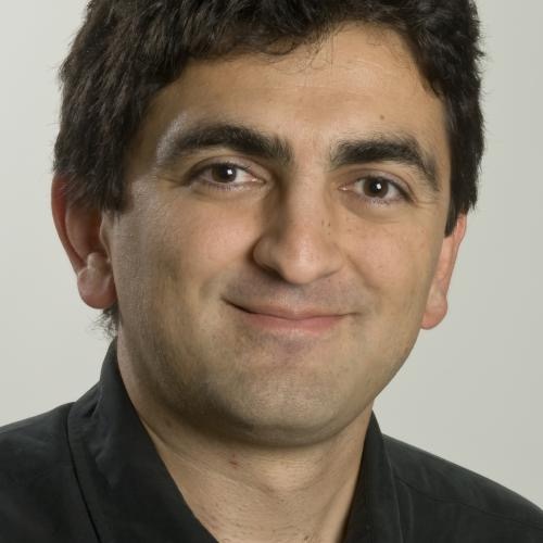 Rod Rassekh合作论文数British Columbia's Children's Hospital32
 Andy Mungall合作论文数Canada's Michael Smith Genome Sciences Centre, BC Cancer31
 Gelareh Zadeh合作论文数Department of Surgery, Temerty Faculty of Medicine, University of Toronto;Toronto Western Hospital, University Health Network31Serge Makarenko合作论文数Department of Surgery, Faculty of Medicine, The University of British Columbia29
 Juliette Hukin合作论文数29
 David Huntsman合作论文数Department of Pathology and Laboratory Medicine, Faculty of Medicine, The University of British Columbia;Department of Obstetrics & Gynaecology, Faculty of Medicine, The University of British Columbia;Department of Molecular Oncology, BC Cancer Research Centre29
 Karen A. Gelmon合作论文数Vancouver Coastal Health Research Institute, The University of British Columbia29
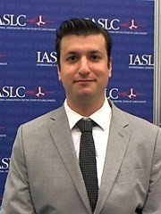 Sheffield Brandon S合作论文数Dept Pathol & Lab Med, Univ British Columbia27Yongjun Zhao合作论文数25Ma Yussanne P合作论文数24
 Stephen K L Chia合作论文数Division of Medical Oncology, Department of Medicine, Faculty of Medicine, The University of British Columbia;British Columbia Cancer Agency23
 Farshad Nassiri合作论文数Division of Neurosurgery,Department of Surgery,University of Toronto/MacFeeters Hamilton Center for Neuro-Oncology,Princess Margaret Cancer Center23
 Cynthia Hawkins合作论文数Department of Laboratory Medicine & Pathobiology, Temerty Faculty of Medicine, University of Toronto;The Hospital for Sick Children23
 Laura Williamson合作论文数Canada's Michael Smith Genome Sciences Centre, BC Cancer22Emma Titmuss合作论文数Canada's Michael Smith Genome Sciences Centre, BC Cancer22
 Kenneth Aldape合作论文数Laboratory of Pathology, Center for Cancer Research, National Cancer Institute, National Institutes of Health22
 Ian Bosdet合作论文数Department of Pathology and Laboratory Medicine, Faculty of Medicine, The University of British Columbia22Lum Amy合作论文数22
 Justin Z. Wang合作论文数21
 Caralyn Reisle合作论文数Canadas Michael Smith Genome Sci Ctr BC Canc, Vancouver, BC, Canada21
 Rebecca J Deyell合作论文数21
 Vikas Patil合作论文数From the Department of Microbiology and Cell Biology, Indian Institute of Science21
 Eric Zhao合作论文数University Health Network, University of Toronto20Levine Adrian B合作论文数20Chloe Gui合作论文数19A. Fok合作论文数19Derek Wong合作论文数Univ British Columbia, Pathol & Lab Med, Vancouver, BC, Canada19
 Alexander D Rebchuk合作论文数Div Neurosurg, Univ British Columbia19Peter Eirew合作论文数19
 Deirdre Weymann合作论文数School of Population and Public Health, University of British Columbia18
 Tony Ng合作论文数Department of Pathology and Laboratory Medicine, Faculty of Medicine, The University of British Columbia18
 Dean Regier合作论文数Faculty of Medicine, School of Population and Public Health, University of British Columbia;BC Cancer;Academy of Translational Medicine - Faculty of Medicine, University of British Columbia18
 Andrew Gao合作论文数Department of Laboratory Medicine and Pathobiology, Temerty Faculty of Medicine, University of Toronto17
 Tabatabai Ghazaleh合作论文数Universitätsklinikum und Medizinische Fakultät Tübingen;Department of Neurology & Interdisciplinary Neuro-Oncology, University Hospital Tübingen17
 Sandra E. Dunn合作论文数Child and Family Research Institute, University of British Columbia17




