The urokinase-type plasminogen activator receptor (uPAR) is currently gaining momentum as a promising molecular target for treatment of various solid cancers. For patient stratification, we developed a high-affinity uPAR-targeting peptide (AE105) detecting primary cancer lesions as well as occult metastasis by positron emission tomography (PET) imaging. uPAR-targeting by AE105 is also used for optical imaging in fluorescence-guided surgery of, for example, head-and-neck cancers. Recently, we showed that a monoclonal anti-uPAR antibody (FL1), in the form of an antibody-drug conjugate (FL1-ADC), efficiently eradicate pancreatic ductal carcinomas in surrogate mouse models leading to long-term remissions. In the current study, we solved high-resolution cryo-EM structures of FL1 in complex with two different conformational states of uPAR. Combined with comprehensive kinetic data from surface plasmon resonance studies, our cryo-EM structures provide essential insights into how FL1 binding impacts the interdomain flexibility of uPAR by restricting the movement of its N-terminal LU domain. This constraint from the bound FL1 drives uPAR into its open conformation, which leads to a pronounced reduction in the binding affinity for both its natural protease ligand (300-fold) and the PET imaging probe AE105 (25-fold). Collectively, these consequences of FL1-binding on uPAR conformation are considered beneficial for both targeted cancer treatment with FL1-ADCs and for the accompanying evaluation of treatment efficacy by longitudinal AE105-based PET imaging.
BACKGROUND:Thrombosis is a major contributor to morbidity and mortality in cardiovascular diseases. Although current antiplatelet therapies reduce thrombotic complications, they are associated with systemic bleeding complications. We previously developed a single-chain variable fragment (scFv) that specifically binds and blocks the activated platelet integrin αIIbβ3 (GPIIb/IIIa [glycoprotein IIb/IIIa]), preventing thrombosis without impairing hemostasis. We hypothesized that an mRNA-based therapeutic encoding scFvαIIbβ3 would offer prolonged expression and enhanced therapeutic durability while maintaining a favorable safety profile. METHODS:We designed and synthesized both conventional mRNA and self-amplifying mRNA constructs encoding scFvαIIbβ3. The constructs were first validated in vitro by confirming their expression of a functional scFvαIIbβ3. For in vivo experiments, mRNAs were encapsulated in a novel lipid nanoparticle formulation comprising of γ-oryzanol and DLin-KC2-DMA (OryKL) for systemic delivery. Their biosafety was assessed by a series of biochemical and histological examinations. The murine FeCl3-induced arterial thrombosis model was used to assess the preventive effect of scFvαIIbβ3-encoding mRNAs on thrombus formation. RESULTS:In vitro assays confirmed the efficient expression and secretion of a functional scFvαIIbβ3 that selectively bound to and blocked activated GPIIb/IIIa, thereby inhibited platelet aggregation. In mice with induced thrombosis, OryKL-delivered scFvαIIbβ3 mRNA significantly prolonged occlusion time at 24 hours posttreatment, while self-amplifying mRNA provided sustained thromboprotection for over 7 days. Tail bleeding times were unchanged across all groups, and no systemic toxicity or histopathologic abnormalities were observed. CONCLUSIONS:Our findings demonstrate that scFvαIIbβ3 mRNA and self-amplifying mRNA therapeutics enable safe, effective, and long-acting antithrombotic protection in vivo, offering a promising strategy for thromboprophylaxis that prevents the bleeding risks associated with current antiplatelet therapies.
mRNA‐loaded lipid nanoparticles (LNPs) offer significant therapeutic potential for various diseases, yet their structural characteristics and component distribution remain incompletely understood. This study utilizes small‐angle neutron scattering (SANS) to explore the internal architecture of KC2 LNPs, employing deuterated lipid substitutions in contrast‐varied media. Our analyses reveal that KC2 LNPs have diameters of 50–60 nm, consistent with cryogenic transmission electron microscopy (cryoTEM) and dynamic light scattering. Core–shell modeling indicates a core radius of 16–17 nm and a shell thickness of 6–8 nm, details often overlooked by cryogenic electron microscopy. Interestingly, results demonstrated that LNPs contain 48%–55% water (total particle volume), which is highly exchangeable with the dispersing medium. This water exchange is crucial during structural monitoring under conditions simulating endosomal environments, as acidification leads to an internal structural reorganization, involving solvent influx into the LNPs and potentially reorganization of components within and across the core and shell phases. Invariant analysis further confirms the high water content of LNPs, validating the model fitting results, affirming KC2 LNPs’ compositional complexity, which are pivotal for understanding LNP stability and responsiveness. These insights can guide the future design of mRNA nanotherapeutics, enhancing their therapeutic efficacy.
In electron microscopy, sparse imaging consists in the collection of a limited subset of the image pixels, which can be used to reduce electron beam damage. Scanning transmission electron microscopy (STEM) is particularly adapted to sparse imaging owing to the scanning nature of the method, scan patterns can be designed where fewer sample locations are targeted. However, since some of the pixels are not scanned, there is an inherent loss of information. Several algorithms were developed to reconstruct missing pixels with high fidelity. Whereas sparse imaging and missing pixel reconstruction in 2D experiments are mature methods, the application of sparse imaging in 3D scanning transmission electron tomography (STET) is rare and still under development. The main difficulty encountered in tomography studies is the tilt-series alignment, which must be accurate to ensure high-quality 3D reconstruction. Because sparse images contain only a certain portion of the original information, the images constituting sparse tilt-series might not share enough mutual information to guarantee an accurate alignment, even after missing pixel reconstruction. This work presents for the first time a thorough analysis of the fiducial alignment and reconstruction of sparse (cryo-)STET tilt-series. Furthermore, the limits of sparse imaging are explored to estimate the minimum amount of information required to obtain good-quality 3D reconstructions. The use of a cryo-fixed biological sample is motivated by the fact that cryo-samples are typical highly beam-sensitive samples, and that the intricate nature and structure complexity of biological samples place them among the most difficult ones to reconstruct with high details.
mRNA nanotherapeutics hold immense potential for treating a wide range of diseases, but their widespread clinical adoption is limited by current lipid nanoparticle (LNP) delivery platforms, which frequently face challenges such as limited biocompatibility, immunogenic response, insufficient mRNA delivery efficacy and stringent cold-chain requirements. In this study, we systematically screened a 20-member lipid mixture library by substituting ionizable lipids and sterol components to identify formulations with improved physicochemical and biological profiles. A lead candidate combining γ-oryzanol and DLin-KC2-DMA as LNPs, termed OryKL (or KO 12 LNPs), was identified, exhibiting spherical bleb-type and core-shell nanostructures (∼150 nm), high mRNA encapsulation, and significantly enhanced in vitro transfection efficiency compared to cholesterol-based controls. Intravenous administration of OryKL delivered Cre recombinase mRNA effectively across multiple organs in Ai9 reporter mice, resulting in distinct cell-level tropism, and no detectable toxicity or inflammation, as confirmed via qPCR, organ histology, hematological assessment and liver function tests. Additionally, OryKL retained transfection potency for at least 60 days in lyophilized form with 20% (w/v) sucrose, supporting ambient-stable storage. These findings establish γ-oryzanol as a promising sterol alternative and position OryKL as a biocompatible, effective, and storage-stable platform for next-generation mRNA therapeutics.
The compaction of chromatin is a prevalent paradigm in gene repression. Chromatin compaction is commonly thought to repress transcription by restricting chromatin accessibility. However, the spatial organization and dynamics of chromatin compacted by gene-repressing factors are unknown. Here, using cryo-electron tomography, we solved the three-dimensional structure of chromatin condensed by the polycomb repressive complex 1 (PRC1) in a complex with CBX8. PRC1-condensed chromatin is porous and stabilized through multivalent dynamic interactions of PRC1 with chromatin. Mechanistically, positively charged residues on the internally disordered regions of CBX8 mask negative charges on the DNA to stabilize the condensed state of chromatin. Within condensates, PRC1 remains dynamic while maintaining a static chromatin structure. In differentiated mouse embryonic stem cells, CBX8-bound chromatin remains accessible. These findings challenge the idea of rigidly compacted polycomb domains and instead provide a mechanistic framework for dynamic and accessible PRC1–chromatin condensates. Here the authors show that a gene-inactivating protein complex packs inactive genes into a dynamic and accessible structure. The study challenges the traditional views that restricted accessibility and low dynamics cause gene repression.
The environmental fragmentation of plastic waste leads to the formation of micro- and nanoplastics (MNPs), which pose serious ecological and human health concerns. Despite increasing interest in their biological effects, many studies rely on artificial, uniform particles that fail to mimic the diverse physical and chemical characteristics of real-world MNPs. To address this limitation, we developed the Accelerated Plastic Aging in Suspension (APAS) system—a scalable, reproducible method that mimics natural aging processes by combining ultraviolet (UV) radiation, thermal stress, and mechanical shear to generate environmentally relevant MNPs from commonly used polymers. We used APAS to fragment polyethylene terephthalate (PET), polyamide 6 (Nylon), and polyacrylonitrile (PAN), and observed time-dependent degradation, including the spontaneous formation of nanoplastics (<100 nm). Flow cytometry revealed substantial increases in particle number and reductions in average particle size over 12 weeks. Imaging flow cytometry confirmed consistent generation of heterogeneous, irregular particles across replicate batches. High-resolution imaging via AFM, TEM, and SEM confirmed the presence of nanoplastics with textured and irregular morphologies. Chemical characterization showed APAS aging altered particle surface charge and induced polymer-specific changes in autofluorescence and Raman spectral profiles, consistent with oxidative surface modifications. Laser Direct Infrared (LDIR) imaging further confirmed structural and chemical changes in polymer spectra post-aging. Functionally, under physiologically relevant shear flow conditions, endothelial cells internalized APAS-generated PET MNPs at significantly higher levels than polystyrene (PS) beads of similar size. Uptake was enhanced particularly under oscillatory flow, highlighting the influence of particle physicochemical properties on cellular interactions. Together, these findings demonstrate the ability of the APAS system to produce complex and realistic MNPs for use in environmental and toxicological studies. The system enables generation of nanoplastics and supports more accurate modelling of biological exposure scenarios compared to conventional synthetic particles. ### Competing Interest Statement The authors have declared no competing interest.
Blood‐ and blood cell‐derived colloidal materials are gaining traction as platforms for targeted therapeutics, imaging, and theranostics. To fully exploit their potential, high‐resolution insight into their structural and compositional features is critical. Here, small‐ and ultra‐small‐angle neutron scattering (SANS/USANS) with contrast variation (CV) is applied to characterize red blood cell (RBC) ghosts, RBC‐derived extracellular vesicles (RBCEVs), vesicle‐depleted human plasma (vdplasma), and human serum albumin (HSA). SANS identifies key contrast match points for lipids (0.5 × 10⁻⁶ Å−2), proteins (2 × 10⁻⁶ Å−2), and cell membrane mixed lipid–protein phases (≈1.4 × 10⁻⁶ Å−2). The compositions, shapes, sizes, and thicknesses of these materials are revealed via structural analyses using model fitting. RBCEVs are resolved as spherical vesicles with a 257 nm diameter and a 3 nm lipid bilayer, supported by data from cryogenic electron microscopy, dynamic light scattering, and nanoparticle tracking analysis. For the first time, SANS reveals differences in structural organization between RBCEV membranes and RBC ghosts, as well as insights into the internal protein and water content of RBCEVs, offering new perspectives on their nanoarchitecture. This study highlights the power of SANS/USANS in resolving complex, hierarchical structures in bioinspired colloids, paving the way for rational design of functional blood‐derived nanomaterials.
Messenger RNA (mRNA) therapeutics provide promising opportunities in cardiovascular diseases. However, effective vascular mRNA delivery requires precise delivery, controlled release, and efficient transfection. To address these challenges, the study utilizes phase‐change perfluorocarbon nanodroplets (PFC NDs) as a unique theranostic platform, integrating multimodal imaging with ultrasound‐triggered mRNA release for enhanced vascular transfection. Lipid‐coated PFC NDs are engineered using perfluoro‐crown‐ether (PFCE), perfluorohexane (PFH), and perfluoropentane (PFP) as core materials. These nanodroplets (200–300 nm) are optimized for mRNA loading and cellular uptake, exhibiting strong ultrasound contrast in tissue‐mimicking phantoms and in vivo. Each PFC generated distinct fluorine‐19 magnetic resonance imaging signals, allowing tri‐spectral imaging capabilities. In vitro, PFH and PFP NDs increased enhanced green fluorescent protein mRNA transfection in CHO cells ( p < 0.0001), with ultrasound stimulation further improving efficiency compared to unstimulated controls ( p < 0.05). In vivo ultrasound‐guided activation of PFH NDs resulted in higher mCherry protein expression in murine carotid arteries ( p < 0.05), demonstrating site‐specific gene therapy. This study establishes PFH NDs as an advanced theranostic platform for vascular mRNA delivery, integrating diagnostics and therapeutics into a single system. By leveraging ultrasound‐responsive activation, these nanodroplets overcome existing delivery limitations, offering a new avenue for precision medicine in cardiovascular disease.
Cryo-FIB milling of biological specimens is a critical and limiting step in the cryo-electron tomography workflow. Preparing electron-transparent cryo-lamellae is a serial, low-throughput process. Even with automation, a skilled operator can typically only produce 15-25 lamellae in a single cryo-FIB session. During sample handling, milling and transfer, the cryo-fixed cells as well as the supporting film layer face various mechanical forces and thermal stresses due to temperature fluctuations. Moreover, after cells are cryo-FIB milled, the resulting thin lamellae continue to endure external forces from mechanical handling and thermal stress. We propose a simple, yet highly effective modification to the standard rectangular milling pattern by implementing "fillets" or corner smoothing providing better mechanical stability. This adjustment helps to avoid sharp corners at the lamella edges, thereby reducing stress concentration. As a result, this modification decreases the likelihood of lamella breakage and improves the overall yield of ready-for-TEM lamellae by over 40 % as verified experimentally.
Utilizing cell membranes from diverse cell types for biointerfacing has demonstrated significant advantages in enhancing colloidal stability and incorporating biological properties, tailored specifically for various biomedical applications. However, the structures of these materials, particularly emulsions interfaced with red blood cell (RBC) or platelet (PLT) membranes, remain an underexplored area. This study systematically employs small- and ultra-small-angle neutron scattering (SANS and USANS) with contrast variation to investigate the structure of emulsions containing perfluorohexane within RBC (RBC/PFH) and PLT membranes (PLT/PFH). The findings reveal that the scattering length density of RBC and PLT membranes is 1.5 x 10(-6) & Aring;(-2), similar to 30% (w/w) deuterium oxide. Using this solvent as a cell membrane-matching medium, estimated droplet diameters are 770 nm (RBC/PFH) and 1.5 mu m (PLT/PFH), based on polydispersed sphere model fitting. Intriguingly, calculated patterns and invariant analysis reveal native droplet architectures featuring entirely liquid PFH cores, differing significantly from the observed bubble-droplet core system in electron microscopy. This highlights the advantage of SANS and USANS in differentiating genuine colloidal structures in complex dispersions. In summary, this work underscores the pivotal role of SANS and USANS in characterizing biointerfaced colloids and in uncovering novel colloidal structures with significant potential for biomedical applications and clinical translation.
In this work, we present a pair of tools to improve the fiducial tracking and reconstruction quality of cryo-scanning transmission electron tomography (STET) datasets. We then demonstrate the effectiveness of these two tools on experimental cryo-STET data. The first tool, GoldDigger, improves the tracking of fiducials in cryo-STET by accommodating the changed appearance of highly defocussed fiducial markers. Since defocus effects are much stronger in scanning transmission electron microscopy than in conventional transmission electron microscopy, existing alignment tools do not perform well without manual intervention. The second tool, Checkers, combines image inpainting and unsupervised deep learning for denoising tomograms. Existing tools for denoising cryo-tomography often rely on paired noisy image frames, which are unavailable in cryo-STET datasets, necessitating a new approach. Finally, we make the two software tools freely available for the cryo-STET community.
Since its inception in the 1930’s, transmission electron microscopy (TEM) has been a powerful method to explore the cellular structure of parasites. TEM usually requires samples of < 100 nm thick and with parasites being larger than 1 µm, their study requires resin embedding and ultrathin sectioning. During the past decade, several new methods have been developed to improve, facilitate and speed-up the structural characterisation of biological samples, offering new imaging modalities for parasitology. In particular, scanning transmission electron microscopy (STEM) can be used to observe sample sections as thick as 1 µm thus becoming an alternative to conventional TEM. STEM can also be performed under cryogenic conditions in combination with cryo-electron tomography providing access to the study of thicker samples in their native hydrated states in 3D. This method, called cryo-scanning transmission electron tomography (cryo-STET), was first developed in 2014. This review presents the basic concepts and benefits of the STEM methods and provides examples to illustrate the potential for new insights into the structure and ultrastructure of parasites.
In photodynamic therapy (PDT), the uses of nanoparticles bearing photosensitizers (PSs) can overcome some of the drawbacks of using a PS alone (e.g., poor water solubility and low tumor selectivity). However, numerous nano-formulations are developed by physical encapsulation of PSs through Van der Waals interactions, which have not only a limited load efficiency but also some in vivo biodistribution problems caused by leakage or burst release. Herein, polymersomes made from an amphiphilic block copolymer, in which a PS with aggregation-induced emission (AIE-PS) is covalently attached to its hydrophobic poly(amino acid) block, are reported. These AIE-PS polymersomes dispersed in aqueous solution have a high AIE-PS load efficiency (up to 46% as a mass fraction), a hydrodynamic diameter of 86 nm that is suitable for in vivo applications, and an excellent colloidal stability for at least 1 month. They exhibit a red/near-infrared photoluminescence and ability to generate reactive oxygen species (ROS) under visible light. They are non-cytotoxic in the dark as tested on Hela cells up to concentration of 100 mu m. Benefiting from colloidal stability, AIE property and ROS generation capability, such a family of polymersomes can be great candidates for image-guided PDT.
Since its inception in the 1930s, transmission electron microscopy (TEM) has been a powerful method to explore the cellular structure of parasites. TEM usually requires samples of <100 nm thick and with protozoans being larger than 1 mu m, their study requires resin embedding and ultrathin sectioning. During the past decade, several new methods have been developed to improve, facilitate, and speed up the structural characterisation of biological samples, offering new imaging modalities for the study of protozoans. In particular, scanning transmission electron microscopy (STEM) can be used to observe sample sections as thick as 1 mu m thus becoming an alternative to conventional TEM. STEM can also be performed under cryogenic conditions in combination with cryo-electron tomography providing access to the study of thicker samples in their native hydrated states in 3D. This method, called cryo-scanning transmission electron tomography (cryo-STET), was first developed in 2014. This review presents the basic concepts and benefits of STEM methods and provides examples to illustrate the potential for new insights into the structure and ultrastructure of protozoans.
Biocatalytic transformation has attracted increasing attention in the green synthesis of chemicals due to the diversity of enzymes, their high catalytic activities and specificities, and environmentally benign conditions. Most redox enzymes in nature are dependent on nicotinamide cofactors like β-nicotinamide adenine dinucleotide (NAD+)/reduced nicotinamide adenine dinucleotide (NADH). The use of solar energy, especially visible light, in the regeneration of cofactors through the combination of photocatalysis and biocatalysis provides an extraordinary opportunity to make complete green processes. However, the combination of photocatalysts and enzymes has been challenged by the rapid degradation and deactivation of the enzymatic material by photogenerated reactive oxygen species (ROS). Here, we design core-shell structured polymer micelles and vesicles with aggregation-induced emission (AIE) as visible-light-mediated photocatalysts for highly stable and recyclable photobiocatalysis under aerobic conditions. NAD+ from NADH can be efficiently regenerated by the photoactive hydrophobic core of polymer micelles and the hydrophobic membrane of polymer vesicles, while the enzymatic material (glucose 1-dehydrogenase) is screened from the attack of photogenerated ROS by the hydrophilic surface layer of polymer colloids. After at least 10 regeneration cycles, the enzyme keeps its active state; meanwhile, polymer micelles and vesicles maintain their photocatalytic activity. These polymer colloids show the potential to be developed for the implementation of industrially relevant photobiocatalytic systems.
Electromagnetic radiation-triggered therapeutic effect has attracted a great interest over the last 50 years. However, translation to clinical applications of photoactive molecular systems developed to date is dramatically limited, mainly because their activation requires excitation by low-energy photons from the ultraviolet to near infra-red range, preventing any activation deeper than few millimetres under the skin. Herein we conceive a strategy for photosensitive-system activation potentially adapted to biological tissues without any restriction in depth. High-energy stimuli, such as those employed for radiotherapy, are used to carry energy while molecular activation is provided by local energy conversion. This concept is applied to azobenzene, one of the most established photoswitches, to build a radioswitch. The radiation-responsive molecular system developed is used to trigger cytotoxic effect on cancer cells upon gamma-ray irradiation. This breakthrough activation concept is expected to expand the scope of applications of photosensitive systems and paves the way towards the development of original therapeutic approaches.
Bacterial chromosomal DNA is packed within a non-membranous structure, the nucleoid, thanks to nucleoid associated proteins (NAPs). The role of bacterial amyloid has recently emerged among these NAPs, particularly with the nucleoid-associated protein Hfq that plays a direct role in DNA compaction. In this chapter, we present a 3D imaging technique, cryo-soft X-ray tomography (cryo-SXT) to obtain a detailed 3D visualization of subcellular bacterial structures, especially the nucleoid. Cryo-SXT imaging of native unlabeled cells enables observation of the nucleoid in 3D with a high resolution, allowing to evidence in vivo the role of amyloids on DNA compaction. The precise experimental methods to obtain 3D tomograms will be presented.
 Sergio Marco合作论文数28
 Min-Hui Li合作论文数15
 Veronique Arluison合作论文数11
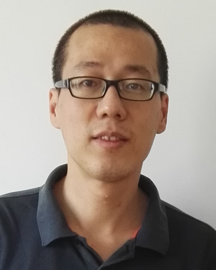 Hui Chen (陈辉)合作论文数Institute of Chemistry, Chinese Academy of Sciences10
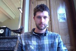 Olivier Lambert合作论文数Laboratoire d’Imagerie Moléculaire et Nano-Bio-Technologie, Université Bordeaux 19Karlheinz Peter合作论文数7
 Mark Louis P. Vidallon合作论文数7
 Cedric Messaoudi合作论文数7Frank Wien合作论文数Institute of Technical Chemistry;University of Karlsruhe7
 Rico F Tabor合作论文数6Wang Xiaowei合作论文数From Atherothrombosis and Vascular Biology Laboratory, Baker IDI;Department of Cardiology and Angiology, University Hospital Freiburg;Fibrinolysis and Gene Regulation Laboratory, Australian Centre for Blood Diseases;and Central Clinical School, Monash University;University Hospital Freiburg, Monash University6
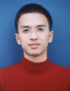 Jun Ling (凌君)合作论文数Department of Polymer Science and Engineering, Zhejiang University6
 Yujiao Fan合作论文数5
 Yangwei Deng合作论文数MOE Key Lab Macromol Synth & Functionalizat, Zhejiang Univ5
 Antoine Malabirade合作论文数Laboratoire Léon Brillouin, CEA-Saclay5
 Xinfeng Tao (陶鑫峰)合作论文数School of Materials Science and Engineering, East China University of Science and Technology5
 Jean-Christophe Taveau合作论文数Université de Bordeaux5Liliana De Campo合作论文数ANSTO, Australian Ctr Neutron Scattering5
 Antoine Cossa合作论文数4David Partouche合作论文数4
 Florian Turbant合作论文数4
 Nian Zhang合作论文数Institut de Recherche de Chimie Paris, PSL Université Paris4Mitchell J Moon合作论文数4
 Georg Ramm合作论文数Department of Biochemistry and Molecular Biology, School of Biomedical Sciences, Faculty of Medicine, Nursing and Health Sciences, Monash University4
 Philippe Bastin合作论文数and MNHN USM 0503, Muséum National d'Histoire Naturelle4
 Arnaud Ducruix合作论文数4
 Mornet Stéphane合作论文数Institute of Condensed Matter Chemistry of Bordeaux, French National Centre for Scientific Research4
 Houssain Benabdelhak合作论文数4Soumia Sid Ahmed合作论文数3
 Jia Guo合作论文数Beijing Adv Innovat Ctr Soft Matter Sci & Engn, Beijing Univ Chem Technol3
 Aziz Taghbalout合作论文数*Department of Molecular Biology and Biophysics, University of Connecticut Health Center3Tao Zou合作论文数3
 Abdeldjalil Ouahabi合作论文数3Vincent Semetey合作论文数3
 Annemarie Wehenkel合作论文数3
 Valeria Militello合作论文数Dipartimento di Scienze Fisiche ed Astronomiche, Università di Palermo3
 Andrea Somogyi合作论文数European Synchrotron Radiation Facility3Zoubeida Messali合作论文数Laboratoire Signaux et Systèmes de Communication, Université de Constantine 250103Alexis I. Bishop合作论文数Centre for Biophotonics and Laser Science;Department of Physics;University of Queensland;Centre for Biophotonics and Laser Science, University of Queensland3Emilie Fortas合作论文数2
 Achilleas S Frangakis合作论文数2Ahmed Refaat合作论文数2
 Omar El Hamoui合作论文数2Lu Zhou合作论文数2Xia Yu合作论文数2Laurène Donati合作论文数EPFL Center for Imaging (imaging.epfl.ch), Lausanne, Switzerland2
 Alexis Voegele合作论文数Inst Pasteur, CNRS, UMR 3528, Chem & Struct Biol Dept, F-75724 Paris 15, France2
 Christophe Sandt合作论文数2
 Michael Unser合作论文数School of Engineering, Ecole Polytechnique Federale de Lausanne;Biomedical Imaging Laboratory, Institute of Imaging and Applied Optics, École polytechnique fédérale de Lausanne;Institute of Electrical and Micro Engineering, Ecole Polytechnique Federale de Lausanne2




