Early recurrence after curative resection remains a major challenge in hepatocellular carcinoma (HCC), particularly in hepatitis B virus (HBV)-related cases. Liquid biopsy using circulating microRNAs (miRNAs) offers a non-invasive approach to identify molecular markers predictive of recurrence. We prospectively enrolled 30 patients with HBV-related HCC presenting with a single tumor (< 5 cm) and no vascular invasion or metastasis. Blood samples were collected preoperatively and on postoperative day 7. Expression of 20 selected miRNAs from circulating cell-free DNA/RNA and exosomes was analyzed. Participants were categorized into early recurrence (within 1 year, n = 6) and non-recurrence (n = 24) groups. Differentially expressed miRNAs were identified, and target genes of significant miRNAs were retrieved from miRTarBase. Protein–protein interaction (PPI) networks were constructed using STRING and visualized in Cytoscape. Enrichment analysis was performed using Gene Ontology and KEGG pathway databases. On postoperative day 7, expression of miR-184 and miR-206 was significantly lower in the early recurrence group than in the non-recurrence group (p < 0.05). Other miRNAs showed no significant differences. Target gene analysis revealed 16 key hub proteins—CCND1, CCND2, KLF4, NOTCH3, BDNF, MET, CDK4, BCL2, AKT2, IGF1R, MYC, HDAC4, ESR1, KRAS, SMARCB1, and AGO2—enriched in cancer-related pathways and involved in HCC progression. Reduced postoperative expression of miR-184 and miR-206 may predict early recurrence in patients with HBV-related HCC. Their associated regulatory networks suggest possible mechanisms of recurrence and represent potential biomarkers for postoperative surveillance. Further studies are needed to validate their prognostic value.
Despite the potential impact of sorafenib on gadolinium-ethoxybenzyl-diethylenetriamine pentaacetic acid (Gd-EOB-DTPA)-mediated contrast enhancements, attempts to assess these effects are rare. This study aimed to investigate the interaction between Sorafenib and Gd-EOB-DTPA by quantifying the T1 and T2 relaxation times and the relative enhancement rates (RERs) of dynamic contrast-enhanced magnetic resonance imaging (DCE-MRI) in the liver. The effects on contrast enhancement were assessed using MRI with Chang liver cells and SD rats. MR phantom images were obtained after treating cells with varying dosages of Gd-EOB-DTPA (5, 10 mM) and sorafenib (10, 30 μM) to evaluate MR relaxivities. For the animal study, DCE-MRI was performed following intravenous administration of 25 µmol/kg Gd-EOB-DTPA and two different doses of sorafenib (10, 30 mg/kg). RER was analyzed to evaluate sorafenib's effects on Gd-EOB-DTPA uptake in the liver. Phantom experiments demonstrated alterations in T1 and T2 values, with a tendency towards shortening disrupted by the addition of sorafenib to Gd-EOB-DTPA-treated Chang liver cells. The RERs of DCE-MRI in the liver exhibited a dose-dependent decrease following sorafenib administration, with recovery observed after 4 h. Our results provide quantitative information on sorafenib-mediated interference with Gd-EOB-DTPA-induced contrast enhancement and offer experimental evidence suggesting the possibility of drug-drug interactions between sorafenib and Gd-EOB-DTPA. Although further research is needed to fully elucidate the impact of these interactions, caution is warranted when using these two agents concurrently for liver MRI.
BACKGROUND Recent research has highlighted DNA methylation as a promising diagnostic biomarker for hepatocellular carcinoma (HCC). Fatty Acyl-CoA Reductase 1 (FAR1) exhibits a high propensity for methylation in HCC. This study aimed to evaluate diagnostic and prognostic potential of FAR1 methylation in liver transplantation (LT) recipients with HCC. MATERIAL AND METHODS This analysis used droplet digital polymerase chain reaction to quantify FAR1 methylation levels in stored pretransplant blood samples. The study cohort (n=48) comprised 25 liver cirrhosis patients with HCC, 13 with cirrhosis but no HCC, and 10 healthy donors. RESULTS Median and mean methylation levels of FAR1 in these groups were 4 copies, zero copies, and zero copies, and 31.6±74.5, 1.5±3.5, and 0.1±0.4 copies, respectively (p<0.001). Receiver operating characteristic curve analysis revealed area under the curve of 0.832 for FAR1, outperforming a-fetoprotein (AFP; 0.737) and protein induced by vitamin K absence or antagonist-II (PIVKA-II; 0.732). A cut-off value of 1 copy for FAR1, defined by Youden's Index (J=0.599), yielded sensitivity of 82.6% and specificity of 77.3%, surpassing diagnostic capacities of AFP and PIVKA-II. Combining FAR1 >1 copy with AFP >7.5 ng/mL or PIVKA-II >40 mAU/mL increased the sensitivity to 91.3%, with specificity of 72.7% and overall accuracy of 82.2%. There was no significant correlation between FAR1 methylation levels and tumor recurrence or overall survival when using a cut-off of 1 copy. CONCLUSIONS These findings suggest that FAR1 methylation is a valuable biomarker for diagnosing HCC in patients with advanced liver disease awaiting transplantation. Further large-scale investigations are necessary to validate clinical efficacy.
Soft-tissue sarcoma (STS) is a rare and heterogeneous group of cancers with more than 100 histological subtypes, which makes biological understanding and therapeutic development particularly challenging. Patient-derived tumor organoid models have transformed cancer research by providing patient-representative preclinical platforms, yet their application in STS has been limited because of low establishment efficiency. To address this problem, a gelatin-based culture protocol was developed to enhance critical cellular processes, including mitochondrial function and cell adhesion, which are essential for organoid self-organization. Using this optimized system, patient-derived tumor organoids were successfully established from representative STS subtypes, such as dedifferentiated liposarcoma and leiomyosarcoma. These organoids retained the histopathological architecture and molecular characteristics of the original tumors and reflected subtype-specific oncogenic pathways, mitochondrial dynamics, and lipid metabolic signatures. Our established gelatin-based organoid culture system enables efficient establishment of patient-derived organoids from representative STS subtypes, faithfully preserving their histopathological and molecular characteristics. These models recapitulate subtype-specific oncogenic pathways, mitochondrial dynamics, and lipid metabolic signatures, providing a robust and clinically relevant preclinical platform for investigating sarcoma biology and developing personalized therapeutic strategies.
Background : DNA methylation is under investigation as an early diagnostic biomarker for cancers such as hepatocellular carcinoma (HCC). p21-activated protein kinase 1 (PAK1) demonstrates a high methylation tendency in HCC. We assessed the diagnostic and prognostic performance of PAK1 methylation in liver transplantation (LT) recipients with and without HCC. Methods : To assess PAK1 methylation, stored pretransplant blood samples from LT recipients were analyzed by droplet digital polymerase chain reaction. Results : This study included 274 patients with HCC and 100 control patients without the disease. Ten-year survival rates in the HCC and control groups were 60.5% and 80.6%, respectively (P=0.001). The 10-year HCC recurrence rate was 39.0%. Ten-year survival rates in the HCC recurrence and nonrecurrence groups were 13.3% and 91.2%, respectively (P<0.001); median PAK1 methylation levels in the control and HCC groups were 50.0 and 108.0 copies (P=0.102). The area under the receiver operating characteristic curve was 0.599, and the Youden J index was 0.199, indicating 57.9% sensitivity and 62.0% specificity at a cutoff of 78 copies. With a cutoff of 5 copies, sensitivity was 94.1% and specificity was 14.0%. The positive likelihood ratio was 1.09 and the negative likelihood ratio was 0.42, indicating diagnostic accuracy below α-fetoprotein and protein induced by vitamin K absence or antagonist-II. PAK1 cutoffs of 5 and 10 copies did not affect HCC recurrence, but a cutoff of 2 copies appeared associated with lower recurrence. Conclusion : s: These data suggest that PAK1 methylation is not a clinically useful biomarker for HCC in LT recipients. New DNA methylation biomarkers are required for early diagnosis.
Technology has been developed to monitor the differentiation process of human mesenchymal stem cells (hMSCs) into hepatocyte-like cells (HLCs) and hepatic progenitor cells (HPCs). These cell lineages, differentiated from MSCs, are ethically unproblematic and are gaining attention as promising cell-based therapies for treating various liver injuries. High-sensitivity, label-free, real-time monitoring technologies integrated with artificial intelligence have been used to evaluate and optimize cell differentiation for enhancing the efficiency of cell therapy delivery. Using an Au-ZnO nanorod array-based surface-enhanced Raman scattering (SERS) sensing chip, cell differentiation from hMSCs to HPCs and HLCs was nondestructively monitored through spectral analysis of cell secretions. Principal component extraction was employed to reduce variables, followed by discriminant analysis (DA). The application of principal component-linear discriminant analysis (PC-LDA), an artificial intelligence algorithm, to spectral data enabled clear grouping of hMSCs, HPCs, and HLCs, with monitoring accuracies of 96.3%, 98.8%, and 98.8%, respectively. Spectral changes observed during the differentiation from hMSCs to HPCs and from HPCs to HLCs over several days demonstrated the effectiveness of SERS combined with machine learning algorithm analysis for differentiation monitoring. This approach enabled real-time, nondestructive observation of cell differentiation with minimal sample labeling and preprocessing, making it useful for sensing differentiation validation and stability. The machine learning- and nanostructure-based SERS evaluation system was applied to the differentiation of ethically sourced MSCs and demonstrated substantial potential for clinical applicability through the use of patient-derived samples.
Background:A decellularized liver scaffold (DLS) is a three-dimensional acellular extracellular matrix created by removing cellular components from liver tissue. Hepatocellular carcinoma (HCC) organoids represent a useful experimental model. Methods:HCC organoids from patient-derived xenografts (PDX), liver organoids, and HepG2 cells were expanded by cultivation within a murine DLS. Results:HCC and liver organoids were generated from HCC PDX and human liver tissues, respectively. Expression levels of hepatocyte paraffin 1 (HepPar1), epithelial cell adhesion molecule (EpCAM), alpha-fetoprotein, keratin-7, and keratin-19 were detected in normal liver tissue, HCC tissue, HCC PDX, and HCC organoids. Fifteen murine DLSs were created, and the complete absence of liver cells was confirmed histologically by Masson trichrome and periodic acid-Schiff staining. Culture of HCC organoids in the DLS resulted in the expansion of numerous HCC cells scattered throughout the DLS framework. Keratin-7 expression was abundant, indicating widely dispersed progenitor cells. In contrast, cultivation of HepG2 cells within the DLS resulted in sparse cell distribution throughout the scaffold. Human hepatocyte organoids could not be cultivated successfully within the murine DLS framework. Conclusions:The DLS collagen framework facilitates the proliferation of HCC organoids. Thus, cultivating HCC organoids within a DLS appears to represent an effective method for rapid cell expansion, although it was ineffective for HepG2 cells and liver organoids. Further studies are required to validate the biological and oncological equivalence between seeded HCC organoids and those rapidly expanded within a DLS.
BRAF-mutated colorectal cancer correlates with poor prognosis and limited response to standard treatments. Combining immune checkpoint inhibitors with BRAF/MEK inhibitors shows promise against BRAF-mutant melanoma in both preclinical and clinical trials. Therefore, we hypothesized that the treatment would be effective against BRAF-mutant colorectal cancer. In this study, we assessed the efficacy of combining immune checkpoint inhibitors with BRAF and/or MEK inhibitors in BRAF-mutant colorectal cancers. We treated BRAF V600E colorectal cancer cells HT-29 and SNU-1235 with encorafenib (BRAF inhibitor) and binimetinib (MEK inhibitor) and assessed the degrees of MAPK inhibition, JAK/STAT inhibition, cell viability, apoptosis, and the expression of antigen presenting machinery. We also inoculated HT-29 cells into mice and treated them with an immune checkpoint inhibitor (durvalumab), encorafenib, and binimetinib for 4 weeks. We found that treatment with BRAF inhibitor, MEK inhibitor, or their combination led to significant tumor growth reduction, along with the MAPK and JAK/STAT pathway inhibition, antigen presenting machinery induction, and cytotoxic T cell activation. Our study demonstrates the potential effectiveness of combining immune checkpoint inhibitors with BRAF or MEK inhibitors for BRAF-mutated colorectal cancers.
Compared to overall survival, conditional survival is a more relevant measure of prognosis in surviving patients over time. This study developed and validated a nomogram-based dynamic prognostic model to predict the conditional survival estimates of patients with hepatocellular carcinoma (HCC) through an analysis of a nationwide cancer registry. This retrospective cohort study included 2492 patients with HCC registered in the Korea Liver Cancer Registry. Patients underwent hepatic resection (HR) from 2008 to 2017, were followed up until December 2019, and were divided into development and validation cohorts. Univariate and multivariate Cox regression analyses were conducted to determine the risk factors for conditional survival of patients who underwent HR. The patients were scored based on the Cox regression coefficients; the nomogram was predicted by calculating the survival probability with Cox model. Our dynamic prognostic model nomogram for predicting conditional overall survival demonstrated Harrell’s C-index of 0.622 and 0.674 in the development and validation sets; for conditional disease-specific survival, it was 0.623 and 0.686 in the development and validation sets. The prediction power of the model is applicable in clinical practice. Factors incorporated in our nomogram included age, albumin, the ADV score, lymph node metastasis, and T stage in American Joint Commission on Cancer staging system. We developed and validated a nomogram to predict conditional survival estimates for overall survival and disease-specific survival. The proposed nomogram incorporating the ADV score presents a more accurate and useful prognostic prediction for patients with HCC who received HR.
Background & Aims The global pandemic caused by the highly contagious SARS-CoV-2 virus led to the emergency approval of COVID-19 vaccines to reduce rising morbidity and mortality. However, limited research exists on evaluating the impact of these vaccines on immunocompromised individuals, such as recipients of living donor liver transplantation, highlighting the need for further studies to better understand their effectiveness in this specific population. Methods From June 2021, we followed up on the effectiveness of the vaccine for patients taking immunosuppressive drugs after living-donor liver transplantation (LDLT). A total of 105 immunocompromised individuals participated, of which 50 patients with hepatitis B were taking antiviral drugs. Patients were assessed to analyze how the combination of immunosuppressive and antiviral drugs affected the efficacy of the BNT162b2, mRNA-1273, and ChAdOx1 nCoV-19 COVID-19 vaccines. Results Before and after the vaccinations, patients were monitored to establish differences between immunosuppressed patients and those additionally taking antiviral drugs. In immunocompromised patients taking antiviral drugs for hepatitis B, we confirmed that the effect of the COVID-19 vaccine was reduced when compared to immunocompromised patients. Interestingly, 23 patients (11 without and 12 additionally with hepatitis B drug administration) encountered breakthrough infections, and although there was a minor discrepancy in vaccine efficacy among the patients taking antiviral drugs for hepatitis B, it did not reach statistical significance. Conclusions Additional COVID-19 vaccination is recommended for patients taking immunosuppressive drugs and hepatitis B antiviral drugs after LDLT.
Hepatitis B Virus (HBV) infection significantly elevates the risk of hepatocellular carcinoma (HCC), with the HBV X protein (HBx) playing a crucial role in cancer progression. Sorafenib, the primary therapy for advanced HCC, shows limited effectiveness in HBV-infected patients due to HBx-related resistance. Numerous studies have explored combination therapies to overcome this resistance. Sodium diethyldithiocarbamate (DDC), known for its anticancer effects and its inhibition of superoxide dismutase 1 (SOD1), is hypothesized to counteract sorafenib (SF) resistance in HBV-positive HCCs. Our research demonstrates that combining DDC with SF significantly reduces HBx and SOD1 expressions in HBV-positive HCC cells and human tissues. This combination therapy disrupts the PI3K/Akt/mTOR signalling pathway and promotes apoptosis by increasing reactive oxygen species (ROS) levels. These cellular changes lead to reduced tumour viability and enhanced sensitivity to SF, as evidenced by the synergistic suppression of tumour growth in xenograft models. Additionally, DDC-mediated suppression of SOD1 further enhances SF sensitivity in HBV-positive HCC cells and xenografted animals, thereby inhibiting cancer progression more effectively. These findings suggest that the DDC-SF combination could serve as a promising strategy for overcoming SF resistance in HBV-related HCC, potentially optimizing therapy outcomes.
Metabolic imbalance leading to inflammatory hypoxia and stabilization of hypoxia-inducible transcription factors (HIFs) is a hallmark of inflammatory bowel diseases. We hypothesize that HIF could be stabilized in CD4+ T cells during intestinal inflammation and alter the functional responses of T cells via regulation of microRNAs. Our assays reveal markedly increased T cell-intrinsic hypoxia and stabilization of HIF protein during experimental colitis. microRNA screen in primary CD4+ T cells points us towards miR-29a and our subsequent studies identify a selective role for HIF-2α in CD4-cell-intrinsic induction of miR-29a during hypoxia. Mice with T cell-intrinsic HIF-2α deletion display elevated T-bet (target of miR-29a) levels and exacerbated intestinal inflammation. Mice with miR-29a deficiency in T cells show enhanced intestinal inflammation. T cell-intrinsic overexpression of HIF-2α or delivery of miR-29a mimetic dampen TH1-driven colitis. In this work, we show a previously unrecognized function for hypoxia-dependent induction of miR-29a in attenuating TH1-mediated inflammation.
BACKGROUND:Human mesenchymal stem cells originating from umbilical cord matrix are a promising therapeutic resource, and their differentiated cells are spotlighted as a tissue regeneration treatment. However, there are limitations to the medical use of differentiated cells from human umbilical cord matrix-mesenchymal stem cells (hUCM-MSCs), such as efficient differentiation methods. METHODS:To effectively differentiate hUCM-MSCs into hepatocyte-like cells (HLCs), we used the ROCK inhibitor, fasudil, which is known to induce endoderm formation, and gelatin, which provides extracellular matrix to the differentiated cells. To estimate a differentiation efficiency of early stage according to combination of gelatin and fasudil, transcription analysis was conducted. Moreover, to demonstrate that organelle states affect differentiation, we performed transcription, tomographic, and mitochondrial function analysis at each stage of hepatic differentiation. Finally, we evaluated hepatocyte function based on the expression of mRNA and protein, secretion of albumin, and activity of CYP3A4 in mature HLCs. RESULTS:Fasudil induced endoderm-related genes (GATA4, SOX17, and FOXA2) in hUCM-MSCs, and it also induced lipid droplets (LDs) inside the differentiated cells. However, the excessive induction of LDs caused by fasudil inhibited mitochondrial function and prevented differentiation into hepatoblasts. To prevent the excessive LDs formation, we used gelatin as a coating material. When hUCM-MSCs were induced into hepatoblasts with fasudil on high-viscosity (1%) gelatin-coated dishes, hepatoblast-related genes (AFP and HNF4A) showed significant upregulation on high-viscosity gelatin-coated dishes compared to those treated with low-viscosity (0.1%) gelatin. Moreover, other germline cell fates, such as ectoderm and mesoderm, were repressed under these conditions. In addition, LDs abundance was also reduced, whereas mitochondrial function was increased. On the other hand, unlike early stage of the differentiation, low viscosity gelatin was more effective in generating mature HLCs. In this condition, the accumulation of LDs was inhibited in the cells, and mitochondria were activated. Consequently, HLCs originated from hUCM-MSCs were genetically and functionally more matured in low-viscosity gelatin. CONCLUSIONS:This study demonstrated an effective method for differentiating hUCM-MSCs into hepatic cells using fasudil and gelatin of varying viscosities. Moreover, we suggest that efficient hepatic differentiation and the function of hepatic cells differentiated from hUCM-MSCs depend not only on genetic changes but also on the regulation of organelle states.
BackgroundThere are few studies on the time to return to activities of daily living (ADL) after craniotomy in patients with brain tumors. This study aimed to investigate the duration before returning to ADLs after craniotomy for brain tumors and present data that can provide information and guidelines on the appropriate time needed.MethodsPatients (n = 183 of 234) who underwent craniotomy for brain tumors between April 2021 and July 2021 capable of self-care upon discharge were enrolled, and data of 158 were collected. The start time of 85 ADL items was prospectively investigated for 4 months postoperatively, using the self-recording sheet.ResultsOver 89% and 87% of the patients performed basic ADL items within a month and instrumental ADL items within 2 months (medians: within 18 days), except for a few. Regarding work, 50% of the patients returned within 4 months. Washing hair with a wound was performed at 18 days of median value, after 4 months of dyeing/perming hair, 6 days of drinking coffee/tea, after 4 months of air travel, and 40 days of complementary and alternative medicine. In patients with infratentorial tumors or surgical problems, return times were much later for various items.ConclusionsIt is possible to provide practical information and guidelines on the duration to return to ADL after craniotomy in brain tumor patients. These study findings also reduce uncertainty about recovery and daily life and help patients return to their daily life at the appropriate time, thereby maintaining function and daily well-being after surgery.
Background : Solid organ transplant recipients exhibit decreased antibody responses, mainly due to their weakened immune systems. However, data are limited on antibody responses after the primary series of coronavirus disease 2019 (COVID-19) vaccines among recipients of various solid organ transplant types. Thus, we compared the antibody responses after three COVID-19 vaccine doses between liver transplant (LT) and kidney transplant (KT) recipients. Methods : We prospectively enrolled solid organ transplant recipients who received three COVID-19 vaccine doses from June 2021 to February 2022 and measured S1-specific immunoglobulin G antibodies using an enzyme-linked immunosorbent assay. Results : Seventy-six LT and 17 KT recipients were included in the final analysis. KT recipients showed consistently lower antibody responses even after the third vaccine dose (86.2% vs. 52.9%, P=0.008) and lower antibody titers (median, 423.0 IU/mL [interquartile range, 99.6–2,057 IU/mL] vs. 19.7 IU/mL [interquartile range, 6.9–339.4 IU/mL]; P=0.006) than were observed in LT recipients. Mycophenolic acid was a significant risk factor for a seropositive antibody response after the third vaccine dose in the multivariable analysis (odds ratio, 0.06; 95% confidence interval, 0.00–0.39; P=0.02). Conclusions: We found a weaker antibody response despite the completion of the primary series of COVID-19 vaccines in KT recipients than in LT recipients. Mycophenolic acid use in KT recipients might be the main contributor to this observation.
Backgrounds The anatomy of the left hepatic vein (LHV) is variable; thus, it should be considered for graft hepatic vein (GHV) venoplasty for left lateral section (LLS) and left liver grafts. This study assessed the incidence of superficial LHV (sLHV) branches according to LHV anatomy and its usability for GHV venoplasty in pediatric liver transplantation (LT). Methods This study consisted of three parts: (1) anatomical classification of LHV variations and the incidence of sLHV branches; (2) morphometric simulative analysis of GHV reconstruction and (3) clinical application based on LHV anatomy. Results The LHV anatomy of 248 potential LLS graft donors was classified into four types according to the number and location of GHV openings: one single opening (type 1; n = 186 [75.0%]), two large openings (type 2; n = 35 [14.1%]), one large and one small adjacent opening (type 3; n = 14 [5.6%]), and two large widely-separated openings (type 4; n = 13 [5.2%]). An sLHV branch was identified in 87 of 248 (35.1%) donor livers. Morphometric analysis of simulative GHV venoplasty with an sLHV branch increased GHV diameters by 30% in type 1 LLS grafts and 20% in type 2/3 LLS grafts. An analysis of 50 consecutive patients who underwent pediatric LT showed that the 2-year rates of GHV obstruction were 2.0% with LLS grafts and 0% with left liver grafts. Conclusions The GHV orifice can be enlarged through LHV anatomy-based unification venoplasty. Unification venoplasty with an sLHV branch provided sufficient enlargement of the GHV orifice.
 Gi-Won Song合作论文数36Shin Hwang合作论文数33
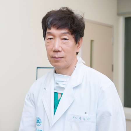 Sung-Gyu Lee (李承奎)合作论文数韩国蔚山大学医学院Asan医学中心26
 Holger Eltzschig合作论文数Department of Anesthesiology, Critical Care and Pain Medicine, McGovern Medical School, The University of Texas Health Science Center;Center for Perioperative Medicine, Department of Anesthesiology, McGovern Medical School, The University of Texas Health Science Center19Young-In Yoon合作论文数17Almut Grenz合作论文数16
 Donghwan Jung合作论文数Asan Medical Center, University of Ulsan College of Medicine15
 Nayoung Kim合作论文数College of Medicine, Seoul National University14Gilchun Park合作论文数14Jooyoung Lee合作论文数11Chul-Soo Ahn合作论文数11Michael A. Zimmerman合作论文数Department of Surgery, University of Colorado Health, Sciences Center11Ji-Ye Kim合作论文数Yonsei Univ, Coll Med, Severance Hosp, Dept Pathol, 50-1 Yonsei Ro, Seoul 03722, South Korea10Tae-Yong Ha合作论文数10Maria Kaplan合作论文数9
 Varvara Kirchner合作论文数Schulze Diabetes Institute, University of Minnesota9Sung Soo Kim合作论文数8Douglas Ridyard合作论文数8Deok-Bog Moon合作论文数8
 Michael R. Blackburn合作论文数Pulmonary Center of Excellence, McGovern Medical School, University of Texas Health Science Center at Houston8
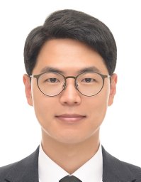 Kihun Kim合作论文数Asan Medical Center, Ulsan University College of Medicine8Igal Kam合作论文数8
 Doo-Sup Choi合作论文数Department of Molecular Pharmacology and Experimental Therapeutics, Mayo Clinic8Namgoong Jung-Man合作论文数7Brodsky Kelley S合作论文数7Jae Hyun Kwon合作论文数Biomaterials Research Center, Korea Institute of Science and Technology7Radu Moldovan合作论文数6
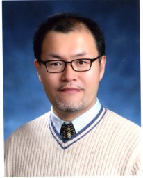 Chan-Gi Pack合作论文数ASAN Inst Life Sci, Univ Ulsan6Joohun Ha合作论文数6
 Jisun Lee合作论文数School of Materials Science and Engineering, Seoul National University6Eric T. Clambey合作论文数6Seonmin Lee合作论文数6Ryunjin Lee合作论文数5Eun-Kyoung Jwa合作论文数5Antasia Giebler合作论文数5
 Seok-Hwan Kim合作论文数Coll Med, Chungnam Natl Univ5Tae Won Kim合作论文数5
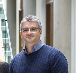 Eoin N Mcnamee合作论文数Mucosal Inflammat Program, Univ Colorado5Badulak Alexander合作论文数5Bauerle Jessica D合作论文数5
 Kim Inki合作论文数5Uwe Christians合作论文数5
 Volker H. Haase合作论文数Volker Haase Lab, Division of Nephrology and Hypertension, Department of Medicine, Vanderbilt University Medical Center5
 Jost Klawitter合作论文数5
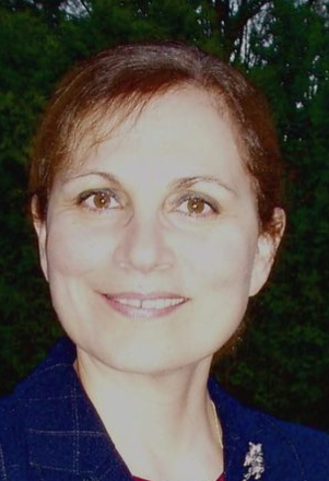 Katya Ravid合作论文数Evans Center for Interdisciplinary Biomedical Research, Boston University;Department of Biology, College of Arts & Sciences, Boston University;Chobanian & Avedisian School of Medicine, Boston University5Kyung Jin Lee合作论文数4Dae Young Jun合作论文数4Julee H. Dalton合作论文数4Dmitriy Lukashev合作论文数4




