Background. Ifapractical, reliable, noninvasive markeroffailed reperfusion was available inreal time, thebenefits offurther therapy inthis patient subgroup could betested. Wedeveloped amethodof12-lead ST-segment recoveryanalysis usingcontinuously updated reference points toprovide sucha marker. Methods andResults. Inthisstudy, our methodwas prospectively tested in144patients given thrombolytic therapy earlyinmyocardial infarction. Allpatients had12-lead continuous ST-segment monitoring andacute angiography, eachanalyzed inanindependent, blinded corelaboratory. ST-segment recoveryandreelevation wereanalyzed up tothemomentofangiography, atwhichtimepatency was predicted. Predictions were correlated toangiographic infarct artery flow, withTIMIflow 0 to1 as occluded andTIMIflow2to3aspatent. Infarct artery occlusion was seenon first injection in27%of patients. Thepositive predictive value ofincomplete STrecoveryorSTreelevation byourmethod was 71%, negative predictive value 87%,with90%ospecificity and64%sensitivity for coronaryocclusion. STrecovery analysis predicted patency in94%ofpatients withTIMI3flow versus81%ofpatients withTIMI2flow andpredicted occlusion in57%ofpatients withcollateralized occlusion versus72%ofpatients with noncollateralized occlusion. Ina regression modelincluding other noninvasive clinical descriptors, ST recoveryalone contained thevastmajority ofpredictive information about patency. Conclusions. Ina blinded, prospective, angiographically correlated studydesign, 12-lead continuous ST-segment recoveryanalysis showspromise asapractical noninvasive markeroffailed reperfusion that may contribute substantially tocurrently available bedside assessment. Ourdataalsosuggest that patients withTIMI2 flowor withcollateralized occlusions may represent a physiological spectrum definable withST-segment recoveryanalysis. (Circulation 1993;88:437-446)
Background Optimal treatment of acute myocardial infarction (AMI) depends on the duration of the ischemia. The Anderson Wilkins (AW) electrocardiographic acuteness score has been shown to complement the historical timing in estimating the time interval from acute thrombotic coronary occlusion in patients presenting with chest pain and evolving myocardial infarction. The purposes of this study were to (1) compare the distributions of the previously developed AW acuteness score in a training population with either anterior or inferior AMI and (2) propose modifications to the formula to achieve distributions similar to the observed distributions of historical times from onset of pain.Methods Two hundred three and 177 patients were included as training and testing population, respectively. All patients had an anterior or an inferior AMI and were without confounding factors on the electrocardiogram.Results The training population had similar distributions of historical times from onset of pain, but differences in distributions of AW acuteness scores, between patients with anterior and inferior AMI (P<.0001). Eighty percent of the inferior AMI group had the highest possible AW acuteness score. Modification of a Q-wave criterion from >= 30 to >= 20 ms resulted in similar distributions in patients with anterior and inferior AMI both in the training and an independent testing population.Conclusions These results suggest that a modified AW acuteness score using a lower Q-wave duration criterion provides similar AMI timing information in patients with anterior and inferior locations. Clinical use of the AW acuteness score will only be practical if the calculation is automated.
The purpose of this study was to determine paramedics' true-positive rate of ST elevation acute myocardial infarction diagnosis. The paramedics' true-positive rate was 49% versus 88% by expert cardiologists (p=0.010), with the difference primarily due to electrocardiograms with confounding factors such as bundle branch block, ventricular hypertrophy, and previous acute myocardial infarction.
This report details a prospectively randomized clinical trial comparing mechanical clamp compression to hand applied pressure for attaining vascular hemostasis after coronary intervention. Effectiveness was determined by comparing the incidence of femoral vascular complications resulting from each of the 2 techniques. Eligible participants included 778 consecutive patients scheduled for percutaneous coronary intervention over an 8-month period. An unselected cohort of the eligible patients (n = 592), determined by the availability of cross-trained clinicians, underwent follow-up serial physical examinations by blinded observers for the duration of their hospital stay. A second, similarly determined cohort (n = 390), underwent color-duplex ultrasonography within 24 hours of sheath removal. Baseline demographic and clinical characteristics, sheath removal parameters, and subsequent outcomes were collected prospectively. The primary end point was a composite of ultrasound-defined femoral vascular complications: femoral artery thrombosis, echogenic hematoma, pseudoaneurysm, or arteriovenous fistulae formation. Complications diagnosed by physical examination constituted the second fundamental end point and included: persistent oozing, ecchymosis, hematoma, bruit, and pulsatile mass. Compared to manual compression, mechanical clamp hemostasis reduced the primary adverse end point by 63% (p = 0.041). Physical examination detected ecchymosis, oozing, and hematomas at equally high frequencies in the two cohorts. Although 65% of the patients in both treatment groups encountered at least one of these cosmetic complications, the diagnoses made by physical examination did not correlate with ultrasound-defined pathology. Multivariable stepwise logistic regression analysis identified a relationship of advanced age and lower body weight to vascular complications. Utilization of a mechanical clamp rather than conventional hand pressure to attain vascular hemostasis significantly reduces ultrasound-defined femoral vascular pathology. Discrepancies between physical examination and ultrasound diagnoses challenge the utility of clinical assessment alone and establish ultrasound as the diagnostic modality of choice.
Objectives. The purpose of this study was to evaluate the importance of time to reperfusion for outcomes after primary angioplasty for acute myocardial infarction.
 Richard Stack合作论文数3Galen S. Wagner合作论文数Duke University3
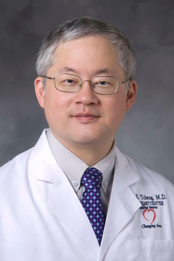 James Enlou Tcheng合作论文数Department of Medicine, School of Medicine, Duke University3
 Robert M. Califf合作论文数Food and Drug Administration3Christopher M. O'Connor合作论文数3Charles Maynard合作论文数3Jonathan A. Lipton合作论文数2
 Eric Topol合作论文数Department of Molecular Medicine, The Scripps Research Institute;Division of Cardiovascular Diseases, Scripps Clinic;Skaggs Graduate School of Chemical and Biological Sciences, The Scripps Research Institute2Maria Sejersten合作论文数The Heart Center, Copenhagen University Hospital2
 Stephen Ellis合作论文数Department of Cardiovascular Medicine, Cleveland Clinic Foundation2Paul N. Leibrandt合作论文数2Debra S. Versteeg合作论文数2
 Kerry L. Lee合作论文数Department of Biostatistics & Bioinformatics, School of Medicine, Duke University;Duke Clinical Research Institute2Dwayne R. Young合作论文数2R. Ripa合作论文数1P Clemmensen合作论文数1Frank D Tice合作论文数1Jack Smith合作论文数1John B Pracyk合作论文数1Je Pope合作论文数1Cindy L. Grines合作论文数1Sanz Mark L合作论文数1j peter longabaugh md合作论文数1
 Samuel J. Bell合作论文数Meta1RT KENNEY合作论文数1William W. O'Neill合作论文数1
 Harry Rissler Phillips合作论文数Department of Medicine, School of Medicine, Duke University School of Medicine1Joseph Andrew Kisslo合作论文数Department of Medicine, Duke University School of Medicine1
 Cindy L. Green合作论文数Department of Biostatistics and Bioinformatics, Duke University School of Medicine1Rendall David合作论文数1
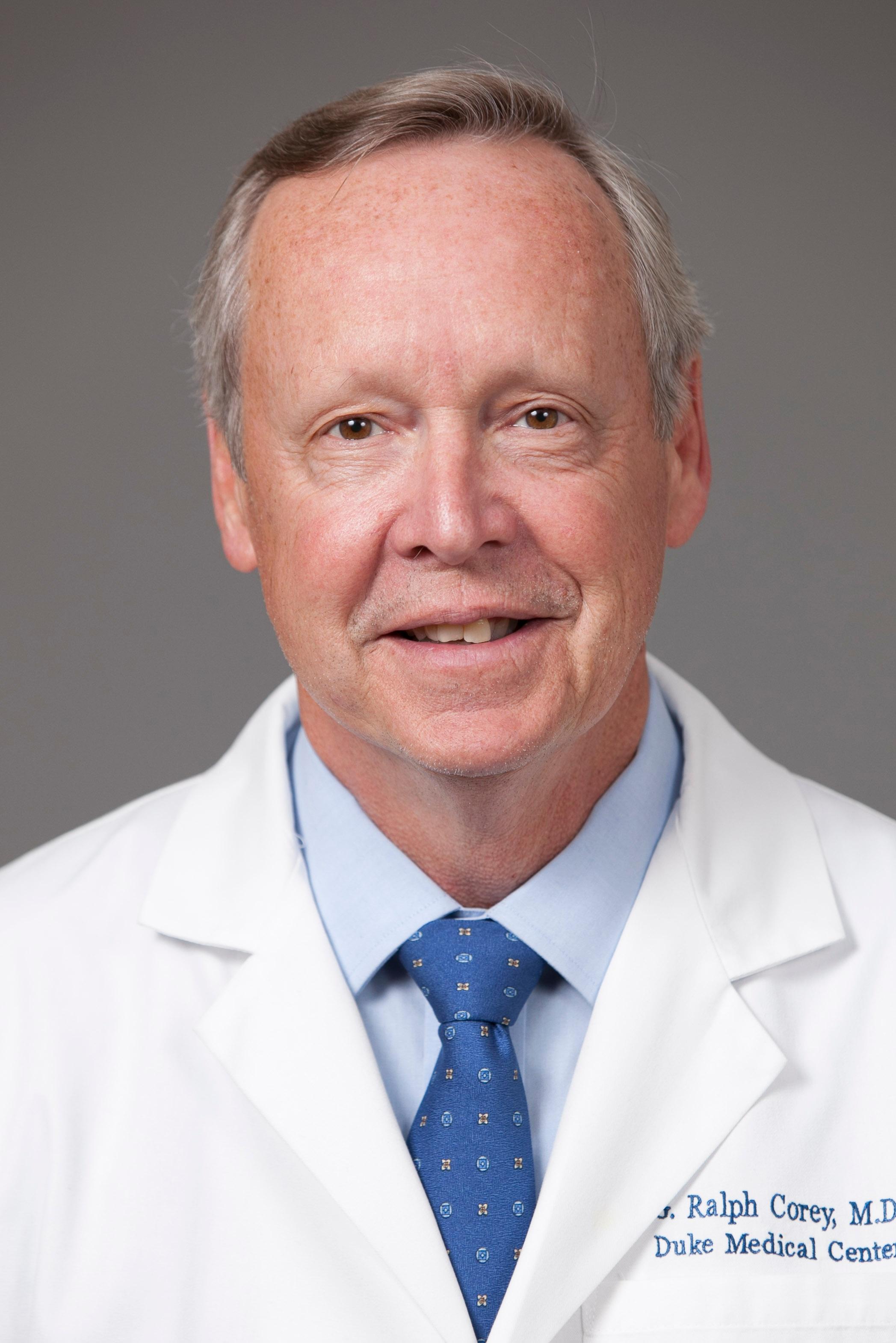 Gordon Ralph Corey合作论文数Department of Medicine, School of Medicine, Duke University1
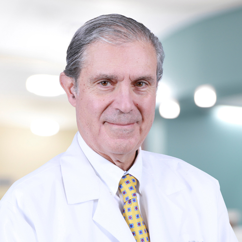 Dean J. Kereiakes合作论文数The Christ Hospital Heart and Vascular Institute, The Christ Hospital Health Network;The Christ Hospital Research Institute, The Christ Hospital Health Network;The Ohio State University;Medpace1Timothy F. Christian合作论文数Rochester1
 Mitchell Wolfe Krucoff合作论文数Duke Clinical Research Institute, Duke University School of Medicine;Department of Medicine, Duke University School of Medicine1
 Michael Hugh Sketch合作论文数Department of Medicine, School of Medicine, Duke University1Olle Pahlm合作论文数Department of Clinical Physiology (Lund),Lund University1
 Barry George合作论文数1
 Christopher Bull Granger合作论文数Department of Medicine, School of Medicine, Duke University;School of Nursing, Duke University1Thomas D. Stuckey合作论文数1William R. Harlan合作论文数1Gail Cox合作论文数1Seth J. Worley合作论文数1Bruce R. Brodie合作论文数1Steven R. Sawchak合作论文数1Richard A. Weintraub合作论文数1Charles Hansen合作论文数1Samuel Jacubowitz合作论文数1
 Stephen C. Hammill合作论文数Mayo Clinic and Mayo Foundation1Eva Persson合作论文数1




