Receptor tyrosine kinase inhibitors improve cancer survival but their cardiotoxicity requires investigation. We investigated these inhibitors’ effects on human cardiac progenitor cells in vitro and rat heart in vivo. We applied imatinib, sunitinib or sorafenib to human cardiac progenitor cells, assessing cell viability, proliferation, stemness, differentiation, growth factor production and second messengers. Alongside, sunitinib effects were assessed in vivo. Inhibitors decreased ( p < 0.05) cell viability, at levels equivalent to ‘peak’ (24 h; imatinib: 91.5 ± 0.9%; sunitinib: 83.9 ± 1.8%; sorafenib: 75.0 ± 1.6%) and ‘trough’ (7 days; imatinib: 62.3 ± 6.2%; sunitinib: 86.2 ± 3.5%) clinical plasma levels, compared to control (100% viability). Reduced ( p < 0.05) cell cycle activity was seen with imatinib (29.3 ± 4.3% cells in S/G2/M-phases; 50.3 ± 5.1% in control). Expression of PECAM-1, Nkx2.5, Wnt2, linked with cell differentiation, were decreased ( p < 0.05) 2, 2 and 6-fold, respectively. Expression of HGF, p38 and Akt1 in cells was reduced ( p < 0.05) by sunitinib. Second messenger (p38 and Akt1) blockade affected progenitor cell phenotype, reducing c-kit and growth factor (HGF, EGF) expression. Sunitinib for 9 days (40 mg/kg, i.p.) in adult rats reduced ( p < 0.05) cardiac ejection fraction (68 ± 2% vs . baseline (83 ± 1%) and control (84 ± 4%)) and reduced progenitor cell numbers. Receptor tyrosine kinase inhibitors reduce cardiac progenitor cell survival, proliferation, differentiation and reparative growth factor expression.
Detection of protein biomarkers is an important tool for medical diagnostics, typically exploiting concentration of particular biomarkers or biomarker release from tissues. We sought to establish whether proteins not normally released by living cells can be extracted without harming cells, with a view to extending this into biomarker harvest for medical diagnosis and other applications. Styrene maleic acid (SMA) is a polymer that extracts nanodiscs of biological membranes (containing membrane proteins) from cells. Hitherto it has been used to harvest SMA-lipid-membrane protein particles (SMALP) for biochemical study, by destroying the living cellular specimen. In this study, we applied SMA at low concentration to human primary cardiovascular cells and rat vascular tissue, to ‘biopsy’ cell proteins while avoiding significant reductions in cell viability. SMA at 6.25 parts per million harvested proteins from cells and tissues without causing significant release of cytosolic dye (calcein) or reduction in cell viability at 24 and 72 hours post-SMA (MTT assay). A wide range of proteins were recovered (20–200 kDa) and a number identified by mass spectrometry: this confirmed protein recovery from plasma membrane, intracellular membranes and cell cytosol without associated cell death. These data demonstrate the feasibility of non-lethally sampling proteins from cells, greatly extending our sampling capability, which could yield new physiological and/or pathological biomarkers.
Introduction: Receptor tyrosine kinase inhibitors (RTKIs) improve cancer survival but have cardiotoxic side effects, the mechanisms for which warrant further investigation. Endogenous cardiac stem ...
Background: Peritoneal carcinomatosis results when tumour cells implant and grow within the peritoneal cavity. Treatment and prognosis vary based on the primary cancer. Although therapy with intention-to-cure is offered to selective patients using cytoreductive surgery with chemotherapy, the prognosis remains poor for most of the patients. Photodynamic therapy (PDT) is a cancer-therapeutic modality where a photosensitiser is administered to patients and exerts a cytotoxic effect on cancer cells when excited by light of a specific wavelength. It has potential application in the treatment of peritoneal carcinomatosis. Methods: We systematically reviewed the evidence of using PDT to treat peritoneal carcinomatosis in both animals and humans (Medline/EMBASE searched in June 2017). Results: Three human and 25 animal studies were included. Phase I and II human trials using first-generation photosensitisers showed that applying PDT after surgical debulking in patients with peritoneal carcinomatosis is feasible with some clinical benefits. The low tumour-selectivity of the photosensitisers led to significant toxicities mainly capillary leak syndrome and bowel perforation. In animal studies, PDT improved survival by 15-300%, compared to control groups. PDT led to higher tumour necrosis values (categorical values 0-4 [4 = highest]: PDT 3.4 +/- 1.0 vs. control 0.4 +/- 0.6, p < 0.05) and reduced tumour size (residual tumour size is 10% of untreated controls, p < 0.001). Conclusion: PDT has potential in treating peritoneal carcinomatosis, but is limited by its narrow therapeutic window and possible serious side effects. Recent improvement in tumour-selectivity and light delivery systems is promising, but further development is needed before PDT can be routinely applied for peritoneal carcinomatosis.
We examined the effect of several recently-introduced anti-oncogenic agents acting against the tyrosine kinase (Trk) receptor family on the survival and biological characteristics of human endogenous cardiac stem cells (eCSCs) in vitro. Three of these Trk inhibitor (Trk-I) drugs (imatinib mesylate; sunitinib malate; sorafenib tosylate) were examined, due to their contrasting ranges of Trk targets, using concentrations comparable to the ‘peak’ and ‘trough’ levels seen in clinical plasma samples (for 24 hours and 7?days respectively). The cardiotoxicity associated with Trk-Is may involve their damaging effects on the resident eCSCs through inhibition of the ability of the CSCs to: maintain their stemness and self-renewal; generate new cardiomyocytes and other cells needed for cardiac tissue homeostasis; produce pro-survival growth factors supporting other injured cells in the myocardium. We identified an impact of all three drugs upon eCSC survival, using a fluorescein diacetate viability assay, with effects seen with ‘peak’ and ‘trough’ concentrations. Expression levels of several genes (Akt, HGF, Wnt2, Nkx2.5) linked with eCSC-to-cardiomyocyte lineage differentiation were significantly reduced by Trk-I application. Further to this, examination of the percentages of eCSCs which committed to the cardiomyocyte lineage during differentiation for 14 days was significantly reduced by sorafenib tosylate. In addition, the Trk-Is’ impacts upon a range of pro-survival growth factors (previously identified as being expressed by eCSCs), and second messenger systems linked with the targeted Trk receptors were examined. In summary, we identified that Trk-Is impact upon human eCSC biology and their ability to contribute to myocardial tissue repair.
Background: Peritoneal carcinomatosis is the dissemination of cancer in the peritoneal cavity secondary to abdominal or extra-abdominal malignancies. Accurate assessment of the disease's burden is a challenge because of the complexity of the peritoneal cavity and the small size of the metastatic nodules. Photodynamic diagnosis (PDD) is an emerging technology in tumor diagnosis. A photosensitizer is administered, which is preferentially taken up by cancer cells. The photosensitizer emits fluorescence when exposed to a light of a specific wavelength. This helps distinguish cancer from normal tissues.Methods: We systematically reviewed the evidence for using PDD in detecting peritoneal carcinomatosis in both animal and human literature. Both Medline and EMBASE databases were searched (November 2014). The titles and the abstracts of all retrieved citations were inspected, and the full articles of the relevant articles were obtained.Results: A total of 12 human and 18 animal studies were included. Clinical studies have shown PDD to be a safe modality with no significant adverse effects. It increases the detection of malignant peritoneal nodules by 21%-34% in comparison with white light alone. The sensitivity and specificity of PDD were reported at 83%-100% and 95%-100%, respectively. These findings were supported by multiple animal studies, which have shown an increase in the sensitivity of tumor detection when using PDD (72%-91%) in comparison with white light alone (39%).Conclusions: PDD is a promising modality, which improves the detection of peritoneal carcinomatosis lesions. Further research, however, should investigate the impact of PDD on the patients' therapeutic management and final outcomes. (C) 2015 Elsevier Inc. All rights reserved.
BACKGROUND:Oncolytic viral therapy and photodynamic therapy are potential therapies for inoperable or advanced pancreatic cancer. Our aim was to investigate the anti-cancer killing effects of reovirus therapy combined with protoporphyrin IX (PpIX)-mediated photodynamic therapy on a variety of human pancreatic cancer cell lines.METHODS:Pancreatic cancer cell lines (PsPC-1 and BXPC-3) and a non-cancer control cell line (HEK293) were infected with reovirus serotype 3 strain Dearing (T3D) at 0, 0·1, 1, and 10 plaque-forming units (PFU) per cell for 48 h. Cells were incubated with PpIX pro-drug 5-aminolevulinic acid (5-ALA) at 0, 1, 2, 3, and 4 mM for 4 h. Then, cells were photo-irradiated for 15 min with visible red light-emitting diodes with a light-fluence of 0·54 J/cm(2) of 653 nm (PpIX optimal excitation wavelength). The killing effects of reovirus combined with PpIX-mediated photodynamic therapy were analysed in methylthiazoltetrazolium (MTT) and trypan blue assays. The effect of adding reovirus after photodynamic therapy was also assessed. The statistical significance of the difference between groups was assessed with the two-tailed Student's t test. p<0·05 was considered statistically significant.FINDINGS:Reovirus monotherapy induced cell death in the two pancreatic lines (mean 57% [SE 10·2] at 10 PFU per cell). PpIX-mediated PDT monotherapy induced cell death in a dose-dependent manner (mean 10% [SE 2·2], 30 [6·4], 50 [8·2], and 70 [13·2] after 1, 2, 3, and 4 mM 5-ALA, respectively). Reovirus with PpIX-mediated photodynamic therapy resulted in a significantly increased cytotoxic effect compared with reovirus monotherapy and photodynamic therapy (p=0·042) with 100% cell death observed across pancreatic cell lines with 10 PFU per cell combined with 1 and 2 mM 5-ALA. There was no difference in cytotoxicity observed between added reovirus before or after photodynamic therapy.INTERPRETATION:To our knowledge, this is the first in-vitro study to combine reovirus oncolytic viral therapy with PpIX-mediated photodynamic therapy to treat pancreatic cancer. These results show a significant additive effect in cell killing and they provide initial evidence for a novel combined therapeutic intervention.FUNDING:National Institute for Health Research.
It has shown that altering crosslink density of biopolymers will regulate the morphology of Mesenchymal Stem Cells (MSCs) and the subsequent MSCs differentiation. These observations have been found in a wide range of biopolymers. However, a recent work published in Nature Materials has revealed that MSCs morphology and differentiation was unaffected by crosslink density of polydimethylsiloxane (PDMS), which remains elusive. To understand such unusual behaviour, we use nanoindentation tests and modelling to characterize viscoelastic properties and surface adhesion of PDMS with different base:crosslink ratio varied from 50:1 (50D) to 10:1 (10D). It has shown that lower crosslink density leads to lower elastic moduli. Despite lower nanoindentation elastic moduli, PDMS with lowest crosslink density has higher local surface adhesion which would affect cell-biomaterials interactions. This work suggests that surface adhesion is likely another important physical cue to regulate cell-biomaterials interactions.
Since the brain is naturally inefficient in regenerating functional tissue after injury or disease, novel restorative strategies including stem cell transplantation and tissue engineering have to be considered. We have investigated the use of such strategies in order to achieve better functional repair outcomes. One of the fundamental challenges of successful transplantation is the delivery of cells to the injured site while maintaining cell viability. Classical cell delivery methods of intravenous or intraparenchymal injections are plagued by low engraftment and poor survival of transplanted stem cells. Novel implantable devices such as 3D bioactive scaffolds can provide the physical and metabolic support required for successful progenitor cell engraftment, proliferation, and maturation. In this study, we performed in situ analysis of laminin-linked dextran and gelatin macroporous scaffolds. We revealed the protective action of gelatin-laminin (GL) scaffolds seeded with mesenchymal stem cells derived from donated human Wharton's jelly (hUCMSCs) against neuroinflammatory reactions of injured mammalian brain tissue. These bioscaffolds have been implanted into (i) intact and (ii) ischemic rat hippocampal organotypic slices and into the striatum of (iii) normal and (iv) focally injured brains of adult Wistar rats. We found that transplantation of hUCMSCs encapsulated in GL scaffolds had a significant impact on the prevention of glial scar formation (low glial acidic fibrillary protein) and in the reduction of neuroinflammation (low interleukin-6 and the microglial markers ED1 and Iba1) in the recipient tissue. Moreover, implantation of hUCMSCs encapsulated within GL scaffolds induced matrix metalloproteinase-2 and -9 proteolytic activities in the surrounding brain tissue. This facilitated scaffold biodegradation while leaving the remaining grafted hUCMSCs untouched. In conclusion, transplanting GL scaffolds preseeded with hUCMSCs into mammalian brain tissue escaped the host's immune system and protected neural tissue from neuroinflammatory injury. This manuscript is published as part of the International Association of Neurorestoratology (IANR) supplement issue of Cell Transplantation.
A complex array of cellular and molecular events underlies the (patho)physiology of bone and cartilage. Tissue culture approaches have provided great insight into the mechanisms that regulate the activity of bone cells (osteoclasts, osteoblasts, osteocytes) and cartilage cells (chondrocytes). These approaches include assays that have evolved to investigate the differentiation processes of progenitor cells as well as characterising the functionality of mature cells. What has become increasingly apparent is that the three-dimensional environment is an important regulator in the control of cell activity, and tissue culture approaches have been developed to evaluate the role of these substrate-derived cues.
The effect of photodynamic therapy (PDT) on neurons is of critical importance when treating cancers within or adjacent to the nervous system. Neurons show reduced sensitivity to meta-tetrahydroxyphenyl chlorin (mTHPC) mediated PDT, so the aim of this study was to investigate whether neuron sparing is due to endogenous cellular antioxidant activity. Dorsal root ganglion (DRG) neurons and their associated satellite glia were subjected to mTHPC-PDT in a 3D co-culture system following incubation with antioxidant inhibitors: diethyl dithiocarbamate (DDC, SOD-1 inhibitor), 2-methoxyestradiol (2-MeOH2, SOD-2 inhibitor) and l-buthionine sulfoximine (l-BSO, glutathione synthase inhibitor). Sensitivity of each cell type was assessed using a combination of live/dead staining and immunofluorescence. Pretreatment with DDC and with l-BSO significantly increased the sensitivity of neurons to mTHPC-PDT and also affected satellite glial cell viability, whereas 2-MeOE2 caused only a small increase in neuron sensitivity (not significant). Pretreatment using a combination of DDC and l-BSO caused a near total loss of neuron and glial cell viability in treatment and control conditions. These findings suggest that the SOD-1 and glutathione pathways are likely to be involved in the neuronal sparing associated with mTHPC-PDT.
Several stem cell-based therapeutic tools are currently being investigated for the regeneration of central nervous system (CNS) injuries. This review focuses on innovative approaches for CNS tissue repair via the use of implantable cellular devices. These devices are supported by biopharmaceuticals and conventional physiotherapy for the restoration of lost neuronal circuits and CNS function. This paper further reviews new and promising tools currently in pre-clinical and clinical tests for the treatment of CNS diseases where substantial loss of cellular and extracellular components of neural tissue has occurred such as stroke, encephalopathy and traumatic neural injuries. We also discuss selected 3D bioscaffolds co-cultured with clinically applicable human mesenchymal stem cells. Recent advances in neural tissue engineering and stem cell differentiation methods have shown promise for their clinical application in treating yet incurable CNS deficits.
Fragile X syndrome is the most common inherited form of mental retardation. It is caused by expansion of a trinucleotide (CGG)n repeat sequence in the 5′ untranslated region of the FMR1 gene, resulting in promoter hypermethylation and suppression of FMR1 transcription. Additionally, pre-mutation alleles in carrier males and females may result in Fragile X tremor ataxia syndrome and primary ovarian insufficiency, respectively. Fragile X is one of the most commonly requested molecular genetic tests worldwide. Quality assessment schemes have identified a wide disparity in allele sizing between laboratories. It is therefore important that clinical laboratories have access to characterized reference materials (RMs) to aid accurate allele sizing and diagnosis. With this in mind, a panel of genotyping RMs for Fragile X syndrome has been developed, which should be stable over many years and available to all diagnostic laboratories. Immortalized cell lines were produced by Epstein–Barr virus transformation of lymphocytes from consenting patients. Genomic DNA was extracted in bulk and RM aliquots were freeze-dried in glass ampoules. Twenty-one laboratories from seventeen countries participated in a collaborative study to assess their suitability. Participants evaluated the samples (blinded, in triplicate) in their routine methods alongside in-house and commercial controls. The panel of five genomic DNA samples was endorsed by the European Society of Human Genetics and approved as an International Standard by the Expert Committee on Biological Standardization at the World Health Organization.
Background: The effect of photodynamic therapy (PDT) on neural cells is important when tumours are within or adjacent to the nervous system. The purpose of this study was to investigate PDT using the photosensitiser, meta-tetrahydroxyphenyl chlorin (mTHPC), on rat neurons and satellite glia, compared with human adenocarcinoma cells (MCF-7). Methods: Fluorescence microscopy confirmed that mTHPC was incorporated into all three cell types. Sensitivity of cells exposed to mTHPC-PDT (0–10 μ g ml –1 ) was determined in a novel 3-dimensional collagen gel culture system. Cell death was quantified using propidium iodide and cell types were distinguished using immunocytochemistry. In some cases, neuron survival was confirmed by measuring subsequent neurite growth in monolayer culture. Results: MCF-7s and satellite glia were significantly more sensitive to PDT than neurons. Importantly, 4 μ g ml –1 mTHPC-PDT caused no significant neuron death compared with untreated controls but was sufficient to elicit substantial cell death in the other cell types. Initially, treatment reduced neurite length; neurons then extended neurites equivalent to those of untreated controls. The protocol was validated using hypericin (0–3 μ g ml –1 ), which caused neuron death equivalent to other cell types. Conclusion: Neurons in culture can survive mTHPC-PDT under conditions sufficient to kill tumour cells and other nervous system cells.
Photodynamic therapy (PDT) is a promising treatment modality for cancer which involves administration of a photosensitising agent that can be activated subsequently within a patient's cells, resulting in cell death from oxidative damage. Peripheral nerve sparing has been reported following PDT with the photosensitiser meta(tetra-hydroxyphenyl) chlorin (mTHPC) [1 & 2]. Dorsal root ganglia (DRG) neurons have been shown to be relatively insensitive to mTHPC-PDT doses that killed other cell types in a 3D collagen culture system [3]. The aim here was to determine the extent to which 'surviving' neurons were able to sprout neurites as an indication of functional recovery following PDT.
 James Phillips合作论文数Department of Pharmacology, School of Pharmacy, Faculty of Life Sciences, University College London6
 Andrew Smith合作论文数School of Medicine, Dentistry and Nursing, University of Glasgow4
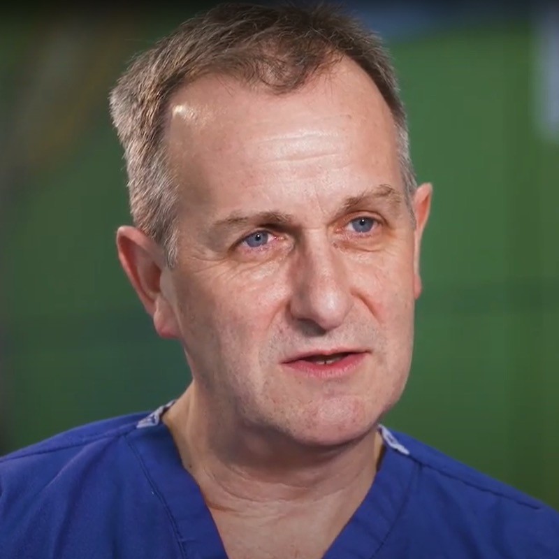 David Jayne合作论文数School of Medicine, University of Leeds;National Institute for Health and Care Research4
 E. Liniker合作论文数3
 Georgina Ellison-Hughes合作论文数School of Basic & Medical Biosciences, Faculty of Life Sciences & Medicine, King’s College London3Alexander J. Macrobert合作论文数3Gemma Gossedge合作论文数2Prashant Ruchaya合作论文数2Aj Macrobert合作论文数2Alexander J MacRobert合作论文数2
 Robert Walmsley合作论文数2
 Karen E Porter合作论文数2
 Qutayba Almerie合作论文数2
 Forraz Nico合作论文数2
 Andrew J. Smith合作论文数Institute of Membrane and Systems Biology, University of Leeds2Schumann Sophie合作论文数2
 Stephen Muench合作论文数School of Biomedical Sciences, University of Leeds2John Colyer合作论文数2M. Jill Saffrey合作论文数Department of Anatomy & Embryology, University College London2M.A. Birch合作论文数2Adrian Whitehouse合作论文数Molecular Medicine Unit;University of Leeds;St James ' s University Hospital2G. Gossedge合作论文数1G. M. Ellison-Hughes合作论文数1M Jurga合作论文数1ross hawkins合作论文数1Malcolm Hawkins合作论文数1Michael Sweeney合作论文数1Anuj Tripathi合作论文数1Melanie Moore合作论文数1B Nadal-Ginard合作论文数1J. Boyle合作论文数Natl Inst Biol Stds & Controls1
 Fiona Lewis-McDougall合作论文数William Harvey Research Institute, Barts and The London School of Medicine and Dentistry, Queen Mary University of London, London, UK1CP McGuckin合作论文数1
 Marilena Loizidou合作论文数UCL, London, England1
 Krystyna Domanska-Janik合作论文数Mossakowski Med Res Ctr, Polish Acad Sci1Elaine Gray合作论文数1Jablonska Anna合作论文数1Drela Katarzyna合作论文数1O'Grady Anna合作论文数1
 Yazan S Khaled合作论文数Department of Hepato-Pancreato-Biliary Surgery, North Manchester General Hospital1Lewis Fiona C合作论文数1Strojek Lukasz合作论文数1
 Jinju Chen (陈金菊)合作论文数School of Materials and Energy, University of Electronic Science and Technology of China1Stacey Glyn合作论文数1
 Colin P. Mcguckin合作论文数Cell Therapy Research Institute, Parc Technologique de Lyon Saint-Priest1
 Chris J. Burns合作论文数1
 Anna Sarnowska合作论文数Mossakowski Medical Research Centre, Polish Academy of Sciences1Jacquelyn Bond合作论文数Molecular Medicine Unit, University of Leeds1
 Alan Melcher合作论文数Division of Radiotherapy and Imaging, The Institute of Cancer Research1




