Cancer cell invasion through physically confined spaces is governed by the biophysical interplay between morphological plasticity and mechanical constraints. A fundamental but unresolved question is how cells sustain efficient movement under such confinement without compromising their global volume homeostasis. Here, using a Matrigel-overlay quasi-three-dimensional system, we show that confinement-enhanced migration does not arise from static structural changes, but from a reorganization of volume-surface dynamics accompanied by changes in cellular mechanical state. To quantify this reorganization, we introduce a volume-surface dynamic correlation coefficient, . This metric decreases significantly during confined migration, indicating a state of weak coupling between volume regulation and surface remodeling. Perturbations of processes essential for both motility and volume homeostasis consistently increase , despite affecting cell behavior through distinct routes. Together, these findings identify weak volume-surface coupling as a dynamic descriptor of efficient migration under confinement.
BACKGROUND:Emery-Dreifuss muscular dystrophy (EDMD) is a rare genetic disorder characterized by early-onset joint contractures, progressive muscle atrophy, and cardiac abnormalities. Patients with EDMD carrying LMNA sequence variations often exhibit severe cardiac manifestations, including frequent atrioventricular block and ventricular tachycardia. Approximately 20% of those patients may ultimately require heart transplantation. The molecular mechanisms by which LMNA sequence variations lead to EDMD remain unknown. METHODS:Five clinically diagnosed patients with EDMD carrying LMNA sequence variations were recruited. Patient-specific induced pluripotent stem cells (iPSCs) were generated using a nonintegrating Sendai virus. Previously generated iPSCs, derived from 2 healthy donors, were used as controls. The LMNA L204P sequence variation was corrected by genome editing in EDMD iPSC lines to generate isogenic controls. All iPSC-derived cardiomyocytes (iPSC-CMs) were generated using a monolayer-based differentiation protocol. Three-dimensional, strip-format, and force-generating human engineered heart tissues were generated from iPSC-CMs. A knock-in mouse model carrying the Lmna L204P sequence variation was also generated. RESULTS:EDMD-specific iPSC-CMs exhibited a variety of deleterious phenotypes, including disorganized sarcomeres, abnormal nuclear envelope structure, arrhythmias, and contractile dysfunction, when compared with control and gene-corrected iPSC-CMs. Multi-omics analysis further revealed that LMNA directly binds the WNT5A promoter and the Leu204Pro sequence variation reduces chromatin accessibility and WNT5A transcription in EDMD iPSC-CMs. WNT5a (Wnt family member 5a)/RhoA (Ras homolog family member A) signaling inactivation was shown to lead to actin depolymerization and inhibition of actin polymerization in EDMD iPSC-CMs. This results in a deformed nuclear envelope, contractile dysfunction, and impaired trafficking of Cx43 (connexin 43). The impairment of Cx43 trafficking causes reduced distribution of Cx43 at cell-cell borders, contributing to the arrhythmic phenotype in EDMD iPSC-CMs. Pharmacological interventions of exogenous WNT5a supplementation, RhoA activator, or an actin polymerization stabilizer effectively rescued the pathogenic phenotypes of EDMD iPSC-CMs. EDMD engineered heart tissues displayed dysfunctional contractile force generation, which was significantly alleviated by RhoA activator. Lmna L204P heterozygous knock-in mice exhibited impaired cardiac function and developed cardiac arrhythmias in response to sympathetic stress. CONCLUSIONS:We present WNT5a-mediated aberrant actin filament dynamics as a novel mechanism underlying cardiac pathogenic phenotypes in LMNA-related EDMD. Our findings indicate that activating WNT5a/RhoA and stabilizing actin assembly may serve as novel therapeutic strategies for this condition.
Transient receptor potential vanilloid-1 (TRPV1) plays a critical role in noxious heat sensation and pain hypersensitivity development in chronic pain. Sustained or repeated exposure to capsaicin, a classic TRPV1 agonist, induces TRPV1 desensitization. This partially accounts for the analgesic effect of capsaicin. However, the regulatory mechanisms of TRPV1 desensitization remain poorly understood. In this study, we found that capsaicin acts on TRPV1 to induce the activation of calpain, a Ca2+-sensitive cysteine protease, in a manner unrelated to cellular injury. Calpain cleaves rTRPV1 at the carboxyl terminus of G819/S820. Lack of the distal carboxyl terminus leads to reduced TRPV1 localization on the plasma membrane, potentially due to increased receptor internalization and impaired subunit assembly. This finding was corroborated by whole-cell patch clamp recordings. Additionally, the Δ820-838 mutant of rTRPV1 shows resistance to tachyphylaxis as induced by repetitive capsaicin stimulation. This study reveals a pivotal role for calpain in TRPV1 desensitization where its activation constrains TRPV1 channel function while simultaneously increasing its resistance to tachyphylaxis, thereby acting to maintain TRPV1 activity within an appropriate range. ### Competing Interest Statement The authors have declared no competing interest. Ministry of Science and Technology of China STI 2030-Major Projects, , 2021ZD0203202 National Natural Science Foundation of China, , 32271190, 31771295, 82370354, 82400372 LingYan Research and Development Project, , 2024C03155
Sunitinib is a receptor tyrosine kinase inhibitor used for the treatment of renal cell carcinoma and imatinib-resistant gastrointestinal stromal tumors. Clinical data have shown that patients receiving sunitinib develop reduced cardiac function, arrhythmia and heart failure, thereby largely limiting its clinical use. However, the molecular mechanisms underlying sunitinib-induced arrhythmogenesis remain unclear. Here, utilizing the human induced pluripotent stem cell-derived cardiomyocyte (iPSC-CM) model, we found that sunitinib caused a variety of deleterious phenotypes, including cardiomyocyte death, sarcomeric disorganization, irregular Ca2+ transients, impaired ATP2A2a/SERCA2a (ATPase sarcoplasmic/endoplasmic reticulum Ca2+ transporting 2a) activity, arrhythmia, and excessive macroautophagy/autophagy. Mechanistically, SQSTM1/p62 (sequestosome 1) interacts with MYLK3 (myosin light chain kinase 3) and drives excessive autophagic degradation of MYLK3 in sunitinib-treated iPSC-CMs. Downregulation of MYLK3 suppresses the phosphorylation of CAMK2/CAMKII (calcium/calmodulin dependent protein kinase II), thereby reducing the phosphorylation level of its downstream substrate PLN (phospholamban), leading to impaired ATP2A2a/SERCA2a activity and subsequent Ca2+ dyshomeostasis and arrhythmia. Moreover, pharmacological intervention of the cardiac myosin activator omecamtiv mecarbil (OM) or overexpression of MYLK3 significantly restored the expression of MYLK3 and reversed pathogenic phenotypes in sunitinib-treated iPSC-CMs. Nanoparticle delivery of OM effectively prevented sunitinib-induced cardiac dysfunction in mice. Our findings suggest that sunitinib-induced MYLK3 degradation causes the inhibition of the CAMK2-PLN-ATP2A2a signaling pathway and leads to sunitinib-induced arrhythmogenesis, and that MYLK3 can act as a novel cardioprotective target for sunitinib-induced cardiotoxicity.Abbreviation: ACTN:actinin alpha;APD:action potential duration; ATG:autophagy related;ATP2A2a/SERCA2a:ATPase sarcoplasmic/endoplasmicreticulum Ca2+ transporting 2a;BafA1:bafilomycin A1;Caff: caffine; CAMK2/CAMKII:calcium/calmodulin dependent protein kinase II;CASP3:caspase 3;CQ, chloroquine;DADs:delayed afterdepolarizations; EAD:early afterdepolarization; ECG: electrocardiogram; EF: ejectionfraction; FS: fractional shortening; iPSC:inducedpluripotent stem cell;iPSC-CM: inducedpluripotent stem-cell-derived cardiomyocyte;ISO: isoprenaline; LVIDs: left ventricular end systolic diameter;LVIDd: left ventricular end diastolic diameter;MAP1LC3/LC3:microtubuleassociatedprotein 1 light chain 3;MYL2v/MLC2v:myosin light chain 2 v;MYLK3:myosin light chain kinase 3;OE: overexpression; OM:omecamtiv mecarbil; PLN: phospholamban;SIC:sunitinib-induced cardiotoxicity; SR:sarcoplasmic reticulum; TUNEL:TdT-mediated dUTP nick end labeling.
Correction for 'In vitro vascularized liver tumor model based on a microfluidic inverse opal scaffold for immune cell recruitment investigation' by Pingwei Xu et al., Lab Chip, 2024, 24, 3470-3479, https://doi.org/10.1039/D4LC00341A.
Acute kidney injury (AKI) is a clinical syndrome with high mortality, and its pathogenesis involves complex inflammatory regulatory mechanisms. As core components of the cytokine network, interleukins (ILs) exert pleiotropic effects in the development of AKI, participating in processes such as inflammation, fibrosis, tissue damage repair, and remote organ injury. Moreover, ILs influence the progression of AKI by mediating the crosstalk among renal resident cells, immune cells, and fibroblasts. Pro-inflammatory ILs primarily accelerate the progression of AKI by recruiting neutrophils and inducing renal cell apoptosis, whereas anti-inflammatory ILs alleviate AKI by inhibiting the release of inflammatory cytokines and enhancing regulatory T cell function. Dual-function ILs may either promote disease progression or facilitate tissue repair depending on their cellular origin or the specific pathological stage. In terms of therapeutic strategies, monoclonal antibodies targeting ILs and their receptors, as well as advancements in extracellular vesicle technology, have shown promising potential. Future research should focus on elucidating the specific signaling networks of ILs and their intercellular interactions in order to promote precision medicine approaches for AKI and to block the transition from AKI to chronic kidney disease (CKD).
With the rapid development of wearable technology, multifunctional sensors have demonstrated immense application potential. However, the limitations of traditional rigid materials restrict the flexibility and widespread adoption of such sensors. Hydrogels, as flexible materials, provide an effective solution to this challenge due to their excellent stretchability, biocompatibility, and adaptability. This study developed a multifunctional flexible sensor based on a composite hydrogel of polyvinyl alcohol (PVA) and sodium alginate (SA), using poly(3,4-ethylenedioxythiophene)/polystyrene sulfonate (PEDOT:PSS) as the conductive material to achieve multifunctional detection of strain, temperature, and physiological signals. The sensor features a simple fabrication process, low cost, and low impedance. Experimental results show that the prepared hydrogel exhibits outstanding mechanical properties and conductivity, with a strength of 118.8 kPa, an elongation of 334%, and a conductivity of 256 mS/m. In strain sensing, the sensor demonstrates a rapid response to minor strains (4%), high sensitivity (gauge factors of 0.39 for 0–120% and 0.73 for 120–200% strain ranges), short response time (2.2 s), low hysteresis, and excellent cyclic stability (over 500 cycles). For temperature sensing, the sensor achieves high sensitivities of −27.43 Ω/K (resistance mode) and 0.729 mV/K (voltage mode), along with stable performance across varying temperature ranges. Furthermore, the sensor has been successfully applied to monitor human motion (e.g., finger bending, wrist movement) and physiological signals such as electrocardiogram (ECG), electromyogram (EMG), and electroencephalogram (EEG), highlighting its significant potential in wearable health monitoring. By employing a simple and efficient fabrication method, this study presents a high-performance multifunctional flexible sensor, offering novel insights and technical support for the advancement of wearable devices.
Dear Editor, Human pluripotent stem cell-derived cardiomyocytes(hPSC-CMs)are of great values for developmental studies,disease modeling,drug screening,cell therapy,and beyond[1].Achieving high-purity of hPSC-CMs is essential for promoting their application.
The decrease of peritubular capillaries, a pathological feature of acute kidney injury (AKI), is a critical part that promotes the transition of AKI to chronic kidney disease (CKD). Ginsenoside Rb1 (Rb1) has various pharmacological effects on multiple systemic diseases. However, whether Rb1 delays the transition of AKI to CKD and the mechanism are unclear. Here, we discovered that Rb1 could alleviate kidney pathological damage in mice with unilateral ischaemia/reperfusion injury (uIRI), and it enhanced kidney function, reduced renal fibrosis while increasing microvessel density. By virtual screening and molecular docking approaches, we identified vascular endothelial growth factor receptor 2 (VEGFR2) as the principal molecular target of Rb1. Furthermore, we uncovered that Rb1 activated AKT phosphorylation-mediated angiogenesis through binding to VEGFR2, which promoted endothelial tube formation and nitric oxide (NO) release in human umbilical vein endothelial cells (HUVECs) and then relieved the endothelial dysfunction induced by hypoxia-reoxygenation (H/R). Also, we found that Rb1 activated VEGFR2/AKT signalling in the kidney tissue of uIRI mice. Knocking down VEGFR2 or inhibiting the AKT signalling pathway can impair the proangiogenic effect of Rb1. Taken together, we demonstrated that Rb1 facilitated renal angiogenesis by activating the VEGFR2/AKT pathway of endothelial cells, thereby arresting the transition from AKI to CKD, and providing a potential therapeutic strategy for AKI.
The gut microbiota of wild animals is characterized by both stability and adaptive shifts in composition and prevalence in response to variation in food availability, nutrient intake, host physiology, temperature, and rainfall. Here, over a 12-month period, we investigated seasonal interactions between diet, weather, and gut microbiota in a wild group of Tibetan macaques in Huangshan by recording feeding behavior, monitoring weather, and analyzing 209 fecal samples using plant DNA metabarcoding (trnL region) and 16S rRNA gene sequencing. Based on the field observations and plant DNA metabarcoding, results revealed marked seasonal shifts in plant types and species consumed by Tibetan macaques. Despite dietary variability, only two enterotypes were presented throughout the year and gut microbiota composition exhibited lower dissimilarity within and across seasons compared to diet, except in autumn when low dietary diversity correlated with reduced microbial diversity. In addition, we also found that the enrichment of seasonal indicator bacterial genera and functions was related to the temperature or the nutrients of the food consumed by Tibetan macaques during that season. This study highlights the microbiota's resilience and metabolic plasticity in buffering seasonal dietary shifts, underscoring its role in maintaining host energy homeostasis under fluctuating resource availability.
Cell-scale curvature is a key regulator of cell migration, yet its quantitative effects and underlying mechanisms remain elusive. Here, we combine controlled in vitro experiments with a phenomenological theoretical framework to investigate the migration of fibroblasts (NIH3T3) and epithelial cells (MCF10A) on the inner concave surfaces of polydimethylsiloxane microcylinders across a wide range of cell-scale curvatures (∼0.01 per micrometer). We find that migration persistence positively correlates with mean speed across all curvatures, consistent with the universal speed-persistence coupling relation previously observed for cells migrating on 1D and 2D planar substrates, as well as for cells embedded in 3D environments. Cell migration inside microcylinders is stochastic and anisotropic, as quantified by the nematic order parameter, and exhibits a biphasic dependence on curvature. At small curvatures, cells remain fully adhered to the surface, with anisotropy and speed both increasing while persistence decreases. When curvature exceeds a threshold of approximately 1/75 per micrometer, cells detach by forming stress-fiber chords, leading to reduced anisotropy and speed but increased persistence. This adhered-to-chord transition is followed by a shift in preferred orientation: migration initially favors the lateral direction and progressively aligns toward the axis at larger curvatures. These findings demonstrate that cells can actively reorient their stress fibers and migration in response to local cell-scale curvature sensed by the entire cell, even on cylindrical surfaces with constant mean curvature and vanishing Gaussian curvature. A modified persistent random walk model, incorporating persistent randomness and curvature-dependent directionality via bending and adhesion energetics, quantitatively captures these behaviors and predicts the transition threshold in close agreement with experiments. Together, this work establishes a quantitative framework for biphasic, curvature-dependent migration and provides new insight into how local geometry regulates mesenchymal motility.
Xiaochen Wang studied plant biology as a Ph.D. student at Peking University, China, and worked on programmed cell death as a post-doctoral fellow at University of Colorado at Boulder. Wang set up her own research group to initially investigate the clearance of apoptotic cells by lysosomes and later redirected her research to decipher lysosome dynamics and functions in a multicellular organism. Lysosomes are major degradative organelles and signaling centers in the cell that play important roles in a wide variety of processes to maintain cell and tissue homeostasis. Lysosome dysfunction is associated with metabolic disorders, neurodegenerative diseases, and age-related pathologies. As the burier of dead cells, lysosomes degrade apoptotic cells delivered via phagocytosis to enable a safe funeral without stimulating inflammatory responses. The Wang lab has systematically dissected the regulatory pathways by which apoptotic cells are recognized and engulfed by phagocytes, and delivered to and digested by lysosomes. Intrigued by the highly changeable morphology and versatile functions of lysosomes, Wang and colleagues developed C. elegans as a multicellular model to investigate how lysosome dynamics and functions are regulated to maintain animal development and longevity.
Chronic kidney disease (CKD) is an important disease affecting human health, especially in developing countries where CKD has high prevalence and mortality rates.[1] According to the latest CKD epidemiological data, the prevalence of CKD among Chinese people is 8.2%, of which 1.8% are patients with CKD stage 5 (known as end-stage renal disease [ESRD]).[2] Hemodialysis (HD) is the main treatment for ESRD patients. According to the China National Renal Data System (CNRDS), the number of people receiving HD treatment in 2022, was as high as 844,000. Although dialysis can partially replace the function of the kidneys, distant complications in dialysis patients, such as cardiovascular disease (CVD), renal bone disease, and infections, seriously affect the quality of life and prognosis. Among these conditions, CVD is the leading cause of death in dialysis patients.[3] In addition to traditional cardiovascular risk factors such as hypertension and diabetes, the development of CVD has been associated with alterations in the gut microbiota and its metabolites. HD patients and healthy subjects were varied in terms of the number and composition of the gut microbiota. In 2013, Vaziri et al[4] first reported that Firmicutes (new name, Bacillota), Proteobacteria, and Actinobacteria were significantly enriched in HD patients. Later, Wu et al[5] and Shivani et al[6] also reported similar results. Wu et al[5] reported an increased abundance of Bacillota and Proteobacteria and a decreased abundance of Bacteroidetes in HD patients compared to controls. Another study showed a relative increase in Bacillota, Actinobacteria, and Fusobacteria and a relative decrease in Bacteroidetes and Proteobacteria.[6] In a study of children with HD, the relative abundance of Bacteroidetes increased, while that of Proteobacteria decreased.[7] At the genus level, the abundance of Bacteroides increased in HD.[6] These results suggest differences in the abundance of the gut microbiota between ESRD patients who underwent HD and healthy subjects. However, little is known about the changes in the microbiota of ESRD patients before and after dialysis. He et al[8] and Luo et al[9] compared changes in the gut microbiota before and after dialysis in ESRD patients. There was increased abundance of Bifidobacteria and Lactobacillus acidophilus and decreased abundance of Escherichia coli and Enterococcus faecalis in HD patients compared to pre-dialysis ESRD patients.[8] In addition, the abundances of Blautia, Akkermansia, Coprococcus, Proteus, Pseudomonas, and Acinetobacter increased, while the abundances of Prevotella and Paraprevotella decreased in HD patients.[9] Significant alterations in the gut microbiota of HD patients are thought to be related to the uremic environment, especially the HD process itself. Some other factors specific to this population, such as alterations in diet and medication composition, are also thought to contribute. In uremia patients with renal-excretion function decreased, urea translocated into the intestinal lumen increases; urea is broken down by urease-containing bacteria into ammonia (CO[NH2]2 + H2O → CO2 + 2NH3); ammonia is further converted to NH4OH, which can damage the intestinal mucosa and cause gut microbiota dysbiosis. HD patients are unique in that they undergo dialysis at least three times a week (3–5 h each time); and the effects of vascular access, hemodynamic instability, and complications during dialysis can affect the gut microbiota. The special dietary structure of HD patients, such as low intake of dietary fiber and the restriction of potassium-, sodium-, and phosphorus-containing foods, are known to be important factors affecting the gut microbiota. Furthermore, drug interventions, such as phosphate binders, potassium binders, iron supplements, and antibiotic interventions, can cause changes in the flora. All these factors can lead to differences in the gut microbiota between HD patients and healthy people. These studies tended to include patients who were undergoing maintenance HD (dialysis for at least 3 months), who had not used antibiotics or immunosuppressants within 3 months, or who had excluded cancer.[4–6] Interestingly, even for the same bacterium, results varied in different studies, with Bacteroidetes being reduced in adult HD patients[5,6] and increasing in children with HD.[7] This can be attributed to differences in age, race, geographic location, lifestyle, primary disease, dialysis adequacy, and vascular access. A shift in the host gut environment and microbiome from a symbiotic to a dysbiotic state is associated with increased production of uremic toxins. Indoxyl sulfate (IS), p-cresol sulfate (pCS), and trimethylamine N-oxide (TMAO) accumulate in CKD patients. The IS and pCS concentrations in predialysis patients were 116 and 41 times greater than those in healthy individuals, respectively.[10] Due to the tight binding of IS and pCS to albumin, the dialysis clearance of IS and pCS was only 0.21-fold and 0.39-fold, whereas the dialysis clearance of urea and creatinine was up to 4.2-fold and 1.3-fold, respectively.[10] Serum TMAO concentrations were approximately 30-fold greater in HD patients than in healthy individuals.[11] These protein-bound uremic toxins have been shown to be important contributors to the development of CVD.[12] IS, a metabolite of tryptophanase-containing bacteria, induces endothelial dysfunction through oxidative stress, induces endothelial cell senescence through increased reactive oxygen species (ROS) and p53 activity, and is also involved in thrombosis and vascular calcification.[13]pCS, a metabolite of tyrosine, promotes ROS production by increasing nicotinamide adenine dinucleotide phosphate (NADPH) oxidase activity and is associated with impaired left ventricular diastolic function and cardiac apoptosis.[14] IS and pCS are considered markers of endothelial cell injury, and both have proinflammatory effects and are involved in vascular injury.[15] TMAO, a metabolite of cholines or trimethylamines, has been identified as a predictor of CVD and is involved in the formation of atherosclerosis. TMAO can be mediated by the inhibition of reverse cholesterol transport, the induction of the macrophage expression of scavenger receptor A (SRA) and CD36, and the promotion of foam cell formation.[16] At present, interventions to regulate gut microbiota imbalances and remove gut-derived uremic toxins include probiotics and prebiotics. The use of probiotics/prebiotics has been shown to be effective at restoring the gut microbial composition and reducing uremic toxins. However, there is a lack of clear guidelines for informing HD patients when to take these dietary supplements, as well as the types and dosages that must be taken, which can be confusing for clinicians when prescribing them to their patients. Fecal microbiota transplantation (FMT), an emerging therapy for modulating the gut microbiota, has therapeutic potential for recurrent Clostridioides difficile infections (rCDIs), inflammatory bowel disease, and gastrointestinal tract tumors.[17] However, there is a lack of basic and clinical trial-based confirmation of the efficacy of FMT in HD patients. In conclusion, there is still much work to be done on the study of intestinal microecological status in HD patients. In addition, the special and complex pathophysiological status of HD patients brings challenges to related research. Funding This research was funded by grants from the National Natural Science Foundation of China (Nos. 62271506 and61971441), the National Key R&D Program of China (No. 2021YFC1005300), and the Jinzhongzi project of Beijing Chao-yang Hospital (No. CYJZ202203). Conflicts of interest None.
LMNA mutation related Emery-Dreifuss muscular dystrophy (LMNA-related EDMD), is a rare genetic disorder often involving life-threatening cardiac complications. However, the molecular links between LMNA mutations and their related EDMD cardiac phenotypes have remained unclear. Here, using EDMD patient-specific and genome-edited induced pluripotent stem cell-derived cardiomyocytes (iPSC-CMs), we link the LMNA L204P mutation with the pathogenic phenotypes of arrhythmia and contractile dysfunction. Using multi-omics analysis, we then show that LMNA L204P results in decreased chromatin accessibility, leading to the downregulation of JAK2 in EDMD iPSC-CMs. Mechanistically, JAK2/STAT3 signaling pathway suppression in EDMD iPSC-CMs is shown to cause mitochondrial dysfunction and oxidative stress, ultimately resulting in the above phenotypes. Conversely, pharmacological or genetic activation of JAK2/STAT3 signaling effectively rescues both the arrhythmic and contractile dysfunction phenotypes in EDMD iPSC-CMs via improvements in mitochondrial function. In addition, whilst EDMD engineered heart tissues (EHTs) display dysfunctional contractile force generation, this can also be significantly alleviated by STAT3 activation. Taken together, we present chromatin compartment change-mediated JAK2/STAT3 suppression as a novel mechanism underlying cardiac pathogenic phenotypes in LMNA-related EDMD. Our findings indicate that activating the JAK2/STAT3 signaling pathway may hold the potential to serve as a novel therapeutic strategy for this condition.
The maintenance of lysosome membrane integrity is vital for cell homeostasis and viability, but the underlying mechanisms are not well understood. In this study, we identified a novel role of SPHK-1, the sole Caenorhabditis elegans sphingosine kinase, in protecting lysosome membrane integrity. Loss of SPHK-1 affects lysosomal integrity and degradative function, causing cargo accumulation and lysosome membrane rupture. sphk-1(lf) mutants show severe defects in embryonic and larval development and have significantly shortened lifespan. We found that sphk-1(lf) mutants accumulate high levels of sphingosine, predominantly in lysosomes. Accordingly, sphingosine supplementation leads to the appearance of damaged lysosomes in wild-type worms. We identified sptl-1 and sptl-3 mutations that fully suppress the lysosomal integrity defects in sphk-1(lf) mutants. sptl-1 and sptl-3 encode serine palmitoyltransferases that catalyze the first and rate-limiting step of de novo sphingolipid synthesis. Loss of sptl-1 alleviates sphingosine accumulation, reverses lysosomal integrity and degradation defects, and restores normal development and longevity in sphk-1(lf) mutants. Our study indicates that sphingolipid metabolism via sphingosine kinase is important for maintaining lysosome membrane integrity and function, and is essential for animal development and longevity.
Nature Cell Biology turns 25 years old, we asked cell biologists across the globe to share their thoughts on what a productive mentor–mentee relationship looks like and their views on training the next generation of cell biologists.
Lysosomes are degradation and signalling centres crucial for homeostasis, development and ageing1. To meet diverse cellular demands, lysosomes remodel their morphology and function through constant fusion and fission2,3. Little is known about the molecular basis of fission. Here we identify HPO-27, a conserved HEAT repeat protein, as a lysosome scission factor in Caenorhabditis elegans. Loss of HPO-27 impairs lysosome fission and leads to an excessive tubular network that ultimately collapses. HPO-27 and its human homologue MROH1 are recruited to lysosomes by RAB-7 and enriched at scission sites. Super-resolution imaging, negative-staining electron microscopy and in vitro reconstitution assays reveal that HPO-27 and MROH1 self-assemble to mediate the constriction and scission of lysosomal tubules in worms and mammalian cells, respectively, and assemble to sever supported membrane tubes in vitro. Loss of HPO-27 affects lysosomal morphology, integrity and degradation activity, which impairs animal development and longevity. Thus, HPO-27 and MROH1 act as self-assembling scission factors to maintain lysosomal homeostasis and function. The conserved HEAT repeat protein HPO-27 is identified as a lysosome scission factor in Caenorhabditis elegans, and the human homologue MROH1 also serves the same function to maintain lysosomal homeostasis.
The composition of the gut microbiota varies among end-stage renal disease (ESRD) patients on the basis of their mode of renal replacement therapy (RRT), with notably more pronounced dysbiosis occurring in those undergoing hemodialysis (HD). Interventions such as dialysis catheters, unstable hemodynamics, strict dietary restrictions, and pharmacotherapy significantly alter the intestinal microenvironment, thus disrupting the gut microbiota composition in HD patients. The gut microbiota may influence HD-related complications, including cardiovascular disease (CVD), infections, anemia, and malnutrition, through mechanisms such as bacterial translocation, immune regulation, and the production of gut microbial metabolites, thereby affecting both the quality of life and the prognosis of patients. This review focuses on alterations in the gut microbiota and its metabolites in HD patients. Additionally, understanding the impact of the gut microbiota on the complications of HD could provide insights into the development of novel treatment strategies to prevent or alleviate complications in HD patients.
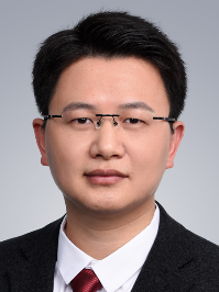 Shixiong Chen (陈世雄)合作论文数School of Medicine, The Chinese University of Hong Kong, Shenzhen19
 Guanglin Li (李光林)合作论文数Research Center for Neural Engineering, Shenzhen Institute of Advanced Technology, Chinese Academy of Sciences;School of Artificial Intelligence, University of Chinese Academy of Sciences19
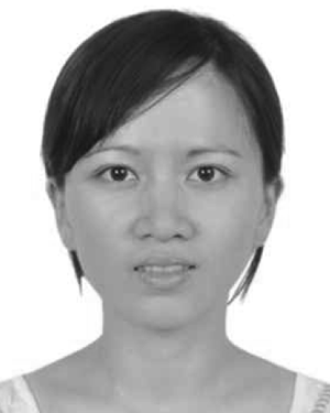 Mingxing Zhu合作论文数19
 Fangfu Ye (叶方富)合作论文数Institute of Physics, Chinese Academy of Sciences;Wenzhou Institute, University of Chinese Academy of Sciences17
 Xin Wang (王昕)合作论文数School of Mechanical Engineering and Automation, Harbin Institute of Technology, Shenzhen17
 Chonglin Yang (杨崇林)合作论文数Center for Life Sciences, School of Life Sciences, Yunnan University17
 Qihui Fan合作论文数13
 Hong Zhang (张宏)合作论文数State Key Laboratory of Biomacromolecules, Institute of Biophysics, Chinese Academy of Sciences10
 Ping Liang (梁平)合作论文数Institute of Translational Medicine, School of Medicine, Zhejiang University9
 Wei Zou (邹炜)合作论文数Institute of Translational Medicine, Zhejiang University9Meijiao Li合作论文数8Zijian Yang合作论文数Chinese Acad Sci, Shenzhen Inst Adv Technol, CAS Key Lab Human Machine Intelligence Synergy Sy, Shenzhen 518055, Peoples R China8
 Ding Xue (薛定)合作论文数Department of Molecular, Cellular & Developmental Biology, College of Arts and Sciences, University of Colorado Boulder8
 Hanyu Zhu (朱晗玉)合作论文数Chinese PLA General Hospital8
 Yan Zhang合作论文数7
 Zhang Haoshi合作论文数7
 Oluwarotimi Williams Samuel合作论文数School of Computing, College of Science and Engineering, University of Derby7
 Shiya Cheng (程世亚)合作论文数School of Basic Medical Sciences, Faculty of Medical Sciences, Wuhan University7
 Yang Jiao (焦阳)合作论文数Department of Physics, School for Engineering of Matter, Transport and Energy, Arizona State University7
 Liyu Liu (刘雳宇)合作论文数Human Phenome Institute, Fudan University7
 Shohei Mitani (三谷昌平)合作论文数Department of Physiology II, Tokyo Women's Medical University;Institute for Integrated Medical Sciences, Tokyo Women's Medical University7Xiaonan Ding合作论文数6Yu Zheng合作论文数6David K Ann合作论文数6
 Yun Yen合作论文数Graduate Institute of Cancer Biology and Drug Discovery, Taipei Medical University;Research Center of Cell Therapy and Regeneration Medicine, Taipei Medical University;International Center for Health Information Technology, College of Medical Science and Technology, Taipei Medical University6
 Qun Lu (陆群)合作论文数School of Life Science and Engineering, Southwest Jiaotong University6
 Xiyong Liu合作论文数6
 Xin Li (李欣)合作论文数College of Engineering, China Agricultural University5Junxia Du合作论文数5BangWei Zhou (周邦维)合作论文数5Yang Liu (柳阳)合作论文数5Qiuxia Han合作论文数5Pengfei Guo合作论文数5
 Baohui Chen (陈宝惠)合作论文数Department of Cell Biology, School of Basic Medical Sciences, School of Medicine, Zhejiang University;The First Affiliated Hospital, Zhejiang University School of Medicine5Dong Zhang (张东)合作论文数Chinese PLA General Hospital5Bingsen Zhou合作论文数5Cheng Wang合作论文数4Jiashuo Zhuang合作论文数4Xiaoyan LI (李小燕)合作论文数4Hanjie Deng合作论文数Chinese Acad Sci, Key Lab Human Machine Intelligence Synergy Syst, Shenzhen Inst Adv Technol, Shenzhen 518055, Guangdong, Peoples R China4Jinlan Shan合作论文数4Yanan Sun合作论文数Coll Life Sci, Beijing Normal Univ4Yan-Wei Wu合作论文数4LeiChun Zhang (张蕾春)合作论文数4WanZhang Yang (杨宛章)合作论文数4Xin Tian (田欣)合作论文数4
 Yigong Shi (施一公)合作论文数School of Life Sciences, Westlake University4
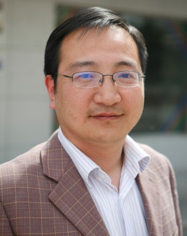 Junwei Zheng (郑军卫)合作论文数Northwest Institute of Eco-Environment and Resources, Chinese Academy of Sciences;University of Chinese Academy of Sciences4
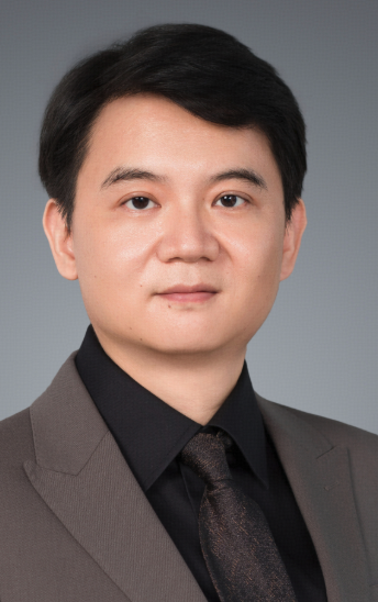 Weida Li (李维达)合作论文数School of Life Sciences and Technology, Tongji University4




