BackgroundDiabetic foot ulcers (DFUs) are among the most severe complications of diabetes, often resulting in prolonged healing, recurrent infections, and a high risk of amputation. Despite comprehensive standard treatments, many patients develop chronic non-healing ulcers due to poor tissue perfusion and immune dysregulation. Traditional Chinese medicine, such as Huafu Shengji ointment (HFSJO), offers a novel topical approach that may promote tissue repair and accelerate healing in refractory cases.Case presentationThis report describes four patients with persistent DFUs that responded poorly to conventional therapies and faced either prior amputation or an imminent risk of amputation. Ulcer sizes ranged from 3.0 cm × 3.0 cm to 7.0 cm × 5.0 cm, with comorbidities including hypertension and cardiovascular disease. In addition to routine wound care, patients received topical HFSJO treatment every other day. Within 30–180 days, all wounds showed substantial granulation, epithelialization, and eventual closure. No clinically significant abnormalities in the liver or renal function tests or routine hematological parameters were observed during treatment or follow-up.ConclusionTopical application of HFSJO appears to be a safe and effective conservative strategy for the management of complex DFUs, particularly in patients unsuitable for surgery or unresponsive to standard care. Through its dual mechanism, i.e., targeted debridement and regenerative stimulation, it may help overcome healing stagnation. These cases offer a promising direction for integrative wound care and warrant further validation in controlled clinical trials.
OBJECTIVE:To investigate the mechanism of Huiyang Shengji unguent (, HYSJ) for improving inflammation and promoting wound healing in patients with diabetic foot. METHODS:The primary components of the HYSJ unguent were analyzed using ultra-performance liquid chromatography-tandem mass spectrometry (UPLC-MS/MS). A total of 20 patients with a diabetic foot wound were divided randomly into either a HYSJ treatment group (10 cases) or control group (10 cases). The HYSJ group was treated for 14 d with Hui Yang Shengji unguent, while the control group was treated for 14 d with basic fibroblast growth factor unguent. Central granulation tissue and wound secretions were collected before treatment and on the 7th and 14th day, respectively. The proteins prospero homeobox protein 1 (PROX-1), lymphatic vessel endothelial hyaluronan receptor 1 (LYVE-1) and vascular endothelial growth factor receptor 3 (VEGFR-3) associated with lymphatic angiogenesis were detected by immunohistochemistry, the levels of inflammatory cytokines in wound exudates including interleukin-1β (IL-1β), interleukin-18 (IL-18), and vascular endothelial growth factor C (VEGF-C) were measured using enzyme-linked immunosorbent assay, while the protein levels of NOD-like receptor family, pyrin domain containing protein 3 (NLRP3), caspase-1, gasdermin D, and N-terminal gasdermin D were measured using immunoimprinting. RESULTS:UPLC-MS/MS analysis revealed that the high-abundance peak compounds in HYSJ unguent included coclaurine, glucoraphanin, citric acid, gallic acid, L-glutamine, and gentianose. Following treatment, there was a significant reduction in IL-1β and IL-18 levels in the HYSJ group's wound exudates on day 14 compared to pre-treatment values (P < 0.05). Conversely, VEGF-C levels showed a significant increase from pre-treatment levels (P < 0.05). In addition, the expression of PROX-1, LYVE-1 and VEGFR-3 in wound tissue increased significantly after 14 d of treatment when compared to pre-treatment levels (P < 0.05). Taken together, the results of these analyses provide insights into the dynamic changes of these factors in wound healing processes. On the 7th and 14th day of treatment, the expression levels of IL-β and IL-18 in wound secretions and NLRP3, caspase-1, gasdermin D, and N-terminal gasdermin D in wound tissue were significantly lower in the HYSJ group than those measured in the control group (P < 0.05). At the same time, the expressions of VEGF-C, PROX-1, and LYVE-1 in wound secretions in the HYSJ group were significantly higher than those in the control group (P < 0.05). CONCLUSIONS:The mechanism of HYSJ to promote wound healing of the Yinsyndrome in patients with diabetic foot may involve inhibition of cell necrosis mediated by the NLRP3/caspase-1/gasdermin D pathway, promotion of lymphangiogenesis, and establishment of a "protective field" for wound healing.
Purpose:To investigate the mechanism by which HYSJ unguent promotes lymphangiogenesis and improves the healing of diabetic chronic wounds (DCWs). Methods:The main components of HYSJ were identified by mass spectrometry. A mouse model with chronic skin ulcers was established. The ultrastructure and lymphatic drainage of lymphatic endothelial cells in wounds were examined. Lymphatic markers in wound tissues were detected, and proteomic and bioinformatics analyses were performed to identify differentially expressed proteins and associated pathways. In vitro, a high-glucose inflammatory environment that mimics DCWs was induced in human lymphatic endothelial cells (HLEC). HYSJ and caspase inhibitors were used for intervention. Diverse assays were conducted to assess HLEC function and activation of inflammatory cell death. Results:The primary constituents of HYSJ unguent included coclaurine, sinapine, and ononin, among others. HYSJ increased healing of DCWs in diabetic mice, protected lymphatic endothelial cells, restored lymphatic drainage in the wound, and upregulated expression of key lymphatic proteins. Numerous proteins, including TLR2, Myd88, STAT1, and inflammatory response-related proteins such as NLRP3, Caspase-1, GSDMD, were expressed differentially in mice. HYSJ unguent also stimulated expression of key lymphatic proteins in HLEC, protected cell function, and suppressed inflammatory cell death. Conclusion:HYSJ unguent enhances lymphangiogenesis, protects lymphatic endothelial cells in a high-glucose inflammatory environment, and accelerates DCW healing by suppression of TLR2/Myd88/caspase-1 signaling pathway. These findings provide important experimental support for the pharmacological mechanism by which HYSJ unguent facilitates healing of DCWs.
BACKGROUND:The Huiyang Shengji Decoction (HYSJD) is a renowned compound herbal formula known for its ability to accelerate the healing of chronic wounds, including diabetic skin ulcers(DSU). However, the precise mechanisms remain to be further investigated. PURPOSE:The primary goal of this investigation was to evaluate the therapeutic impact of HYSJD extract on DSU in a murine model. Additionally, the study sought to decipher the intricate mechanisms driving the wound healing process, leveraging a comprehensive multi-omics analysis coupled with a network pharmacology framework. MATERIALS AND METHODS:The constituents of HYSJD were characterized utilizing liquid chromatography in conjunction with tandem mass spectrometry (LC-MS/MS). A model of DSU was developed in mice,A multi-faceted approach incorporating transcriptomics, pharmacological networking, and metabolomics was employed to investigate the mechanisms by which HYSJD promotes healing of DSU. Additionally, in vivo studies were executed to substantiate the proposed mechanisms of HYSJD. RESULTS:The application of HYSJD has demonstrated efficacy in accelerating the healing process of wounds in a DSU mouse model. Through transcriptomic profiling and pharmacological networking, Sinapine and Arginine were identified as the predominant bioactive constituents exerting their effects on DSU lesions. These elements were found to suppress cellular apoptosis and modulate signaling cascades associated with inflammatory responses. Metabolomic evaluations uncovered a set of 12 distinct metabolites and 7 metabolic routes that are influenced by HYSJD's intervention in DSU. Supplementary experimental data validated the capacity of HYSJD to regulate the NF-κB/STAT3/NLRP3 signaling axis, which in turn, manages inflammatory mediators in both wound tissue and serum, while also curbing cellular apoptosis. CONCLUSION:HYSJD augments the wound healing capability in diabetic mice by mitigating cellular apoptosis and diminishing inflammatory activity, attributable to its regulatory effect on the NF-κB/STAT3/NLRP3 signaling pathway.
Background: Allergic-related skin diseases, including atopic dermatitis (AD), urticaria, and contact dermatitis (CD), are significant global public health challenges. Currently, there is a lack of systematic analysis of allergic-related skin diseases globally. Methods: This study aimed to quantify the global burden of AD, CD, and urticaria and evaluate their global epidemiology patterns. The Global Burden of Diseases (GBD) database was used to assess incidence, prevalence, and disability-adjusted life years (DALYs) for these allergic-related skin diseases. Additionally, the Bayesian Age-Period-Cohort (BAPC) model was employed to predict disease burden for the next 15 years. Results: From 1990 to 2021, cases of AD, CD, and urticaria rose steadily. In 2021, AD prevalence reached 129 million, a 20.02% increase from 1990. However, average annual percentage change (AAPC) values for the age-standardized prevalence rate (ASPR) of AD declined constantly (AAPC = −0.28). CD had the highest incidence, with 253 million new cases in 2021, though AAPC for ASPR of CD showed minimal changes. AD and urticaria peaked in early life, while CD peaked at ages 75–79. Moreover, AD had the strongest positive correlation with the Socio-demographic Index (SDI) (p = 2.2e-16, ρ = 0.626). AD, CD, and urticaria show the highest age-standardized rate in high, middle, and low-middle SDI regions, respectively, with all 3 conditions declining in high SDI. Health inequality analysis showed AD's burden is now more evenly distributed across SDI groups, while the global burden gap for urticaria and CD change limitedly. Conclusion: Although the global disease burden of allergic-related skin diseases continues to rise, the overall age-standardized rates of AD have steadily declined and are projected to decrease further. In contrast, CD and urticaria require increased attention.
Objective: Atopic dermatitis (AD) is an allergic inflammatory skin disease. Changes in circulating inflammatory proteins are reflected in the entire process of AD progression, and its pathophysiology is still unclear. This Mendelian randomization study was conducted to further evaluate the role of circulating inflammatory proteins in AD. Methods: This study investigated the potential causal relationship between circulating inflammatory proteins and AD. We used a two-sample Mendelian randomization (MR) method to analyze data from a large-scale genome-wide association study to explore the relationship between 91 circulating inflammatory proteins, 41 inflammatory factors, and CRP and AD. The inverse variance weighted method was mainly used to evaluate the causal relationship between exposure and outcome based on the effect indicator odds ratio (OR) and 95% confidence interval (CI). In addition, MR-Egger, weighted median, simple model, weighted model and MR-PRESSO multiple sensitivity analyses were applied to strengthen the final results. The leave-one-out method, heterogeneity test, and horizontal gene pleiotropy test were used to verify the stability and reliability of the results. Results: Forward MR analysis showed that there was a significant correlation between AD risk and changes in the levels of multiple inflammatory proteins at different p-value thresholds, among which increased levels of interleukin-18 receptor 1 were found to increase the risk of AD, which was significant in all three groups of analysis (P IVW<0.05); increased levels of C-X-C motif chemokine 9 and Fms-related tyrosine kinase 3 ligand were found to reduce AD risk at P<5×10⁻⁸ and p<5×10⁻⁷ thresholds; increased levels of C-X-C motif chemokine 11 were found to be associated with a reduced risk of AD at P<5×10⁻⁸ and P<5×10⁻⁶ thresholds (P IVW<0.05). Reverse MR analysis showed that increased AD risk was associated with decreased levels of AXIN-1, natural killer cell receptor 2B4, interleukin-1 receptor subunit α, and interleukin-33 (P IVW<0.05). In addition, increased AD risk was associated with increased Cystatin D levels (P IVW<0.05). In the 41 inflammatory factor data sets, increased AD risk may be associated with increased IL18 levels (P IVW=0.036) and MIG levels (P IVW=0.046). No significant heterogeneity and horizontal pleiotropy were observed in the analysis. After verification MR analysis, it was found that there was a significant association between the levels of inflammatory proteins such as Fms-related tyrosine kinase 3 ligand, interleukin 18 receptor 1, C-X-C motif chemokine 9, and tumor necrosis factor ligand superfamily member 14 and AD risk, and there was consistency between different P value thresholds. Bidirectional MR showed that there was a complex bidirectional causal relationship between interleukin 18 receptor 1 levels and AD. The leave-one-out analysis showed that the results were stable, there were no instrumental variables that had a strong impact on the results, and the leave-one-out method verified the robustness of the results. There was heterogeneity test and horizontal pleiotropy in the reverse causal relationship between the level of tumor necrosis factor ligand superfamily member 14 and the AD validation set. Conclusion: The results of MR analysis indicate a potential causal relationship between circulating inflammatory proteins and AD. This study provides a new approach for exploring the biological mechanisms of AD in the future and proposes possible therapeutic targets. Further research is needed to confirm these results and understand the specific role of these proteins in AD, and to provide reference value for future studies on the relationship between circulating inflammatory proteins and AD.
目的:从促进内皮祖细胞(EPCs)动员的角度,研究回阳生肌膏促进糖尿病足溃疡阴证创面愈合的可能机制.方法:BABL/c雄性小鼠随机分为空白组、模型组、回阳生肌膏组,采用链脲佐菌素-激素-创面-塑料环埋置方法建立糖尿病足溃疡阴证创面小鼠模型,空白组和模型组创面予凡士林纱条,回阳生肌膏组创面予回阳生肌膏纱条,每日换药1次.比较给药第7、14天各组创面形态、计算创面面积及愈合率,HE染色观察创面皮损组织形态,流式细胞术检测骨髓中EPCs(BMEPCs)及外周血中EPCs(PBEPCs)数量,硝酸还原酶法检测创面皮损组织中一氧化氮(NO)表达,ELISA法检测外周血中血管内皮生长因子(VEGF)、基质细胞衍生因子-1α(SDF-1α)的表达,对创面愈合率、BMEPCs、PBEPCs、SDF-1 α、VEGF、NO之间进行相关性分析.结果:与空白组同期比较,模型组小鼠第7、14天创面面积显著增大(P<0.01),创面愈合率显著降低(P<0.01),创面皮损组织受炎性浸润程度重时间长、新生血管及肉芽组织出现缓慢,BMEPCs、PBEPCs数量均显著降低(P<0.01),NO、SDF-1α、VEGF水平均显著降低(P<0.01);与模型组同期比较,回阳生肌膏组第7、14天创面面积显著减小(P<0.01),创面愈合率显著提高(P<0.01),降低创面炎症细胞浸润,增加创面新生血管,促进创面组织重塑,BMEPCs及PBEPCs数量显著提高(P<0.01),NO、SDF-1α、VEGF水平显著提高(P<0.01).创面愈合率与BMEPCs、PBEPCs、SDF-1α、VEGF、NO之间呈正相关(P<0.01).结论:回阳生肌膏应用于糖尿病足溃疡小鼠能增加BMEPCs、PBEPCs的数量,增强EPCs动员增殖能力,促进血管新生、创面愈合,这可能与回阳生肌膏上调VEGF、SDF-1α及NO的合成释放,刺激EPCs动员有关.
血栓闭塞性脉管炎为本虚标实之证,本虚可致标实,标实可致本更虚.该病以"脾肾亏虚"为本,"寒、毒、瘀"为标,"瘀"贯穿整个病程,临证时把握标实之间转化与影响.针对标实,治以活血清热解毒,本虚以养血益气为主,兼顾他脏,避免峻补.外治方面通过局部辨证的特点,选择性采用蚕食清创术结合化腐、解毒、生肌等传统制剂以促进疮面愈合.
目的 探讨低分子肝素钙联合α-硫辛酸对糖尿病足的疗效及作用机制.方法 回顾性分析 2019 年 1月—2022 年 12 月在我院接受治疗的糖尿病足患者 89 例,按照治疗方式的不同分为观察组(44 例)和对照组(45 例).两组患者在常规治疗的基础上,对照组采用α-硫辛酸治疗,观察组采用低分子肝素钙联合α-硫辛酸联合治疗,比较两组患者治疗前后的经皮氧分压(TcPO2)值、足背动脉血流速度和半径、血清指标变化、治疗后的溃疡面积、创面肉芽情况、临床疗效及不良反应情况.结果 治疗后,两组患者的 TcPO2 值均较治疗前降低,且观察组显著低于对照组(P<0.05).治疗前,两组足背动脉血流速度和足背动脉内径差异无统计学意义(P>0.05),治疗后,观察组与对照组相比增高明显(P<0.05).治疗前,两组血清 IL-6、基质金属蛋白(MMP)-2 及 MMP-9 差异无统计学意义(P>0.05),治疗后,观察组血清 Il-6、MMP-2、MMP-9 水平均低于对照组(P<0.05).治疗后,观察组溃疡面积、创面肉芽改善优于对照组(P<0.05);观察组的临床治疗总有效率高于对照组(P<0.05);观察组的不良反应发生率与对照组对比差异无统计学意义(P>0.05).结论 给予糖尿病足患者低分子肝素钙与α-硫辛酸联合干预,有利于改善患者肢端局部缺血,增加足部血液循环,促进创面愈合,提高临床治疗效果,且不良反应较少,可供临床借鉴.
目的 探讨2型糖尿病并发糖尿病足患者Wanger分级与红细胞压积(HCT)、白蛋白(ALB)、HCT和ALB差值(|HCT-ALB|)的相关性.方法 回顾性分析2019年1月至2020年1月首都医科大学附属北京中医医院收治的90例2型糖尿病并发糖尿病足患者,按Wanger分级将其分为Wagner1级组30例,Wagner 2~3级组25例,Wagner 4~5级组35例,比较三组临床资料,分析血清HCT、ALB、|HCT-ALB|水平与Wanger分级的相关性.结果 Wagner 2~3级组、Wagner 4~5级组年龄、糖尿病病程高于Wagner 1级组,差异有统计学意义(P<0.05).Wagner 4~5级组钠、血红蛋白、HCT、ALB水平均低于Wagner 1级组、Wagner 2~3级组,白细胞计数、C反应蛋白水平高于Wagner 1级组、Wagner 2~3级组,差异有统计学意义(P<0.05).Wagner 2~3级组C反应蛋白水平高于Wagner 1级组,HCT、ALB低于Wagner 1级组,差异有统计学意义(P<0.05).2型糖尿病并发糖尿病足患者Wagner分级与血清HCT、ALB水平均呈负相关(rs<0,P<0.05),与|HCT-ALB|无相关性(P>0.05).结论 血清HCT、ALB水平与2型糖尿病并发糖尿病足患者Wagner分级密切相关,一定程度上可为该病的病情严重程度评估提供参考依据.
血脉是气血运行的通道,属于奇恒之府,联络脏腑器官.心肝脾多脏腑经络功能失调,可导致血脉病变,而发生心脑血管疾病、周围血管疾病.血脉病变常表现为缺血和出血,其病变包括本虚和标实两个方面,治疗应明辨病因病机.心脑血管疾病常以动脉粥样硬化为基础,其治疗思路,既应强调活血化瘀,又当针对形成血脉瘀滞的病因采取针对性的个体化治疗措施.而周围血管病的治疗,则需要判断疾病的病势及标本缓急,强调活血化瘀法的应用,同时重视局部辨证与整体辨证相结合,必要时手术干预.此外,采用刺络放血技术可治疗多种杂病,尤其在治疗血瘀性疾病疗效确切.活血通脉治法,前景广阔.
坏死性筋膜炎是少见的外科危急重症.徐旭英以阴阳辨证为纲,认为坏死性筋膜炎属阴重阳轻的半阴半阳证.患者正气不足,外伤火毒易侵犯机体,气血阻滞于经络筋膜,郁而化热,热盛肉腐表现于皮肉外导致疾病发生.并根据疾病发展的不同阶段进行分期治疗,遵循早期"给邪以出路"、中期"扶正兼祛邪"和后期"透阴转阳"的治疗原则.内治法以"托里益气、清热解毒""补托气血、清解余毒""温阳通络、养阴益气"为用;外治法则结合局部辨证,灵活运用箍围、祛腐、生肌类中医外科传统制剂,临床疗效显著.
儿童银屑病由多种因素共同诱发,由于小儿脏腑娇嫩,与成人相比治疗方法较为局限.中医药疗法安全有效,在儿童银屑病治疗中发挥重要作用.燕京赵氏流派学术传承人孙丽蕴教授在传承运用燕京赵氏皮科流派银屑病"从血论治"辨证体系基础上,总结出儿童银屑病多为"脾胃失和,血热内郁"发于肌肤所致,以"清热凉血、理脾消导"为基本治则.对孙教授治疗儿童银屑病经验进行了阐述,并附病案一则,以飨同道.
糖尿病足是糖尿病患者最常见的并发症之一,其中阴证疮面又是临床治疗的难点之一.应用回阳生肌法,通过整体辨证和局部辨证相结合,联合中药内治法和外治法,治疗糖尿病足阴证疮面取得了良好疗效.归纳总结回阳生肌法辨治糖尿病足阴证疮面的经验,以为临床治疗提供借鉴思路.
血瘀证银屑病多病情反复,缠绵难愈,对患者生活质量影响较大.孙丽蕴教授在传承燕京赵氏学术流派治疗银屑病"从血论治"思想的基础上,认为血瘀证银屑病是阴阳失调所致的血热与燥、湿、瘀、毒、虚互结之证.复方秦艽丸及四藤饮是燕京赵氏学术流派赵炳南教授的经典方药,有祛风除湿、清热解毒、活血通络、养血滋阴、益气升阳、沟通阴阳之效.孙丽蕴教授在此基础上加减化裁治疗血瘀证银屑病,配合忍冬藤、络石藤、大血藤、青风藤等加强化瘀通络、调节气血阴阳之力;配合刘寄奴、徐长卿、威灵仙等加强除湿通络之效;配合白英、白芷、白花蛇舌草加强解毒之功;配合郁金、生牡蛎安神解郁;配合益母草、泽兰、丹参、元胡、牡丹皮、仙鹤草、秦皮、椿皮等调经理血.本文特举典型病案予以总结.
下肢动脉硬化闭塞症介入术后的主要难题是再狭窄.脂肪酸结合蛋白4(FABP4)主要由巨噬细胞分泌,促进巨噬细胞内脂质积聚,从而使巨噬细胞转化为泡沫细胞,造成动脉粥样硬化.在动脉粥样硬化闭塞介入术后,血管内皮细胞也能特异性分泌FABP4;FABP4作用于血管平滑肌细胞,使之增殖和迁移形成增生内膜,并促进炎症反应,造成介入术后再狭窄.提示FABP4可能是治疗动脉粥样硬化和介入术后再狭窄的一个重要靶点.本文简述FABP4的生物学特性与功能,并就其在动脉粥样硬化及介入术后再狭窄中的作用及机制进行综述.
Exposure of skin to ultraviolet B (UVB) irradiation induces oxidative damage, immune suppression, inflammation, and skin cancer. Recently, an increase in the use of traditional Chinese medicine decoction with antioxidant properties has emerged as protection for skin tissues against UVB-induced damage. The aim of this study was to investigate mechanisms of the protective effect of the Haoqin-Huaban formula (HQHB) on UVB-induced skin damage. First, cell survival, apoptosis, and oxidative stress were evaluated upon UVB irradiation in the presence of HQHB using HaCaT cells and mice as model systems. Subsequently, bioinformatic analyses, RNA pulldown assays, RNA immunoprecipitation, luciferase reporter assays, and chromatin immunoprecipitation were conducted to verify the regulation among HQHB, hypoxia-inducible factor 1α (HIF-1α), HOXA11-AS and enhancer of zeste homolog 2 (EZH2) in HaCaT cells. In this study, we found that administration of HQHB inhibited, in a dose-dependent manner, UVB-induced skin damage by eliminating oxidative stress. HQHB was found to upregulate HOXA11-AS expression by activating HIF-1α. Furthermore, HOXA11-AS stabilized the EZH2 protein by inhibiting its ubiquitination and proteasomal degradation. Consequently, rescue assays demonstrated that HOXA11-AS promoted proliferation and inhibited apoptosis in HaCaT cells by reducing oxidative stress. Taken together, our results help to elucidate the function and regulatory mechanism of HQHB in reducing UVB-induced skin damage.
目的 观察回阳生肌膏对过氧化氢(H2O2)诱导的小鼠骨髓内皮祖细胞(EPCs)功能损伤的影响.方法 通过密度梯度离心法联合差速贴壁法提取、分离、培养小鼠骨髓EPCs,观察细胞形态及采用FITC标记的荆豆凝集素-1联合Dil标记的乙酰化低密度脂蛋白(Dil-ac-LDL)双摄取法鉴定EPCs.制备回阳生肌膏水提取物,采用细胞增殖/毒性试剂盒(CCK-8)筛选出最大无毒质量浓度(C).针对回阳生肌膏水提取物进行化学成分定性分析.以0.12 mL/L H2 O2刺激EPCs模拟糖尿病足溃疡氧化应激状态建立模型,将细胞密度调整至5×10⁸ L-1,分别种于培养板,5孔为1组.将细胞分为空白对照组,模型组,回阳生肌膏高、中、低剂量组.空白对照组与模型组不予干预,各给药组分别予回阳生肌膏水提取物高剂量(C)、中剂量(C/2)、低剂量(C/4).采用CCK-8法、Transwell实验、小管形成实验分别检测细胞的增殖、迁移、小管形成功能,酶联免疫吸附测定(ELISA)及硝酸还原酶法分别检测细胞上清液中血管内皮细胞生长因子(VEGF)、基质细胞衍生因子-1α(SDF-1α)、一氧化氮(NO)的表达情况.结果 提取的EPCs诱导培养至第7天经鉴定细胞为小鼠骨髓EPCs.确定C为50 mg/L,回阳生肌高、中、低剂量组的剂量分别为50、25、12.5 mg/L.回阳生肌膏水提取物中共有黄芪甲苷、人参皂苷、肉桂醛等35种成分.与空白对照组比较,模型组OD值降低(P<0.01),迁移细胞数减少(P<0.05),小管形成数量减少、总分支长度缩短(P<0.01),VEGF、SDF-1α、NO含量均降低(P<0.01);回阳生肌膏中、低剂量组OD值均升高(P<0.01),回阳生肌膏中剂量组迁移细胞数增多(P<0.01),VEGF、SDF-1α含量均降低(P<0.05,P<0.01),回阳生肌膏低剂量组VEGF、SDF-1α、NO含量均降低(P<0.01).与模型组比较,回阳生肌膏高、中、低剂量组OD值均升高(P<0.01),小管形成数量均增多(P<0.01),SDF-1α含量均升高(P<0.01);回阳生肌膏高、中剂量组迁移细胞数均增多(P<0.01),总分支长度延长(P<0.05,P<0.01),VEGF、NO含量均升高(P<0.01).结论 回阳生肌膏水提取物应用于氧化应激下的EPCs可以有效修复细胞受损的增殖、迁移、小管形成功能,促进EPCs积极发挥增殖、迁移、血管新生作用,可能与其上调VEGF、SDF-1α、NO的表达有关.
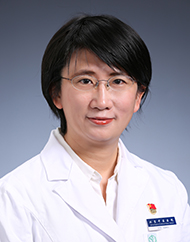 Liyun Sun (孙丽蕴)合作论文数Beijing Traditional Chinese Medicine Hospital18
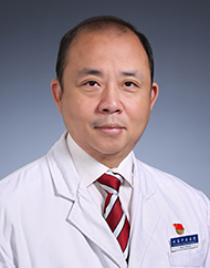 Fusheng Wang (王伏声)合作论文数Beijing Hospital of Traditional Chinese Medicine, Capital Medical University15
 Yu Wang (王雨)合作论文数14
 JianXun Dong (董建勋)合作论文数Beijing University of Chinese Medicine12Yu Dong (董雨)合作论文数11Yi Ding (丁毅)合作论文数11YuDong Kang (康煜冬)合作论文数8XiangLong Jia (贾湘隆)合作论文数6
 Bohua Yang (杨博华)合作论文数Dongzhimen Hospital, Beijing University of Chinese Medicine6Min ZHOU (周敏)合作论文数5
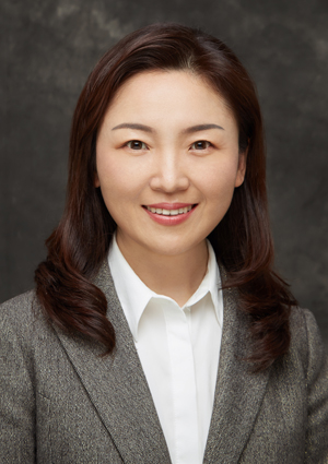 Peijing Rong (荣培晶)合作论文数Institute of Acupuncture and Moxibustion, China Academy of Chinese Medical Sciences5
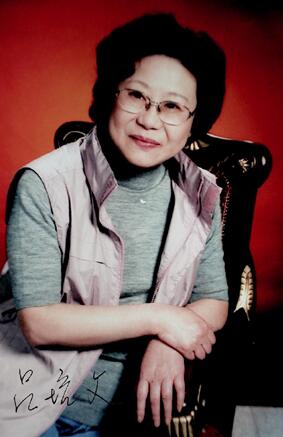 Peiwen Lv (吕培文)合作论文数北京中医医院5Yanping ZHU (朱艳萍)合作论文数4JianXia Meng (孟建霞)合作论文数4LeiYong Wang (王雷永)合作论文数4Wei You (游伟)合作论文数4Feng Huang (黄凤)合作论文数4Yong Li (李勇)合作论文数4
 Xingwu Duan (段行武)合作论文数Dongzhimen Hospital, Beijing University of Chinese Medicine4
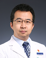 Cang Zhang (张苍)合作论文数Beijing Hospital of Traditional Chinese Medicine, Capital Medical University4
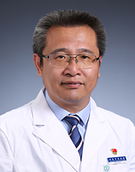 Yuguang Wang (王玉光)合作论文数Beijing Traditional Chinese Medicine Hospital4Jianhua QU (曲建华)合作论文数3TAN Chongfu (谭崇赋)合作论文数3Shurui XIE (谢书睿)合作论文数3Xuhui Zhang合作论文数3PeiWen Lv (吕培文)合作论文数3HuanJie Yang (杨焕杰)合作论文数3
 Jinxi Zhao (赵进喜)合作论文数Dongzhimen Hospital, Beijing University of Chinese Medicine3
 XiuJuan He (何秀娟)合作论文数Beijing Institute of Traditional Chinese Medicine3Ping Wang (王萍)合作论文数Beijing Friendship Hospital, Capital Medical University3
 Mingjing Zhao (赵明镜)合作论文数Dongzhimen Hospital, Beijing University of Chinese Medicine;Institute of Cardiovascular Disease, Beijing University of Chinese Medicine3weichi liu (刘威池)合作论文数2Tiantian Li合作论文数2jian li (李健)合作论文数2GUO Hui (郭卉)合作论文数2WU Li (吴黎)合作论文数2guisheng wang (王贵生)合作论文数2Xun WU (吴珣)合作论文数2Xiaoli KONG (孔晓丽)合作论文数2Lin Li (李琳)合作论文数2JingNa Xu (徐景娜)合作论文数2Yuan Meng (孟元)合作论文数2BingXu Deng (邓丙戌)合作论文数2WeiWen Chen (陈维文)合作论文数2WeiHai Lou (娄卫海)合作论文数2




