目的 研究肝脏硬度对肝细胞癌(HCC)射频消融疗效的影响.方法 2014年10月至2017年2月行超声引导射频消融治疗HCC患者,共106例114个结节.治疗前采用肝纤维化扫描仪(FibroScan)对患者肝脏行硬度测量,根据检测记录,将患者分为高LSM组和低LSM组.统计分析两组患者的疗效差异.结果 治疗后1个月复查,高LSM组与低LSM组分别有67和41个结节完全灭活,两组结节完全灭活率分别为94.4%和95.3%(P=0.88);随访期间,高LSM组和低LSM组分别有13个、3个结节发生局部复发,局部复发率分别为16.9%、7.0%;高LSM组和低LSM组患者分别有18例、4例发生肝内复发,肝内复发率分别为28.4%、10.3%,两组间差异均有统计学意义.结论 肝脏硬度是HCC射频消融治疗后发生局部复发和肝内复发的影响因素.
Objective The study was designed to comparatively analyze ultrasonic appearance,laboratory and pathological findings in autoimmune hepatitis(AIH),and their correlation.The clinical value of ultrasound in diagnosing autoimmune hepatitis was studied.Methods By retrospectively reviewing the ultrasonic images,pathological and laboratory results of 68 patients with autoimmune hepatitis admitted to the PLA 302 Hospital from 2014 to 2015,we tried to reveal the correlation between ultrasonic features and pathological and laboratory findings.Results The ultrasonic diagnosis of liver fibrosis was not statistically correlated with the features of pathological ″interface hepatitis″ and serological liver function test.But it was positively correlated with the features of pathological ″spotty necrosis″(r=0.5099,P < 0.001).The ultrasonic features of ″cord-like structure″ and ″nodular change″ had statistically improved the classification of the degree of AIH fibrosis on ultrasonic diagnosis(t=3.9547,P < 0.01).The ultrasonic feature of the change of ″liver size″ and ″morphology″ also have statistically improved the diagnosis of AIH fibrosis with ultrasonography(t=2.070,2.137,4.584,3.773,all P<0.05).Conclusion Ultrasonic images could provide objective evidence in diagnosis of AIH and evaluation of fibrosis degree.
Objective Through the study of color Doppler ultrasound imaging, to explore the mechanism of portal vein thrombosis for patients with liver cirrhotic portal hypertension splenectomy and its influencing factors. Methods From January 2013 to December 2013, a total of 284 cases of patients with cirrhosis and portal hypertension plenectomy plus pericardial devascularization in 302 Hospital of PLA were reviewed. Color Doppler ultrasound technique was employed to record the thrombosis of portal vein, splenic vein and superior mesenteric vein before and after the surgical procedure. According to the occurrence of portal vein thrombosis after splenectomy, patients were divided into portal vein thrombosis group and non portal vein thrombosis group; the factors that may affect the formation of portal vein thrombus were analyzed by using Logistic regression. Results One hundred and twenty-nine cases of portal vein thrombosis occurred among the 284 patients with splenectomy, the incidence rate was 45.42% (129/284). Logistic regression analysis shows that inner diameters of splenic vein for the group with portal vein thrombosis are significantly different from that of the group without portal vein thrombosis (Z=2.034, P < 0.05), postoperative inner diameter of portal vein (Z=2.037, P<0.05), and prothrombin time (Z=-2.171, P<0.05) are significantly higher in the group with portal vein thrombosis, while preoperative platelet count is significantly lower in the group with portal vein thrombosis (Z=-2.146, P < 0.05); gender, preoperative hepatic artery flow velocity, and blood coagulation time are also the influencing factors of portal vein thrombosis (all P>0.05). Conclusion Color Doppler ultrasound technology can not only monitor, the thrombus formation in portal venous system post splenectomy, it can assess the risk factors of portal vein thrombosis post splenectomy.
目的:探讨肝细胞癌(HCC)患者术前血清 miR-221水平与 R0切除术后患者生存和早期复发的关系。方法选取2009年7月至2011年8月在我院行 R0切除术治疗的78例 HCC 患者,采用实时荧光定量 RT-PCR 法检测血清 miR-221水平,并分析血清 miR-221水平与 HCC 患者临床和病理特征的关系。结果38例高分化 HCC 患者血清 miR-221水平为(6.4±2.2),而39例低分化患者为[(11.5±4.3),P<0.05],13例血清 AFP<20μg/L 患者血清 miR-221水平为(5.9±3.6),而65例血清 AFP>20μg/L 患者为[(9.6±2.9),P<0.05],而在不同性别、年龄、肝硬化有无、肿瘤大小、Child- Pugh 分级间无明显差异(P>0.05);78例 HCC 患者在 R0切除术后血清 miR-221相对水平(3.49±0.73)较术前(8.95±1.57)显著降低(P<0.05);高水平组1 a 生存率为45.3%(24/53),低水平组为72.0%(18/25,P<0.05);高水平组平均疾病进展时间(TTP)和中位生存期(MOS)分别为10.7个月和14.2个月,而低水平组则分别为15.3个月和19.2个月,两组比较差异有统计学意义(P<0.05)。结论血清 miR-221水平与 HCC患者临床和病理特征密切相关,其术前血清高水平提示 HCC 患者在 R0切除术后仍预后不良。
目的:自身免疫性肝病的发病机理至今尚未明确,与多种疾病之间存在着联系,临床诊断具有一定的难度.本研究利用超声弹性成像技术定量分析自身免疫性肝病的病理特征,探讨该技术的诊断价值,为自身免疫性疾病的治疗提供诊断依据.方法:选取我院2011年3月-2013年6月收治的自身免疫性肝病患者182例,随机分为观察组和对照组.观察组98例患者采用弹性成像定量分析的方法进行诊断,对照组84例则采用常规病理学诊断.观察并比较两组患者的诊断准确率及诊断耗时.结果:观察组检出94例,诊断率为95.92%;对照组检出81例,诊断率为96.43%,两组诊断准确率比较无明显差异(P>0.05).观察组平均诊断耗时为5.6h;对照组平均诊断耗时为11.6h,观察组诊断时间比对照组短,差异具有统计学意义(P<0.05).结论:弹性成像定量分析对自身免疫性肝病的诊断准确率与病理活检诊断具有较好的一致性,且诊断耗时短、患者依从性好,值得在临床中进一步推广应用以辅助病理诊断,避免误诊或漏诊现象发生.
Objective: To explore the expression and correlation of XIAP and survivin in primary hepatocellular carcinoma(HCC). Methods: 60 patients with HCC treated in our hospital were selected, and the expression of XIAP and survivin in HCC tissues and adjacent mucosa were detected by immunohistochemistry staining. Results: Comparatively, the positive rate of XAIP and survivin in HCC tissues were significantly higher than that of adjacent mucosa, the difference was statistically significant. The expression intensity of XIAP and survivin had nothing to do with the tumor size, but it expressed highly with the decrease of differentiation degree of tumor, and the differences between different differentiation degree were statistically significant; positive correlation existed between XIAP and survivin. Conclusion: High expression of XIAP and survivin in tumor tissues can promote the incidence, proliferation, metastasis and drug resistance of tumor, as well as reduce the tumor differentiation degree, increase the malignant grade of liver cancer. In addition, there are possible synergies between XIAP and survivin, but their correlation and mechanism needs further exploration.
Objective: To study the dynamic expression characteristics of HIF-1α in HCC tissues and its clinical significance. Methods: WB, IHC and PCR methods were used to determine and compare the positive expression and expression quantity of HIF-1α in different liver tissues. The relationship between the strong positive expression of HIF-1α and clinical pathology was analyzed. Results: The negative rate of HIF-1α in normal liver tissues was much higher than that in other groups; the strong positive rate of HIF-1α in HCC necrosis tissues was the highest. The expression of HIF-1α in normal liver tissues was much less than that in other groups; the HIF-1α expression in HCC necrosis tissues was the highest and clearly higher than HCC liver tissues. The strong positive expression of HIF-1α in group with low differentiation was higher than that in high differentiation group, and the strong positive expression in group with metastasis was higher than that in non metastasis group. The differences were statistically significant(P0.05). Conclusion: The expression of HIF-1α in liver cirrhosis and HCC tissues was all higher than normal liver tissues. HIF-1α expression is associated with tumor differentiation degree and HCC metastasis, but is not related to cancer embolus, expression of HBsAg and prognosis, which provides a new idea for HCC treatment.
<正>患者男,69岁,上腹部胀满不适10余天入院。患者于2011年1月2日无明显诱因出现高热,体温40℃,无寒战,于当地卫生所输液治疗2 d后逐渐出现右上腹部胀满不适,影响进食。患者既往有牧区接触史、不洁饮食史,在当地医院住院期间行腹部CT检查可见直径约20 cm的囊实性组
Objective: To investigate the expression of Hepatitis B Virus(HBV)-specific miRNAs in serum of HBV associated hepatocellular carcinoma(HCC) patients,and to demonstrate its relationship with clinicopathologica1 features and prognosis of HCC.Methods: The expression levels of HBV-specific miRNAs(miR-122 and miR-22) in the serum were measured by reverse transcription polymerase chain reaction(RT-PCR).The correlations between serum HBV-specific miRNAs expression and clinicopathological features and prognosis were analyzed.Results: HBV associated HCC patients had significantly higher miR-122 and miR-22 levels than benign diseases and healthy controls.The expression of miR-122 and miR-22 in postoperative serum was significantly different from that of preoperative serum.The expression level of serum miR-122 and miR-22 had a close correlation with HBV DNA,liver cirrhosis,AFP level,tumor size,tumor thrombi,histological differentiation,TNM classification(P<0.05).The survival time of HCC patients with a lower miR-122 and miR-22 expression levels were longer than those with a higher 1eve1,and the recurrent/metastatic of HCC patients with a lower miR-122 and miR-22 expression levels were also lower than those with higher expression(P<0.01).Conclusion: The up-regulated expression of serum miR-122 and miR-22 are correlated with poor prognosis of HBV associated HCC,indicating that miR-122 and miR-22 can be used as a prognostic marker in HBV associated HCC patients.
Objective: To investigate the expression of miRNA-181 in normal human hepatocytes(L02),hepatpcellular carcinoma cell lines(SMMC722 and Hep3B),normal liver tissues and hepatocellular carcinoma tissues for explore the role of miRNA-181 in hepatocellular carcinoma(HCC).Methods: MiRNA181,including miRNA-181a/181b/181c/181d in normal human hepatocytes(L02),hepatpcellular carcinoma cell line(SMMC722 and Hep3B),normal liver tissues and hepatocellular carcinoma tissues were tested by florescent real-time quantitative PCR.Results: Compared to in the normal hepatocytes(L02),the level of miRNA-19b was markedly increased in SMMC722 and Hep3B(P<0.05 or P<0.001).Compared to in normal liver tissues,the level of miRNA-19b was markedly increased in hepatocellular carcinoma tissues(P<0.05).Conclusions: The expression of miRNA-19b was different in various hepatpcellular carcinoma cell lines,and increased than normal hepatocytes.The expression of miRNA-19b in hepatocellular carcinoma tissues was increased than in normal liver tissues.It probably involved in the progress of HCC and was the marker of diagnosis and prognosis of hepatocellular carcinoma.
Objective: To study the effects of miRNA-19b through observing the expression of miRNA-19b in the liver on hepatic fibrosis(HF) rats.Methods: 20 rats were randomly divided into the normal group,HF group.The HF model in rats was duplicated with Improved Composite Factor Method.The liver tissues were taken after 6 weeks and 8 weeks.MiRNA-19b was tested by florescent real-time quantitative PCR.Results: Compared to the normal control rats,the level of miRNA-19b was markedly decreased in HF rats after 6 weeks(P<0.05).besides that,the level of miRNA-19b was more prominently decreased in HF rats after 7 weeks than 6 weeks(P<0.05).Conclusions: MiRNA-19b was decreased in liver tissues of hepatic fibrosis rats.It probably involved in the progress of HF and was the marker of diagnosis and prognosis of HF.
Objective: To isolate hepatocarcinoma stem cells from MHCC97 cell lines and investigate the expression characteristics of miR-221 in different subpopulations of MHCC97 cell line of human hepatocellular carcinoma(HCC).Methods: By using flow cytometry and Hoechst 33342 dye eglux assay,hepatocareinoma stem cells(HSCs) and non-hepatocareinoma stem cells(non-HSCs) were isolated from MHCC97 human hepatoma cell.The expression levels of miR-221 in different subpopulations of MHCC97 cell line were measured by reverse transcription polymerase chain reaction(Real-time RT-PCR).Results: With the Hoechst 33342 extrusion assay.HSCs were found to account for 1.89% of the MHCC97 cell.The expression of miR-221 in HSCs was significantly higher than in non-HSCs(P<0.01).Conclusion: The significantly up-regulated expression of miR-221 in HSCs of MHCC97 cell line indicated that miR-221 may be involved in the maintenance of the stem cell property in hepatocareinoma stem cells.We could induce differentiation of HSCs through regulation the expression of miR-221,which will offer a possible therapeutic strategy for the treatment of hepatocellular carcinoma.
Objective: To investigate the expression and clinical signficance of Beclin 1 in patients with hepatocellular carcinoma.Methods: The expression of Beclin 1 protein was detected with immunohistochemical analysis in 54 cases of HCC and pericarcinoma tissues and the relationship between the expression of Beclin 1 with the clinical pathological factors were explored.Results: The positive expres-sion rates of Beclin 1 protein in HCC were significantly lower than that in pericarcinoma tissues(x2=7.53,P0.01).The expression of Beclin 1 were positively related to TNM type,tumor differentiation,venous invasion and lymph node metascasis(P0.01).Conclusions: The expression of Beclin 1 protein in hepatocellular carcinoma decreased.Beclin 1 may act as marker predicting tumorigenesis,develop-ment and prognosis of hepatocellular carcinoma.
Objective: To investigate the expression of miR-221 in serum of hepatocellular carcinoma(HCC) patients after liver transplantation,as well as their clinical significance.Methods: The serum samples were collected from 42 patients with HCC that following liver transplantation(LT),26 patients with benign disease and 40 normal subjects.The expression levels of miR-221 in the serum were measured by reverse transcription polymerase chain reaction.All 42 patients were divided in two groups(high expression group and low expression group) according to the mean value of the expression levels of miR-221.Results: HCC patients had significantly higher miR-221 levels than healthy controls(P<0.05).The serum miR-221 levels in patients that received liver transpla-ntation were significantly decreased(P<0.05).The expression of serum miR-221 in preoperative patients had close correlations with the recurrent/metastatic of HCC.The recurrent/metastatic of HCC patients following liver transplantation with a lower miR-221 expression level was also lower than those with higher expression(P<0.05).The serum miR-221 levels of patients that recurred HCC in 6 months postoperative were significantly higher than that of patients without recurrence(P<0.05).Conclusion: The expression of miR-221 in serum had close correlations with the recurrent/metastatic of HCC patients that followed liver transplantation,indicating that miR-221 could be used as a predictive marker for HCC recurrence and prognostic factor of HCC patients after LT.
Objective: To investigate the expression of miRNA-29a in sera of the normal subjects and patients with primary liver cancer,and the expression of miRNA-29a in normal liver tissues and hepatocellular carcinoma tissues in patients.Methods: MiRNA-29a in sera of normal subjects and patients with primary liver cancer.At the same time,the normal liver tissues and hepatocellular carcinoma tissues of patients were collected and detected by using florescent real-time quantitative PCR.Results: 1) Compared with the normal subjects,the level of miRNA-29a had no obvious change in sera of patients with primary liver cancer(P0.05).2) Compared with the normal liver tissues,the level of miRNA-29a was markedly decreased in hepatocellular carcinoma tissues in the patients with primary liver cancer(P0.01).Conclusion: The expression of miRNA-29a in hepatocellular carcinoma tissues was decreased than in normal liver tissues of patients with primary liver cancer.But there was no obvious change in the sera of normal subjects and of patients.
目的:应用血管回声跟踪技术(echo-tracking,ET)评价慢性肾功能衰竭患者股总动脉内皮功能,并探讨该技术治疗慢性肾衰患者心血管并发症的临床意义.方法:选择2012年1月至12月在我院就诊的82例慢性肾功能衰竭患者为观察组,另选47例健康体检者为对照组.应用ET技术检测两组患者的左右侧股总动脉血管弹性指标,包括压力应变弹性系数(Ep),脉搏波传导速度(PWVβ),增大指数(AI),硬度指数(β)和脉顺应性(AC).观察两组患者的检测结果并进行比较.结果:观察组中,压力应变弹性系数(Ep),脉搏波传导速度(PWVβ),增大指数(AI)和硬度指数(β)均高于对照组所对应的值;而脉顺应性(AC)值低于对照组,两组差异有统计学意义(P<0.05).观察组中,需要透析的患者组β、PWVβ、Ep和AI的值均高于非透析患者组对应的值,AC值低于非透析患者组的值,差异有统计学意义(P<0.05).结论:ET对慢性肾功能衰竭患者心血管并发症的早期治疗,改善患者的愈后具有一定的临床指导意义.
Objective: To evaluate the values of liver fibrosis staging of chronic hepatitis B by elastic characteristic quantities and the elasticity ratings in Real-time Tissue Elastography(RTE).Methods: The liver elasticity ratings of 140 patients with chronic hepatitis B patients were detected by RTE.The liver tissue elasticity characteristics: The average relative strain(Mean),the standard deviation of relative strain value(SD),the percentage of blue in solving area(%Area),the degrees of Blue area mixed with other colors(Comp) were determined and compared with liver biopsy pathological staging.Results: There were positive 112 cases and negative 28 cases in the liver elasticity ratings.The sensitivity and specificity of the qualitative diagnosis of liver fibrosis by RTE elasticity ratings were 96.26% and 72.73%,respectively.The correlations of Mean,SD,% Area and Comp contrast with pathological diagnosis were better than those in the conventional ultrasound(Mean: r=-0.838,P0.05;%,Area: r=0.864,P0.05).The sensitivity(RTE: 96.26% VS conventional: 64.49%)and specificity(RTE: 72.73% VS conventional: 33.33%)in the elastic ratings were much higher than those in the conventional ultrasound.Conclusion: RTE provides a new method for the evaluation of liver fibrosis,gave the liver fibrosis diagnosis means a new supplement.
Objective: To investigate the expression of miRNA-492 in normal human hepatocytes(L02),hepatpcellular carcinoma cell lines(HepT1and SMMC-7221),normal liver tissues,hepatocellular carcinoma tissues and metastatic hepatoblastoma tissues for explore the role of miRNA-492 in different types of hepatpcellular carcinoma.Methods: MiRNA-492 in normal human hepatocytes(L02),hepatpcellular carcinoma cell line(HepT1 and SMMC-7221),normal liver tissues,hepatocellular carcinoma tissues and metastatic hepatoblastoma tissues were tested by florescent real-time quantitative PCR.Results: 1) Compared to that in the normal hepatocytes(L02),the level of miRNA-492 was markedly increased in HepT1 cell(P<0.001).But the level of miRNA-492 was in SMMC-7221 cell slightly elevated.There is no statistical difference.2) Compared to that in normal liver tissues,the level of miRNA-492 was markedly increased in metastatic hepatoblastoma tissues.But the level of miRNA-492 was slightly increased in hepatocellular carcinoma tissues,and there was no statistical difference.Conclusion: The expression of miRNA-492 was different in various hepatpcellular carcinoma cell lines.The level of miR-NA-492 was increased in hepatoblastoma cell than normal hepatocytes.Beside that,the expression of miRNA-492 was increased in metastatic hepatoblastoma tissues than in normal liver tissues.But in the primary liver carcinoma tissues,there was no obvious change,which indicated that miRNA-492 was related to the certain types of liver cancer,particularly in the development of metastatic hepatoblastoma.
Objective To discuss the complications of liver biopsy under the guidance of color Doppler ultrasound,and explore its preventive measures.Methods 2 522 cases of patients underwent liver biopsy under the guidance of color Doppler ultrasound in our hospital from January 2009 to March 2012 were selected.The incidences of postoperative complications were surveyed and analyzed.Results Among 2 522 cases of patients with liver biopsy,22 cases(0.87%) were found bleeding,including 2 cases(0.08%) of massive haemorrhage,18 cases(0.71%) of subcapsular hemorrhage,1 case(0.04%) of hemobilia,1 case(0.04%) of delayed hemorrhages.130 cases(5.15%) were found ache.Conclusion Given liver biopsy to patients with liver increase,severe jaundice or long-term cholestasis should be careful.
Objective To investigate the strategies for preventing and avoiding the risks in color Doppler ultrasound-guided liver biopsy for patients with liver diseases.Methods Acuson Sequioa 512 system (Siemens Medical Solutions,Mountain View,USA) with a 3.5-5.0 MHz probe (4V1) was used to obtain sonographic images,and Bard biopsy gun with 16-gauge and 18-gauge biopsy needles was used to obtain tissues for biopsy.Color Doppler ultrasound-guided liver biopsy was performed in the left lateral decubi-tus position,and the incidence of complications and adverse reactions was observed and recorded during and after liver biopsy pro-cedure.Results One-time biopsy success rate reached 99.09% (3397/3428),two-time biopsy success rate was 0.85% (29/3428),and biopsy failure rate was 0.06% (2/3428).Sixty-one (1.78%) patients underwent ultrasound-guided biopsy of space-occupying le-sions in the liver,and 3367 (98.22%) patients underwent ultrasound-guided biopsy of diffuse lesions in the liver.Conclusion Taking proper measures before,during and after performing ultrasound-guided automatic biopsy can effectively avoid the incidence of com-plications and adverse reactions.
 Mei Yang (杨美)合作论文数10Hui Feng (冯卉)合作论文数9Lu Xiong (熊璐)合作论文数7XiaoYu Dong (董晓宇)合作论文数7YueJuan Gao (皋月娟)合作论文数6Zheng Dong (董政)合作论文数6Min Chen (陈敏)合作论文数6Hongping Zhang (张红萍)合作论文数6
 Zhiyan Li (李志艳)合作论文数Chinese People's Liberation Army General Hospital6Jiangke Tian合作论文数5XinLi Zhang (张新力)合作论文数5Meng Li (李猛)合作论文数5Ying Su (苏颖)合作论文数5Yang Liu (刘阳)合作论文数5Xinli Zhang (张新力)合作论文数5ShuHong Liu (刘树红)合作论文数4Jin Li (李进)合作论文数4Yuan Shi合作论文数3FangLi Zhang (章方莉)合作论文数3Song Feng (冯松)合作论文数3
 RuiSheng Li (李瑞生)合作论文数Chinese PLA General Hospital3Lingxiang Yu (余灵祥)合作论文数Chinese People's Liberation Army General Hospital3Yan Wang (王彦)合作论文数2Xi Chen (陈曦)合作论文数2Xia CHEN (陈霞)合作论文数2DaKun Zhang (张大鹍)合作论文数2Yu Tang (汤雨)合作论文数2Zhong Chen (陈重)合作论文数2XiaoDong Guo (郭晓东)合作论文数2Xia Chen (陈霞)合作论文数2FeiFei Liu (刘飞飞)合作论文数2zhong chen (陈重)合作论文数2
 Yongping Yang (杨永平)合作论文数Chinese PLA General Hospital2
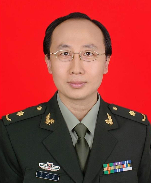 JianYu Li (李建宇)合作论文数Chinese PLA General Hospital2Anjin Zhu (朱安今)合作论文数1Xingang Zhou合作论文数1Xiaodong Guo合作论文数1xia chen (陈霞)合作论文数1Yuanyuan Zhao (赵媛媛)合作论文数1ChaoNan Guo (郭超楠)合作论文数1Ying Zhan (湛瑛)合作论文数1XuePing Yang (杨学平)合作论文数1YuLai Zhao (赵雨来)合作论文数1YongWu Li (李勇武)合作论文数1Qian Wang (王茜)合作论文数1




